

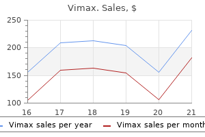
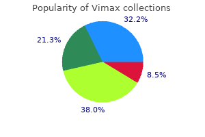
Vimax dosages: 30 caps
Vimax packs: 1 bottles, 2 bottles, 3 bottles, 4 bottles, 5 bottles, 6 bottles, 7 bottles, 8 bottles, 9 bottles, 10 bottles

Vimax 30 caps cheap on line
Nails can be abnormally hypoplastic in chondroectodermal dysplasia and brief and broad in cartilage hair hypoplasia erectile dysfunction treatment muse vimax 30 caps order otc. Club toes or equinovarus could additionally be seen in many problems erectile dysfunction insurance coverage 30 caps vimax best, together with Kniest dysplasia, spondyloepiphyseal dysplasia congenita, Larsen syndrome, more extreme forms of osteogenesis imperfecta, and diastrophic dysplasia. Congenital cardiac defects are seen in chondroectodermal dysplasia (atrial septal defect), the short-rib polydactyly syndrome problems (complex outlet defects, together with isolated ventricular septal defects) and in Larsen syndrome (ventricular septal defect). Gastrointestinal anomalies are uncommon among the many skeletal issues, however congenital megacolon could be seen in cartilage hair hypoplasia, malabsorption syndrome in ShwachmanDiamond syndrome, and omphaloceles in otopalatodigital syndrome and atelosteogenesis I. Diagnostic Tests and Diagnosis After acquiring a radical household history and physical examination, the following step is to acquire a full set of skeletal radiographs or genetic skeletal survey. A full series of skeletal views, together with anterior, lateral, and Towne views of the skull, anterior and lateral views of the entire spine, and anteroposterior pelvis and extremities, with separate views of the hands and feet, is optimal, especially after the new child period. Most of the essential clues to analysis are in skeletal radiographs which are obtained earlier than puberty. Once the epiphyses have fused to the metaphyses, figuring out the precise prognosis could be exceedingly challenging. If an adult is evaluated, all makes an attempt ought to be made to get hold of any available childhood radiographs. A, Distal femur and proximal tibia and fibula showing small irregular epiphyses (single arrow) and irregular metaphyses (double arrows). B, Lateral view of the backbone displaying abnormally rounded vertebral bodies with anterior central beaking (arrow). The defined zones of the growth plate are labeled, H, hypertrophic, P, proliferative, and R, resting. As talked about in preceding text, these radiographic abnormalities can change with age. If potential to obtain, radiographs of patients across a number of ages will assist in analysis. Fractures could be seen in osteogenesis imperfecta (all varieties; see Table 105-1) and extreme hypophosphatasia. In older people, fractures could also be seen in disorders associated with elevated mineralization, such as the osteopetrosis syndromes. Morphologic research of chondroosseus tissue have revealed particular abnormalities in lots of the skeletal dysplasias. These research must be carried out on cartilage growth plate, and though generally carried out on perinatal lethal skeletal problems at time of autopsy, acquiring progress plate histology on people with nonlethal issues is troublesome. If affected people (children) are present process surgery, an iliac crest biopsy can be evaluated, though admittedly this is performed with markedly less frequency than in the past, significantly while our molecular understanding has markedly increased. However, histomorphology studies accomplished on these issues have led to important insights on the pathogenesis of these issues. On morphologic grounds, the chondro- dysplasias can be broadly categorized into those problems that (1) have a qualitative abnormality in endochondral ossification, (2) have abnormalities in cellular morphology, (3) have abnormalities in matrix morphology, and (4) during which the abnormality is primarily localized to the realm of chondroosseous transformation. All of these findings are attribute and diagnostic for these problems and illustrates how morphology research can play in integral half within the investigation of those issues. Further, issues throughout a phenotypic spectrum have been positioned in bone households primarily based on similar cartilage development plate morphologic abnormalities. There has been vital progress in gene identification in these issues, which has impact for affected individuals. As illustrated in Table 105-1, for these issues by which the gene is identified, molecular diagnostic testing is probably obtainable. Molecular prognosis can be utilized to confirm a medical and radiographic analysis, predict provider standing in those households in danger for a recessive dysfunction, and, for some people, allow prenatal prognosis of at-risk fetuses. With commercial availability of whole-exome sequence evaluation, many excessive and low bone mass parallel sequencing panels are available, in addition to whole-exome sequence evaluation for uncommon problems not out there on panels. Treatment the optimum administration of this various set of problems requires an understanding of their medical, skeletal, and psychosocial penalties. Most medical problems in these disorders outcome from orthopedic issues, they usually differ relying on the particular disorder. This causes knee or ankle pain in many people, particularly kids, and consideration should be made for correction by osteotomies. Frequently, sufferers with these issues have vital joint ache and, in some cases, joint limitations. Because most of these disorders end result from mutations in genes critical to cartilage operate, the cartilage on the joint surfaces might not present enough assist and cushioning perform. Last, weight control in adults with quick stature is an ongoing issue and contributes to inactivity, loss of perform, adult-onset diabetes, hypertension, and coronary disease. The majority of those people are of regular intelligence, have a traditional life span, and lead unbiased and productive lives. The mean final height in achondroplasia is one hundred thirty cm for men and a hundred twenty five cm for women, and specific development charts have been developed to doc and track linear development, head circumference, and weight in these people. Clinically, these infants present central apnea, sleep apnea, profound hypotonia, motor delay, or extreme sweating. Other problems embody upper airway obstruction, thoracolumbar kyphosis, and hydrocephalous in a small number of people. Craniofacial abnormalities result in dental malocclusion, and acceptable remedy is critical. As adults, the main potential medical complication is impingement of the spinal root canals, and this might be manifested by lower limb paresthesias, claudication, clonus, bladder, or bowel dysfunction. It is crucial that these complaints are addressed because with out appropriate decompression surgical procedure, paralysis of the spinal twine may result. Biochemical and Molecular Abnormalities Based on clinical, radiographic, and histomorphologic similarities, skeletal dysplasias have been placed into bone dysplasia families. In current years, there was an explosion in our understanding of the essential biology of those problems. This has resulted from advances within the human genome project that improved varied methodology, together with candidate gene method, linkage evaluation, positional cloning, human/mouse synteny, wholeexome and whole-genome evaluation, permitting identification of the responsible disease genes (see Table 105-1). There are nonetheless skeletal dysplasias for which the disease-producing genes are unknown. Below are just some examples of the molecular mechanisms concerned within the skeletal dysplasias. Both these disorders are associated with early-onset osteoarthritis, notably of the hips and the knee, resulting in joint replacements in many people in early maturity. Mutations that lead to a substitution for a triple-helical glycine residue seem to be the most typical sort of mutation. The explosion in delineating molecular defects has shown the complexity of cartilage as a tissue and the massive variety of cellular processes necessary for a traditional skeleton.
Diseases
- Familial opposable triphalangeal thumbs duplication
- Neuroendocrine cancer
- Dementia pugilistica
- Bullous pemphigoid
- Spinal-bulbar muscular atrophy
- Salla disease
Discount 30 caps vimax with mastercard
Therefore impotence natural treatment vimax 30 caps purchase fast delivery, one sees a affected person with a drastically rising bilirubin with comparatively regular liver perform exams erectile dysfunction guide vimax 30 caps cheap on line. Another methodology of management included placement of a constant suction T tube with subsequent resolution. Drain placement at the time of laparotomy is normally indicated with obvious bile staining. It is frequent for liver injuries to have transient early postoperative serosanguinous and bilious drainage. Bilious drainage of at least 50 mL/d that continues after 2 weeks is taken into account a biliary fistula. Major left or right bile duct injury typically requires further intervention for closure. In the previous the surgical strategy was recommended with resection or Roux-en-Y procedures predominating. Percutaneous stenting of accidents and drainage of biloma collections has been utilized. At the Shock Trauma Center, Dabbs et al found that 29 of 30 sufferers encountered with major hepatic necrosis underwent initial operative intervention and 87% underwent damage management laparotomy. A massive variety of these patients then required resection of their necrotic hepatic parenchyma. Both serial debridements and formal lobectomy had been carried out, however lobectomy was associated with fewer procedures general and a decrease complication price. Often the affected person does well initially with resolution of hemothorax, no proof of jaundice, and stabilization of liver damage only to turn into significantly tachypneic per week or extra later. Rothberg et al promote operative intervention to have the ability to consider for significant diaphragmatic damage, liver necrosis, or lung necrosis with potential bronchial involvement. Technical issues including continued hemorrhage, adjacent organ damage, and small duct size can show troublesome. A well timed diagnosis and therapy method might show to be the survival difference in sufferers with these extreme accidents. In a Seattle paper, 38% were a result of blunt mechanisms, much like the 31% with blunt mechanism quoted in a 1995 multi-institutional trial. When analyzing these with both portal vein and hepatic artery injury, the mortality is 99%. Instead, a number of accidents to the liver, porta, vena cava, and surrounding viscera most often happen. The findings of an ill-defined contour of the wall, collapse of the lumen, or intraluminal hemorrhage, highly recommend blunt gallbladder harm. Ultrasound examination in gallbladder injury has not been formally evaluated, but intuitively ought to present useful details about this injury. Despite these diagnostic methods, the prognosis of gallbladder harm is most frequently secured at laparotomy, at which time cholecystectomy is the instructed therapy. The sufferers with late presentation might develop jaundice, stomach distention and pain, intolerance to enteral feeding, fever, or worsening base deficit due to bilious ascites or an infection. There could additionally be some indication of pancreatic head fullness, duodenal thickening, or portal edema but these are nonspecific findings. In the presence of bile staining throughout an operative process and no apparent harm, a cholangiogram via the gallbladder could be helpful. Ninety-seven percent of sufferers had concomitant accidents, thus illustrating the importance of full exploration and belly evaluation. A beneficiant midline incision should be made, followed by evacuation of blood clot and hemoperitoneum with urgent packing of bleeding buildings. Hematoma or bleeding around, or inside, the hepatoduodenal ligament or extreme parenchymal harm leading to the porta hepatis should increase suspicion of a portal triad damage. Bile staining must also be absolutely investigated, as 12% of bile duct accidents may be missed on the initial operation. In order to obtain adequate examination and exposure for restore, a large proper medial visceral rotation ought to be performed, which includes mobilizing the ascending and hepatic flexure areas of the colon, thus exposing the duodenum. Similarly, a full Kocher maneuver should mobilize the duodenum and head of the pancreas medially to expose the inferior vena cava. However, there have been reviews of laparoscopic cholecystectomy in penetrating trauma. Chapter 29 Liver and Biliary Tract 569 process should be done with great reserve, since many gallbladder injuries are related to other intra-abdominal injuries in both penetrating and blunt trauma. Though the laparoscope may give a great superficial examination of the peritoneal cavity, visualization of the duodenum, pancreas, and porta hepatis is, in most arms, not adequate. Minor gallbladder contusions can typically be managed nonoperatively,a hundred and forty,141 however might lead to cholecystitis or delayed rupture. Cholecystectomy must also be carried out on all sufferers with damage to the cystic duct or right hepatic artery that may get rid of the blood supply to the gallbladder. In the affected person who stays in shock and coagulopathic, packing and placement of drains in the area of the biliary injury is adequate until reexploration is performed. Four broad categories of biliary duct harm have been described: (1) avulsion of cystic duct or small laceration, (2) transection without loss of tissue, (3) extensive defect within the wall, and (4) segmental loss of ductal tissue. A T tube with a limb beneath the repair can be used; nonetheless, this can be troublesome to insert in a patient with a normal sized duct. For avulsions during which major repair might slim the lumen, a bit of the cystic duct or proximal gallbladder wall can be utilized for the repair. One should be sure to perform minimal dissection across the duct or the lacerated ends to be able to keep enough blood provide for therapeutic. Ivatury et al reported a 55% stricture price in the end-to-end anastomoses that then required enteric conversion. Saphenous vein grafts have had difficulties with shrinking and fibrosis, which then required stenting. Roux-en-Y hepaticojejunostomy with cholecystectomy and T-tube drainage is probably the most utilized approach to advanced damage. A retrocolic Roux limb of a minimal of forty cm long is created and can be brought as a lot as the common hepatic duct or even to the hilar plate, much like the Kasai procedure. An avulsion of the hepatic ducts at the bifurcation may be managed by suturing the ducts collectively medially earlier than the endto-side hepaticojejunostomy. However, the vascularity in this anastomosis is crucial and any sign of frequent bile duct vascular damage would lead the surgeon to assemble an anastomosis closer to the widespread hepatic duct. Patterns of fluid accumulation on screening ultrasonography for blunt belly trauma. Ultrasound based key medical pathway reduces the use of hospital assets for the analysis of blunt stomach trauma. Abdominal injuries with out hemoperitoneum: a possible limitation of targeted stomach sonography for trauma. Blunt abdominal trauma: emergency contrast-enhanced sonography for detection of stable organ injuries. Appearance of strong organ damage with contrast-enhanced sonography in blunt stomach trauma: preliminary expertise. Blunt abdominal trauma sufferers: can organ harm be excluded with out performing computed tomography
Buy vimax 30 caps on-line
Simultaneous rupture of the pericardium into the left and the proper pleural spaces has also been described erectile dysfunction caused by hernia buy vimax 30 caps on line, as has herniation of the guts inferiorly into the peritoneal cavity impotence workup buy 30 caps vimax amex. The organs most regularly involved in pericardial herniation are the transverse colon, stomach, omentum, liver, and small bowel. Exploratory laparotomy is really helpful as the popular strategy for the acute restore of these injuries. Once the prognosis is made, restore in the acute section can usually be completed using the surgical methods described. In the latent or obstructive phases of presentation, restore or reconstruction of the diaphragm may be a surgical challenge. If gastrointestinal obstruction, perforation or ischemia happen with a persistent post-traumatic diaphragmatic hernia, postoperative morbidity and mortality are significant. Key elements of phrenic motoneuron and diaphragm muscle development in the course of the perinatal period. Penetrating left thoracoabdominal trauma: the incidence and scientific presentation of diaphragm accidents. Occult injuries to the diaphragm: potential analysis of laparoscopy in penetrating injuries to the left decrease chest. Chronic traumatic and congenital diaphragmatic hernias: presentation and surgical management. Thoracoscopic restore of missed diaphragmatic harm in penetrating trauma: case report. The position of laparoscopy within the diagnosis and therapy of missed diaphragmatic hernia after penetrating trauma. The function of laparoscopy within the prognosis and therapy of missed diaphragmatic rupture. Biological materials for diaphragmatic restore: preliminary experiences with the PeriGuard Repair Patch. Latissimus dorsi reverse flap to substitute the diaphragm after extrapleural pneumonectomy. Use of belly wall muscle flap in repair of huge congenital diaphragmatic hernia. The management of liver accidents continues to evolve with improved modes of diagnosis and administration, both operatively and nonoperatively. However, probably the most extreme liver parenchymal and retrohepatic venous injuries as nicely as those involving the portal triad proceed remain a problem and despite technological advances, nonetheless usually lead to demise. Therefore, despite our progress in liver injury administration, many avenues for improvement stay to be explored. The artery then branches into the gastroduodenal, right gastric, and correct hepatic arteries. The correct hepatic is found within the porta hepatis often to the left of the frequent bile duct and anterior to the portal vein. It is discovered by transversely incising the peritoneum overlying the hepatoduodenal ligament, a maneuver facilitated by mobilization of the hepatic flexure of the colon toward the midline. At the hilum of the liver, the artery bifurcates into a proper (the longer branch) and a left hepatic artery. The most frequent (11%) is the aberrant superior mesenteric origin of the right hepatic artery traversing behind the duodenum. Other variants include a left hepatic artery origin from the left gastric artery (8%) and the left and right hepatic arteries arising from a superior mesenteric artery origin (9%). With these multiple variants, great care should be taken when controlling traumatic hemorrhage. The superior, center, and inferior vein branches originating from the right lobe form the right hepatic vein. In 90% of patients, the middle hepatic vein joins the left hepatic vein just earlier than draining into the inferior vena cava. Most essential is the posterior positioning of the vein when dissecting the left coronary ligament; nice warning should be used in this area to keep away from inadvertent damage. The liver is divided into two lobes by a 75� angle traversing from the gallbladder fossa posteriorly to the left facet of the inferior vena cava. Therefore, the left lobe contains the hepatic tissue to the left of the falciform ligament along with the quadrate and caudate lobes. Functional Anatomy the practical anatomy of the liver separates the liver into segments pertinent to resection. In 1953, Couinaud supplied the premise of contemporary resection planes by dividing the liver based on the distribution of the hepatic veins and glissonian pedicles. The portal vein lies posterior to the hepatic artery and bile ducts as it ascends toward the liver. At the parenchyma, the portal vein divides into a short right and a longer left extrahepatic department. Surgically, the portal vein can be approached by division of the pancreas at its neck or with a beneficiant Kocher maneuver of the duodenum toward the midline. The triangular ligaments are at the lateral extensions of the proper and left coronary ligaments. The falciform ligament with the underlying ligamentum teres attaches to the anterior peritoneal cavity. The medial portion of the coronary ligaments is the place the hepatic veins traverse and subsequently dissection on this space should be done with warning. In order to successfully visualize the liver throughout operative remedy, these ligaments must be divided and the liver fully mobilized into the sector of view. It receives the blood of the hepatic veins and in addition multiple small direct hepatic vessels. Exposure of this space can be very difficult, especially when an injury and accompanying hemorrhage make visualization very troublesome. Surgical publicity may be facilitated by extending a midline laparotomy right into a proper thoracoabdominal incision with division of the costochondral cartilage and continuation onto the best chest. The diaphragm is radially taken down with cautery, taking care to go away sufficient of a rim of diaphragm for reapproximation, and control of the inferior vena cava could be obtained in the chest. Out of 26,392 complete trauma encounters at the Shock Trauma Center from 2010 to 2013, 796 sufferers (3%) had liver accidents (252 penetrating and 542 blunt). The American Association for the Surgery of Trauma established an in depth classification system that has been broadly utilized (Table 29-1). Of utmost significance is the preliminary evaluation, including attention to airway, respiratory, and circulation. Other lifethreatening accidents may take precedence over potential internal injury in the main survey.
Vimax 30 caps discount visa
The ultrasound transducer ought to be held within the nondominant hand perpendicular to the skin erectile dysfunction doctor cape town 30 caps vimax buy free shipping. The cannulating needle is held within the dominant hand and directed on the goal vessel in actual time erectile dysfunction treatment shots order 30 caps vimax fast delivery. The tip of needle must be visualized in the image whereas the needle is being advanced. Once the vein is cannulated by the needle, the rest of the procedure is completed utilizing the Seldinger technique. Typically, the transverse view is used to identify puncture of the vessel, whereas the longitudinal view is useful to localize the needle tip and wire. The axillary vein is the distal continuation of the subclavian vein and its cannulation underneath ultrasound-guidance could be more simply carried out. Similar to imaging of the internal jugular vein, transverse and longitudinal photographs of the subclavian vein and artery are obtained before draping the area. Diagnostic paracentesis consists of acquiring a small quantity of peritoneal fluid for culture to rule out an infection and to characterize the fluid as a transudate or exudate. Therapeutic paracentesis is a method that removes a large volume of ascites (typically in extra of 2 L) to scale back intra-abdominal pressure and deal with the resulting stomach discomfort and dyspnea. Ultrasound steering for paracentesis in patients with ascites results in higher success in buying fluid compared to conventional methods, 95% versus 61%. The common area of the puncture site is localized 2�3 cm medial and 2�3 cm cephalad to the anterior iliac backbone within the left decrease quadrant. Skin preparation and sterile draping of the area and use of the ultrasound probe is performed adopted by insertion of the paracentesis needle. The paracentesis catheter is left in place to achieve removing of the specified diagnostic sample or to reduce intraabdominal stress. In the emergency department, the focused ultrasound examination is extra incessantly carried out to evaluate sufferers with suspected intra-abdominal inflammatory processes. Similarly, more advanced ultrasound methods including echocardiography are actually being used to guide the resuscitation of critically unwell patients. Of note, these surgeonperformed ultrasound examinations are currently thought-about as commonplace care in many medical settings. A potential examine of surgeon-performed ultrasound as the primary adjuvant modality for injured affected person evaluation. The status of ultrasonography training and use in general surgery residency packages. Ultrasound steerage improves the success fee of internal jugular vein cannulation. Emergency heart ultrasonography within the analysis of hemoperitoneum: a potential study. Early detection of hemoperitoneum by ultrasound examination of the right higher quadrant: a multicenter examine. Focused stomach sonogram for trauma: the educational curve of nonradiologist clinicians in detecting hemoperitoneum. Prospective proof of the superiority of a sonography-based algorithm within the evaluation of blunt stomach harm. The utility of focused belly ultrasound in blunt belly trauma: a reappraisal. Abdominal ultrasound is an unreliable modality for the detection of hemoperitoneum in sufferers with pelvic fracture. Blunt hepatic trauma: analysis with contrast-enhanced sonography: sonographic findings and medical application. The position of ultrasound in sufferers with attainable penetrating cardiac wounds: a potential multicenter study. A caveat to the efficiency of pericardial ultrasound in patients with penetrating cardiac wounds. Management of sufferers with anterior abdominal stab wounds: a Western Trauma Association multicenter trial. Role of ultrasonography in penetrating abdominal trauma: a prospective scientific examine. Appraisal of early analysis of blunt chest trauma: development of a standardized scoring system for preliminary clinical decision making. Diagnostic utility of cholescintigraphy and ultrasonography in acute cholecystitis. Revised estimates of diagnostic check sensitivity and specificity in suspected biliary tract illness. Ultrasound, computed tomography, and laboratory findings within the prognosis of appendicitis. Feasibility of emergency physician prognosis of hypertrophic pyloric stenosis using point-of-care ultrasound: a multi-center case series. Ultrasound steerage decreases problems and improves the value of care amongst sufferers present process thoracentesis and paracentesis. A practical approach to goal-directed echocardiography in the crucial care setting. Focused bedside echocardiography in the surgical intensive care unit: comparison of three methods to estimate cardiac index. Determination of cardiac output in critically unwell sufferers by twin beam Doppler echocardiography. Pleural ultrasound compared with chest radiographic detection of pneumothorax resolution after chest drainage. Chest sonography: a useful gizmo to differentiate acute cardiogenic pulmonary edema from acute respiratory misery syndrome. Prospective evaluation of a rapid trauma ultrasound examination performed by emergency physicians. Trauma ultrasound examination versus chest radiography in the detection of hemothorax. Sonographic screening of mass casualties for belly and renal accidents following the 1988 Armenian earthquake. Ultrasonographic functions after mass casualty incident caused by Wenchuan earthquake. Screening ultrasonography of two,204 patients with blunt stomach trauma in the Wenchuan earthquake. Ocular examination for trauma; scientific ultrasound aboard the International Space Station. The utility of focused evaluation with sonography for trauma as a triage device in multiple-casualty incidents during the second Lebanon struggle. Portable ultrasound for remote environments, Part I: Feasibility of area deployment. A systematic evaluate and meta-analysis of diagnostic efficiency of imaging in acute cholecystitis. Diagnostic standards and severity assessment of acute cholecystitis: Tokyo Guidelines.
European Black Alder (Black Alder). Vimax.
- Are there safety concerns?
- Sore throat, pharyngitis, and bleeding in the intestines.
- Dosing considerations for Black Alder.
- What is Black Alder?
- How does Black Alder work?
Source: http://www.rxlist.com/script/main/art.asp?articlekey=96585
Proven 30 caps vimax
The receiving hospital is obligated to report whenever a affected person was inappropriately transferred does erectile dysfunction cause premature ejaculation cheap 30 caps vimax otc. Hospitals with specialised capabilities or services are required to settle for transfers of patients who require such specialized companies erectile dysfunction treatment videos buy vimax 30 caps low price. One technique already being employed is for surgeons to refuse to take trauma calls and for hospitals to close their doors to injured sufferers. The moral and authorized ramifications of this approach are debatable and can undoubtedly be subjected to shut scrutiny. The proactive response is for well being care employees and facilities to familiarize themselves with the provisions of the law and to establish, in cooperation with one another, a trauma program that might be in compliance. Properly executed switch agreements ought to, generally, streamline the method. Managed care may also intervene with the orderly transfer of sufferers as established by a regional trauma system. Payments for emergency and after-hours transfers have been denied for lack of prior authorization, although such authorization was inconceivable to get hold of on the time. Triage to a hospital throughout the managed care system may be mandated, even though that facility may not be an authorized trauma middle. The American College of Surgeons has issued an announcement condemning such practices and inspiring cooperation of managed care and trauma methods. A mechanism for making certain early evaluation at the bedside by the suitable surgeon or emergency medication physician three. A small proportion, in all probability lower than 10%, will be either transient responders or nonresponders and will remain hemodynamically irregular despite continued expert resuscitation. If the patient has a number of accidents, suffers from vital medical comorbidity, is on the extremes of age, or has accidents requiring subspecialty (ie, neurosurgical) or complex intensive care, transfer will likely be essential. The more distant the receiving facility is, the greater the danger that the affected person will deteriorate during transfer unless something is completed to tackle the underlying downside, usually hemorrhage. In this circumstance, the native basic or subspecialty surgeon should be ready to operate and stabilize the patient before transfer. Definitive Surgery Under most circumstances a stabilizing procedure could be carried out and completed in typical fashion, such that no further therapy might be required for that particular drawback. Most reviews on harm management surgical procedure and momentary closure strategies have come from urban trauma facilities. Following a interval of rewarming and correction of hypothermia, acidosis, and a coagulopathy within the intensive care unit, the affected person is then returned to surgical procedure for definitive repair of the remaining accidents. Occasional sufferers will have such complex accidents that definitive management exceeds the technical abilities or assets of the native surgeon and hospital, but may be amenable to temporizing maneuvers, adopted by rapid transfer in an aircraft equipped as an airborne intensive care unit. Most had multisystem accidents and required operations by surgical subspecialists, as properly. Under most circumstances, the only choice is to reduce secondary mind harm with applicable ventilatory and pharmacologic maneuvers and expedite switch. Local Definitive Care Most trauma patients may be cared for at a local community hospital able to offering steady surgical care. It is important for surgeons to remember that colleagues, nursing workers, and ancillary providers should also be in a position to provide the necessary adjunctive care. Patients at the extremes of age, with a quantity of organ system involvement, the necessity for prolonged ventilator help and/or intensive care, and severe underlying sicknesses, will in all probability benefit from care in a trauma middle underneath most circumstances. It is necessary to be conscious of pertinent state statutes or switch guidelines used by the regional trauma system and to understand that in the event of misadventures, the burden of proof will rest with the physician who chooses to not transfer a affected person. Most sufferers with accidents to the spleen or liver and normal hemodynamics could be managed with out an operation. When an operation is critical, the well-trained basic surgeon is certainly fully able to performing a splenectomy or splenorrhaphy. Major hepatic injuries have the potential for overwhelming the technical capacity of the surgeon or the sources of a small blood bank, but may be amenable to easy suture repair, resectional debridement, or packing and immediate switch as beforehand noted. The drawback lies with the affected person who deteriorates unexpectedly, turns into hypotensive, and requires pressing surgery. In these circumstances, a lower threshold for early operation or transfer is suitable. Because most rural trauma is motorcar related, insurance protection is healthier than average and supports the financial stability of the hospital. Finally, acceptable native care reduces the burden on busy regional trauma facilities. Hours are inconvenient, and trauma emergencies can wreak havoc with elective schedules. The excessive incidence of substance abuse in association with traumatic occasions is properly documented and should make analysis and administration of injured sufferers tough or disagreeable. A examine out of Canada has developed an attention-grabbing evaluation of the relationship between potential entry to trauma care and actual access to trauma care, thereby distinguishing the relative contributions of trauma heart location from that of implementation of trauma transfer protocols. By pooling efforts with others in the area in frequent purpose, small hospitals profit from educational and prevention companies, improved patient transport, elevated political affect, higher monetary help, and entry to new technology. Failure to take benefit of these opportunities could consign a rural hospital and its sufferers to isolation and insufficient trauma care. Death brought on by simply correctable and comparatively minor injuries still occurs with an alarming frequency in the rural setting. Division of Medical Sciences, National Academy of Sciences/National Research Council. Trauma registry-new computer method from multifactorial analysis of a significant well being problem. The critically injured affected person: a plan for the organization of a statewide system of trauma amenities. Emergency medical providers techniques regionalization grant maps by State and Territory. A Symposium on the Illinois trauma program: a techniques method to the care of the critically injured. Most trauma consultants are convinced that an inclusive and built-in method to trauma will be essential within the Chapter 9 15. Trauma�a controllable disease within the Eighties (Fourth Annual Stone Lecture, American Trauma Society). A population-based study of the association of medical manpower with county trauma dying rates within the United States. Major trauma sufferers transferred from rural and distant Western Australia by the Royal Flying Doctor Service. Rural motorcar crash mortality: the position of crash severity and medical resources. Changing epidemiology of injury-related pediatric mortality in a rural state: implications for harm management. Analysis of preventable trauma deaths and inappropriate trauma care in a rural state.
Discount vimax 30 caps overnight delivery
It can present months after an episode of fungemia erectile dysfunction treatment options injections vimax 30 caps discount with mastercard, and scientific manifestations are inclined to erectile dysfunction treatment supplements buy cheap vimax 30 caps online be milder than bacterial infections at the similar websites. Bone changes of osteomyelitis are generally demonstrated in radiographs of the symptomatic site. The diagnosis is established when culture of involved bone obtained by both open or needle biopsy has identified quite a lot of Candida species. Candida albicans causes the majority of infections, however the position of nonalbicans species has been increasing lately. For example, Candida glabrata frequently demonstrate reduced susceptibility to azole antifungal brokers and amphotericin but remain susceptible to echinocandins; Candida krusei are often immune to azoles and will show reduced susceptibility to amphotericin but in addition stay susceptible to echinocandins; Candida lusitaniae are often resistant to amphotericin; and Candida parapsilosis might demonstrate decreased susceptibility to echinocandins. With vertebral involvement however no neurologic issues, medical remedy alone has been effective. Some concerned joints have been previously affected by arthritis, and an infection has followed arthrocentesis in isolated circumstances. Histologic research of synovium present nonspecific continual inflammation somewhat than granulomas. It mostly involves the pores and skin and lymphatics however may disseminate from the lungs to the central nervous system, eyes, bones, and joints. Sporotrichosis most frequently occurs in individuals with a persistent illness that alters host protection, such as alcoholism or a myeloproliferative dysfunction. Sporothrix arthritis is most often indolent and infects a single joint or a number of joints in equal proportions. The knee, hand, wrist, elbow, and shoulder are most frequently involved; hand and wrist involvement distinguishes this from different fungal arthritides. Articular an infection shows a propensity to spread to adjacent soft tissues, forming draining sinuses. Radiographic modifications differ from juxta-articular osteopenia to the generally noticed punched-out bone lesions. Synovitis is characterized on gross evaluation by destructive pannus and on microscopic examination by granulomatous histologic features or, less incessantly, by nonspecific inflammation. Organisms are tough to establish in tissue, and the prognosis is often made by constructive tradition of joint fluid or involved tissue. In a small variety of circumstances, sporotrichosis could disseminate to trigger a doubtlessly deadly infection characterized by lowgrade fever, weight reduction, anemia, osteolytic bone lesions, arthritis, skin lesions, and involvement of the eyes and central nervous system. In forty four circumstances reported in 1979, remedy was optimum with mixed joint d�bridement and high-dose intravenous amphotericin B (11 of 11 cured) and slightly less efficient with amphotericin alone (14 of 19 cured). In distinction, fluconazole has demonstrated solely modest success in osteoarticular sporotrichosis. In distinction, invasive an infection is an important life-threatening complication in immunocompromised adults and youngsters. A, Aspergillus might unfold immediately from the lung to adjacent vertebrae, disk areas, and ribs (more often in children) or through the bloodstream. Treatment with combined surgical d�bridement and antifungal remedy is an ongoing challenge. In 2012, an outbreak of fungal infections, initially believed to be due to Aspergillus fumigatus but later attributed to the not often pathogenic fungus Exserohilum rostratum, was traced to contaminated injectable methylprednisolone produced by a single compounding pharmacy in Massachusetts. Liposomal amphotericin B followed by itraconazole is the popular therapy for extreme an infection and itraconazole for much less severe cases. They could trigger focally invasive and disseminated infection after cutaneous inoculation. Scedosporium prolificans has a predilection for bone and cartilage, leading to both septic arthritis and osteomyelitis. Infections are troublesome to eradicate with surgical procedure and antifungal brokers, and the organism is proof against amphotericin. More latest advances embrace the event of less poisonous formula- tions of amphotericin B, liposomal amphotericin B, and amphotericin B lipid advanced. Voriconazole and posaconazole, which are broad-spectrum azole antifungals, have demonstrated improved activity against aspergillosis and mucormycosis, respectively. The echinocandin antifungal brokers caspofungin, micafungin, and anidulafungin have emerged as various therapies for aspergillosis and as the remedies of selection for some Candida infections. A loading dose of itraconazole of 200 mg thrice a day for two days is beneficial, adopted by 200 mg to 400 mg day by day. Absorption of itraconazole is unpredictable and blood ranges of itraconazole ought to be measured to guarantee enough drug exposure. Absorption of itraconazole requires abdomen acid, so concurrent administration of medicine that cut back the acidity of the stomach corresponding to proton pump inhibitors and H2 blockers must be avoided. At least 6 months of therapy is required, and some patients may have so long as a year of remedy. Specific treatment protocols and detailed facet effect profiles are offered in critiques,93-102 Infectious Diseases Society of America tips,12,32,forty one,51,seventy five,87 and infection-specific references (see Table 112-2). In the biggest revealed collection, 12 of 13 cases followed remedy with infliximab, and 1 was associated with etanercept. In one medical center included on this collection, the relative threat of coccidioidomycosis infection was 5. Histoplasma antigen could additionally be recognized within the urine in 92% of patients with disseminated histoplasmosis, and this take a look at may facilitate rapid analysis. Whether this pertains to decreased threat with these agents or smaller numbers of sufferers treated remains to be seen. Elevated serum ranges of -D-glucan, a common component of fungal cell walls, may assist within the diagnosis. Radiographic findings may be difficult to distinguish from methotrexate pneumonitis. Patients should be cautioned to reduce publicity to sources of infection during remedy. There is, as but, no sensible position for screening for latent an infection or for prophylactic therapy. Holley K, Muldoon M, Tasker S: Coccidioides immitis osteomyelitis: a case collection evaluate. Coccidioidal synovitis: medical, diagnostic, therapeutic, and prognostic issues. Durkin M, Estok L, Hospenthal D, et al: Detection of Coccidioides antigenemia following dissociation of immune complexes. Lerch K, Kalteis T, Schubert T, et al: Prosthetic joint infections with osteomyelitis as a outcome of Candida albicans. Candida arthritis: diagnostic and prognostic implications and therapeutic considerations.
Generic vimax 30 caps on line
Iatrogenic injuries are commonly successfully managed nonoperatively diabetic erectile dysfunction icd 9 code best vimax 30 caps, whereas surgical repair remains the mainstay of for exterior trauma erectile dysfunction after drug use 30 caps vimax discount with amex. Exposure of the cervical esophagus is achieved by way of the left neck with the incision positioned alongside the anterior border of the sternocleidomastoid muscle. Lateral retraction of this muscle and blunt dissection in the avascular prevertebral aircraft will enable a Penrose drain to encircle the esophagus reaching 360� publicity. As opposed to the trachea, the blood provide to the esophagus programs longitudinally within the submucosa allowing for full mobilization. During dissection care should be exercised to avoid injuring the recurrent laryngeal nerves that lie in the tracheoesophageal grove. It is important to remember the esophagus lacks a serosal layer and due to this fact is extra vulnerable to a postoperative leak. A basic principle in repairing esophageal injuries is visualizing the whole extent of the mucosal injury. The defect in Diaphragm Pericardium the muscular layer is kind of at all times much less in depth than that within the mucosa. The extent of the mucosal defect is exposed by incising the muscular layer till both ends of the mucosal tear are visualized. Intrathoracic esophageal injuries are associated with extra severe sequelae and require extra intensive publicity. The operative principles are preservation of esophageal length, primary restore, buttressing the repair, wide drainage, antibiotics and enteral feeding entry. With the exception of its most distal segment, the intra-thoracic esophagus is approached via a proper posterolateral thoracotomy. The mediastinal pleura broadly opened exposing the esophagus which is mobilized by blunt dissection. Muscularis is incised superiorly and inferiorly to enable visualization of the extent of mucosal defect before two-layer closure of the perforation if possible. A contrast study is carried out approximately 1 week postoperatively, and if no leak is famous, oral feedings are began. There are several surgical approaches to the distal esophagus or esophagogastric junction together with a left sixth or seventh interspace posterolateral thoracotomy, laparotomy, or a thoracoabdominal approach. Injuries to the distal intrathoracic esophagus are best approached by way of thoracotomy. The choice of incision to expose the esophagogastric junction is influenced by the precise location of the damage and related injuries. If there are concomitant stomach injuries, laparotomy alone could also be sufficient, whereas related intrathoracic injuries could additionally be approached by thoracotomy or thoracoabdominal incision. Regardless of the placement, a major buttressed restore and sufficient mediastinal drainage is the optimal treatment. Esophageal excision and resection with diversion ought to be averted, and each effort made to preserve esophageal size. Creating a managed esophageal fistula by using a T-tube is an effective procedure. Devastating harm to the abdomen and esophagogastric junction presents a novel challenge. Continuity is reestablished by an esophagojejunostomy carried out several months later. Management of traumatic lung harm: a western trauma affiliation multicenter evaluate. Penetrating thoracic nice vessel harm: influence of admission hemodynamics and preoperative imaging. Can routine trauma bay chest x-ray be bypassed with an prolonged centered evaluation with sonography for trauma examination Occult pneumothorax within the blunt trauma patient: tube thoracostomy or remark Management of pulmonary contusion and flail chest: An Eastern Association for the Surgery of Trauma apply management guideline. Flail chest accidents: A evaluate of outcomes and treatment practices from the nationwide Trauma Data Bank. Surgical publicity of the left subclavian artery by median sternotomy and left supraclavicular extension. Lung-sparing surgery after penetrating trauma using tractotomy, partial lobectomy, and pneumonorrhaphy. Stapled pulmonary tractotomy: a fast way to management hemorrhage in penetrating pulmonary accidents. Total pulmonectomy in trauma: a still unresolved problem-our expertise and evaluate of the literature. Contemporary endotracheal tubes with high volume, low stress cuffs have virtually eliminated this complex drawback among ventilated patients. Combined airway and esophageal accidents, repaired with out muscle interposition, are in danger to develop a tracheoesophageal fistula. The scientific presentation initially may be refined, with symptoms ascribed to other conditions, particularly respiratory. Recurrent pneumonia and persistent cough with unidentified cause alone warrants analysis in the appropriate patient. Once identified, remedy generally requires surgical intervention, with fistula ligation, restore of the respective tracheal and esophageal defects and muscle interposition. Ideally this is accomplished as quickly as the patient is liberated from mechanical air flow, as the operative restore is more complex on mechanical help. As described earlier, with a mixed airway and esophageal damage, a precise technical restore of every individual construction and muscle interposition will tremendously cut back the change of this complication. Experience with ultraflex expandable metallic stents within the management of endobronchial pathology. Evaluation of penetrating accidents of the neck: potential examine of 223 patients. Role of flexible endoscopy in the evaluation of possible esophageal trauma after penetrating injuries. T-tube intubation in the administration of late traumatic esophageal perforations: case report. Prospective multicenter trial of esophageal Z-stent placement for malignant dysphagia and tracheoesophageal fistula. Utility of removable esophageal lined self-expanding steel stents for leak and fistula management. Survival after trauma pneumonectomy: the pathophysiologic stability of shock resuscitation with proper coronary heart failure. Empyema thoracis in patients present process emergent closed tube thoracostomy for thoracic trauma. Predicting the need for thoracoscopic evacuation of residual traumatic hemothorax: chest radiograph is insufficient. Delayed problems of emergency airway administration: a research of 533 emergency division intubations. Mattox the guts and its tributaries are encased within the thoracic skeleton composed of the manubrium, sternum, clavicles, rib cage, and vertebral our bodies.
Vimax 30 caps purchase
Morning stiff ness is prominent erectile dysfunction pills sold at gnc vimax 30 caps generic with mastercard, and symptoms usually resolve during a quantity of days to 2 weeks erectile dysfunction drugs injection discount 30 caps vimax with visa. Proximal interphalangeal, metacarpo phalangeal, wrist, elbow, ankle, and knee joints are most incessantly affected. Following vaccination, joint signs happen much less frequently and are normally less extreme and of shorter duration than people who occur follow ing naturally acquired rubella. Persistent rubella infection could additionally be established in syno vial cell cultures and, as described, in tissues. Persistence of rubella virus can be suggested by earlier studies, which demonstrated that virusspecific IgM might persist for so lengthy as four years following each natural infection and vaccina tion. It could additionally be clinically confused with parvovirus B19 infection, but serologies differentiate the 2 infections. Predictors of nonrecovery embrace patient age older than 45 years, severe preliminary joint pain, and presence of underlying osteoarthritis. Pain inside or round tendons was additionally a typical trait and developed to tenosynovitis. Some patients with persistent rheumatic signs manifested progressive erosive arthritis. It will stay to be determined whether or not this autoimmune profile is more outstanding in recovered Pogosta sufferers versus those with longterm arthritis. The virus is shed into the saliva of persistently infected individuals who spread the virus to uninfected individuals. Studies of this virus have made huge leaps forward for the explanation that implementation of efficient cell tradition techniques. Acute an infection goes together with delicate and generalized symptoms, and subsequently mostly stays undiagnosed. In greater than half of the sufferers, nevertheless, the infection per sists and, through the years, may cause liver harm similar to fibrosis, cirrhosis, or hepatocellular carcinoma. Viral antigen is detectable throughout the first weeks of publicity along with an increase in serum liver transaminases. Arthritogenic Viruses Use the Innate Interferon-Type I Response to Favor Persistence the role of our immune system (innate and adaptive) is to quickly control infection by viruses, as an example, at the major portals of entry (skin, nasopharynx) and earlier than dis semination to different organs. This coordi nated response ought to in precept restrict viral spreading and persistence through the mobilization of adaptive immune T and B cells. Other adjustments embody the induction of molecules (such as p53) that improve susceptibility to celldeath�inducing stimuli, thus enhancing the killing of virally infected cells by the immune system. Infection and replication of alphavirus normally trigger a common host translational shutoff, leading to severe cytopathic actions in mammalian cells. Macroautophagy, referred herein as autophagy, is a basic homeostatic process that leads to the degradation and recycling of longlived proteins and organelles. Many of the arthritogenic viruses aforementioned could be present in synovial tissue or fluid. The diagnostic significance of those findings depends each on the particular microbe and on the scientific context, whereby the findings should be interpreted with caution. Furthermore, for establishing the etiology of continual arthropathy induced by latent viral an infection, the focus and the placement of the pathogen. One of the rising paradigms is that synovial tissue cells are infected and display an altered phenotype involved in tissue accidents. These contaminated macrophages might recapitulate humoral and mobile inflammatory processes to drive arthritis (angiogenesis, tissue remodeling, and injuries). Synovial cells can be divided into fibroblastlike and macrophagelike syn oviocytes. These cells current several distinct phenotypical traits compared with basic fibroblasts and tissue macrophages. It remains to be addressed whether or not these Tregs are mobilized by other arthritogenic viruses. In humans, articular branches of the tibial nerve that innervate the posterior knee joint capsule comprise 70% to 80% unmyelinated C fibers and sympathetic nerves and which have also been associated with pain. Nociceptors categorical a broad vary of receptors for ligands that can induce action potentials when the receptors are engaged and transmitting nociception to the brain. It is, nevertheless, critically important to research also wholesome controls in order to avoid misinterpreta tions of illness causality. The major operate of those alarmins is to increase innate and adaptive immune cell antiviral actions, however may also contribute to continual inflammation and joint ache. Overproduction of proinflammatory cytokines is believed to be orchestrated predominantly by macrophagelike synoviocytes. Fibroblastlike synoviocytes invade the cartilage and contribute to joint destruction. The activation and differ entiation of resident macrophages into osteoclasts is a key course of leading to bone erosion. Women are thrice extra affected than men, which has been interpreted as the influence of the pro inflammatory estrogens, but in addition extra hormonal influ ences. Several research have already instructed that macrophage recruit ment and subsequent secretion of proinflammatory cyto kines and chemokines play a significant role within the development of arthritis in alphavirus infection. Ideally, vaccines ought to combine low price, singledose efficacy and fast and longlived immunity with negligible risk of serious adverse reactions. Causal hyperlinks will need to be verified in animal models, which may be managed for immune status, immune senescence, or some other comor bidities, such as persistent systemic inflammation. The improvement of animal fashions of persistent and flaring viral arthritis will be instrumental for screening new drug therapies and vaccines and to evaluate the short and longterm effects of immunomodulation therapies of these arthritides. Gene and protein profiling research might be required to tackle the phenotypic polarization of the syno vial cells (fibroblasts and macrophages) that are chronically infected and which can contribute to aggressive destruction of the joints. Corcoran A, Doyle S: Advances in the biology, prognosis and host pathogen interactions of parvovirus B19. Sawada T, Hirohata S, Inoue T, et al: Development of rheumatoid arthritis after hepatitis C virus an infection. Beutler B, Eidenschenk C, Crozat K, et al: Genetic analysis of resis tance to viral an infection. Cong H, Jiang Y, Tien P: Identification of the myelin oligodendrocyte glycoprotein as a mobile receptor for rubella virus. Grahame R, Armstrong R, Simmons N, et al: Chronic arthritis asso ciated with the presence of intrasynovial rubella virus. Schilte C, Staikowsky F, Couderc T, et al: Chikungunya virus associated longterm arthralgia: a 36month prospective longitudinal study. Sissoko D, Malvy D, Ezzedine K, et al: PostEpidemic Chikungunya Disease on Reunion Island: Course of Rheumatic Manifestations and Associated Factors over a 15Month Period.
Order 30 caps vimax otc
Laceration of the gland itself is commonly not reparable l-arginine erectile dysfunction treatment discount 30 caps vimax with mastercard, though an try at closure of the parotidomasseteric fascia may be made erectile dysfunction age 60 30 caps vimax cheap with mastercard. Injury to the parotid gland may end in a salivarycutaneous fistula or sialocele. Treatment options for ductal accidents embody main anastomosis, creation of an oral fistula, ductal ligation, and conservative nonoperative measures. Sialoceles should be aspirated in serial fashion, and a strain dressing may be utilized. Injury to branches of the facial nerve usually accompanies damage to the parotid gland. If proof of paralysis in a quantity of areas of the seventh nerve is found on bodily examination, an try at main microsurgical reanastomosis must be made at the time of initial wound restore. Otherwise, the principles of soft tissue approaches include minimizing (and avoiding) incisions in facial pores and skin and protecting neurovascular structures while achieving maximal exposure. A scalp flap is raised anteriorly in either the subgaleal or the subperiosteal airplane between the temporal traces. If a pericranial flap will be harvested, the subgaleal airplane is commonly adopted, leaving a healthy layer of unfastened areolar tissue down on the pericranium. Alternatively, the pericranium can be raised with the scalp and harvested from the scalp flap secondarily. Extreme care is required to keep away from harm to the temporal branches of the facial nerve. The dissection can then be carried ahead to the superior and lateral orbital rims and inferiorly to the zygomatic arches. After an incision in the superior oral vestibule is made perpendicular to mucosa and then deepened perpendicular to bone, a subperiosteal dissection over the face of the maxilla is carried out, utilizing care to keep away from the infraorbital nerve. When greater publicity is required, a bilateral sublabial approach may be converted to a midface degloving strategy. The nasal vestibule is incised circumferentially, connecting the nasal ground, membranous septum, and intercartilagenous area. The orbits are directly approached via modified forehead and blepharoplasty incisions. In the decrease lid, a subciliary pores and skin incision can provide access to the inferior rim and ground, however it does produce a facial scar (even though fine) and does carry greater danger of lid retraction than does an strategy via the conjunctiva. The transconjunctival method may include a lateral extension, which requires a canthotomy and inferior cantholysis. The conjunctiva could additionally be left open or is closed with a 6-0 fast absorbing gut suture. Subperiosteal dissection exposes the psychological nerves and the anterior two thirds of the mandible. Closure is water-tight, and the delicate tissue of the mentum should be resuspended from the skeleton. An incision alongside the anterior border of the ramus is used to expose the vertical mandibular constructions, together with the coronoid course of, the sigmoid notch, and the condylar neck. This ramus strategy combined with a transbuccal stab incision is usually sufficient for discount of a subcondylar, ramus, or angle fracture. These are positioned in acceptable pores and skin creases (relaxed skin rigidity lines), and care is taken to avoid branches of the facial nerve. The surgeon reduces and fixates fractured skeletal components to have the ability to restore proper type and function and to optimize bony therapeutic. Rigid fixation not only maintains alignment of bone segments, but also eliminates motion in the fracture gap. In the Nineteen Seventies and 1980s, rigid fixation of the facial skeleton with plates and screws began to achieve reputation, and these techniques now predominate. Rigid fixation, because the name suggests, involves correctly making use of fixation units to bone in order that the dynamic forces of distraction in function are overcome. When correctly adapted to bone using screws, a plate offers immobilization and robust, rigid splinting. Miniplate technology reliably achieves full therapeutic with comparable success charges. The heads of locking screws thread-lock to the plate gap, and features extra like an exterior fixator. Lag screws can be used whenever bone fragments overlap or meet in a way that allows fixation of the screw within the second cortex. As the screw is driven towards the ramped screw gap, the plate and bone are displaced in reverse directions, leading to axial compression of the fracture segments. The airplane of dissection is carried deep to the superficial layer of deep temporal fascia thereby protecting the facial nerve. Miniplate fixation along these strains counteracts the predominant forces acting in each area. Note that the proximal section is overdrilled and that a countersink is created in its cortical floor. Still, most maxillofacial repair is began with utility of arch bars and wires. In makes an attempt to save time and keep away from harm to the surgeon, arch bars that can be applied with screws as an alternative of wires have been developed. As noted above, the aim of rigid fixation is to overcome the forces that can are likely to distract the fracture fragments. With tension on the superior border and compressive forces on the inferior border of symphysis, parasymphsysis, and physique fractures, Champy demonstrated the mechanical benefit of inserting a "tension band" plate throughout the superior border. For fractures of the symphysis and parasymphysis, Champy proposed a second plate, placed inferiorly to overcome any rotational forces. This has led to decreased use of open reduction, a choice made extra acceptable by the tolerable results seen with closed treatment. Instead, it should be called "closed management," with the conclusion that management of the occlusion is a type of "compelled adaptation" of the occlusion to a lower than ideal anatomic position of the underlying bone. It is a wonderful research, and the reader is referred to the original publication for further elaboration. A mandibular reconstruction plate is fixated to enough proximal and distal bone stock, incorporating the comminuted fragments between. Comminuted fragments could also be fixed to each other with miniplates or wires or lagged to the reconstruction plate. Note, however, that not only is bending a heavy reconstruction plate harder than bending a miniplate, greater precision in adapting reconstruction plates is required to avoid creating an uncorrectable malocclusion. Inset, inferior traction on the distal fragment allows the proximal fragment to fall into a decreased place. Occlusal forces impart solely compressive forces to the medial and lateral buttress, and the masseter muscle imparts solely mild-to-moderate quantities of shearing and rotation to a fractured zygoma.