

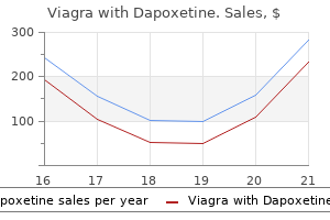
Viagra with Dapoxetine dosages: 100/60 mg
Viagra with Dapoxetine packs: 12 pills, 20 pills, 32 pills, 60 pills, 90 pills, 120 pills, 180 pills, 270 pills, 360 pills, 30 pills, 240 pills, 300 pills

Buy discount viagra with dapoxetine 50/30mg line
Clinical manifestations of cerebral ischemia reflect the features related to the realm of ischemia and embrace paresis erectile dysfunction nutritional treatment viagra with dapoxetine 100/60mg order free shipping, paresthesia how to cure erectile dysfunction at young age buy 50/30 mg viagra with dapoxetine free shipping, imaginative and prescient loss, language disturbances, vertigo, diplopia, ataxia, dysarthria, headache, nausea, and vomiting. Emboli or thrombi brought on by atherosclerosis, hypertension, or diabetes mellitus and located in large, medium, and small arteries account for most strokes. Strokes caused by emboli of cardiac origin account for 20% of the entire ischemic stroke incidence. Atrial fibrillation is the commonest cause of cardioembolic strokes, occurring in up to 20% of such patients. Mitral stenosis and atrial myxoma are different cardiac circumstances related to intracranial embolism. Another reason for cerebral ischemia is elevated viscosity of the blood as a result of being pregnant and the postpartum period, use of oral contraceptives, postoperative and posttraumatic states, hyperviscosity syndromes, polycythemia, and sickle cell illness. Although the presentation of stroke is often attribute, the prognosis should be differentiated from that of other conditions that may mimic strokes, such as a number of sclerosis, subdural hematoma, cranial nerve palsy, encephalitis, hypoglycemia, seizures, brain tumor, hypertensive encephalopathy, syncope, migraine, and useful dysfunction. Also, an assessment of danger components is important for treating a patient with suspected stroke. Modifiable risk elements include diabetes mellitus, hypertension, hyperlipidemia, cardiac arrhythmias, smoking, alcohol use, illicit drug use, migraine, and hypercoagulable states. A scale of 0�42 is used for the assessment, with 0 being normal function and 42 being the most extreme functional impairment. More data on this scale, in addition to training in its use and certification, is out there at Diagnostic studies For practical purposes, diagnostic research may be separated into these done in an acute care setting, corresponding to in the emergency division, and those done in a more subacute setting, similar to in a steady inpatient or steady outpatient setting. Carotid duplex ultrasonography could additionally be used to consider the patency of the extracranial carotid arteries; transcranial Doppler ultrasonography can consider the intracranial arteries. Early detection of these circumstances by such strategies could allow for early therapy, which can be beneficial in salvaging tissue in danger. Cerebral arteriography is usually required only if the trigger is unclear or if intra-arterial thrombolysis or surgical intervention is being strongly thought of. Investigation of the systemic arteries and the center is essential in figuring out the cause for cerebral ischemia. Multiple bruits could counsel widespread arterial disease however may be present without vital occlusion. Evidence of a cardioembolic supply should be pursued aggressively, especially in younger normotensive individuals with cerebral ischemia and in older patients, for whom atrial fibrillation is included within the differential prognosis. Echocardiography is commonly useful in excluding intracardiac emboli; transesophageal Doppler echocardiography is most sensitive in this regard. Treatment the objectives of treating ischemic stroke are to restore blood flow to the mind and to salvage ischemic mind tissue that has not already infarcted. There is a slim window during which to accomplish these aims, typically within 4� hours of the onset of symptoms. Thrombolytic and antithrombotic agents are the first medicine used in the remedy of ischemic stroke. This initiates fibrinolysis at the thrombus website, thereby bettering blood flow to ischemic areas not but infarcted (ischemic penumbra). However, the exclusion criteria for patients treated 3�4� hours from symptom onset (age >80 years, extreme stroke, diabetes mellitus with a previous infarct, and any anticoagulant use) were more restrictive than for these treated at 3 hours or less. Recently, stroke facilities have used intra-arterial catheter-directed remedy for delivering thrombolytic brokers directly to the location of the vascular occlusion. This has been proven to improve recanalization rates and clinical outcomes and is recommended for center cerebral artery infarctions up to 6 hours after stroke onset. Two giant medical trials confirmed a advantage of therapy with aspirin over placebo in short-term mortality and recurrent stroke risk when aspirin is initiated within 48 hours of ischemic stroke onset. Use of heparin could additionally be thought of in the acute care setting for stroke because of postoperative atrial fibrillation in patients with mechanical coronary heart valves or in those with cervicocephalic arterial dissections. Hypertension is an important threat factor for stroke; thus, sufferers with hypertension after a stroke ought to be handled even within the absence of a historical past of hypertension. Hyperlipidemia is also an essential and modifiable danger issue for stroke recurrence. Guidelines for the early management of sufferers with ischemic stroke: 2005 guidelines update a scientific statement from the Stroke Council of the American Heart Association/American Stroke Association. Carotid Occlusive Disease Carotid atherosclerosis occurs most regularly in the proximal inner carotid artery (origin) and at the carotid bifurcation. This is a crucial distinction, because the management recommendations for these 2 situations differ. Asymptomatic carotid bruits occur in 4% of the population older than forty years, and the annual stroke rate in these people is 1. This identical population has an annual mortality price of 4%, primarily from issues of coronary heart disease. The presence of a carotid bruit is a better predictor of arteriosclerotic disease than of stroke. Patients with asymptomatic carotid artery stenosis must be screened for modifiable threat components for stroke, with lifestyle modifications instructed and medical therapy corresponding to antihypertensive and cholesterol-lowering drugs prescribed as needed. The physician ought to assess life expectancy and comorbid situations before contemplating the patient for carotid revascularization, and the patient ought to concentrate on the dangers and benefits of the procedure. Interestingly, the study showed that endarterectomy had a larger benefit in older patients (70 years), whereas stenting was more useful in youthful age groups. Transthoracic echocardiography can establish multiple potential cardiac causes for embolism and, as anticipated, the yield is highest if the scientific historical past and physical examination counsel a cardiac source such as atrial fibrillation, rheumatic mitral stenosis, diffuse atherosclerosis, left ventricular aneurysm, or medical endocarditis. Other modalities that are getting used in the prognosis of cardioembolic sources of stroke include inpatient telemetry, ambulatory Holter monitoring, loop recorders, and surgically implantable cardiac monitors. Otherwise, antiplatelet therapy with aspirin (325 mg/day), aspirin/extended-release dipyridamole mixture, or clopidogrel should be initiated. Intracranial Hemorrhage Intracranial hemorrhage constitutes approximately 15% of acute cerebrovascular issues. Bleeding from aneurysms of the arteries composing the circle of Willis, bleeding from arterioles damaged by hypertension or arteriosclerosis, and trauma are the most typical causes of intracranial hemorrhage. Although there are numerous causes of intracranial hemorrhage, the anatomical location of the bleeding significantly influences the clinical picture. By location, hemorrhages could be broadly categorized as follows: subarachnoid hemorrhage intracerebral hemorrhage intraventricular hemorrhage A number of vascular malformations inside and on the floor of the brain parenchyma might current with seizures and complications. Approximately 85% of congenital saccular, or "berry," aneurysms develop within the anterior a part of the circle of Willis derived from the interior carotid artery in its major branches. The most typical website is at the origin of the posterior communicating artery from the interior carotid artery. Such an aneurysm sometimes presents with headache and third nerve palsy involving the pupil. Vascular malformations within and on the surface of the brain parenchyma constitute roughly 7% of instances with subarachnoid hemorrhage. In addition to meningeal irritation and focal neurologic indicators reflecting bleeding, a bruit may be current over the orbit or skull in approximately 40% of patients. Generalized seizures are common with intracerebral hemorrhage and are much less frequent with subarachnoid hemorrhage or cerebral infarction.
Cheap viagra with dapoxetine 100/60 mg free shipping
The commonest incudostapedial anomalies are fibrous union impotence define discount 50/30mg viagra with dapoxetine with mastercard, absence of the joint guaranteed erectile dysfunction treatment generic 100/60 mg viagra with dapoxetine, or bony fusion. Incudostapedial fusion was the second most common middle ear defect in the Teunissen and Cremer sequence. B: Agenesis of the stapes aside from a remnant of the anterior crus; agenesis of stapedius muscle. Malformations include underdevelopment or absence of the stapes and a variety of structural alterations of this middle ear ossicle. Hypoplasia or aplasia defects of the stapes are diagnosed like the other center ear ossicular chain defects mentioned previously. A number of structural alterations of the shape of the stapes have been described. These include the unicrurate defect, incomplete growth of the crura with out attachment to the footplate, entire absence of the superstructure of the stapes, and the so-called columella or monopodal stapes. These defects additionally may be categorized as part of the group of incudostapedial disarticulation defects or disconnection defects. Most of the information on defects of the stapes involve fixation of the stapes footplate. Sellars and Beighton reported a household with an apparently autosomal dominant condition involving type I microtia and absence of the stapes superstructure. This specific syndrome is of observe, because it appears to comprise a spread of findings including alterations of the lengthy strategy of the incus, lenticular means of the incus, and stapes superstructure. Variability in the reported family suggests a typical denominator in development on this area of the ossicular chain. It is of notice that the stapes superstructure and maybe the long means of the incus come from the second branchial arch. Structural alterations of the stapes also occur in Treacher Collins syndrome, oculoauriculovertebral spectrum, Escher-Hirt syndrome, trisomy 13, microtia with meatal atresia, and numerous much less frequent problems. A laser could additionally be useful in removing an ossified stapedius tendon or irregular stapes superstructure. Once completed, a prosthesis could be placed from the footplate to either the tympanic membrane, incus (if available), or malleus deal with. Louryan S, Vanmuylder N, Resimont S: Ectopic stapes: a case report with embryologic correlations. Sellars S, Beighton P: Autosomal dominant inheritance of conductive deafness as a outcome of stapedial anomalies, external ear malformations and congenital facial palsy. In a collection coping with reconstruction of the middle ear by Sheer, 16 of 17 sufferers had a structurally irregular stapes associated to their conductive hearing loss with out an irregular external ear or meatus, and three of these had congenital fixation. However, there are a variety of exceptions to this assertion, and there has been very little systematic examine of the genetic elements of congenital stapes fixation. Congenital stapes fixation has been seen in individuals with prenatal thalidomide syndrome. About one-fourth of the instances reviewed by Sheer had partial fixation with much less conductive loss. The defect is sometimes related to absence of the oval window, which some otologists really feel is the complete expression of this morphologic defect of the stapedial-vestibular joint. Familial circumstances of stapes with fixation by a bony bar to the pyramid of the middle ear have been described and are summarized in Table 14. Stapedectomy involves full elimination of the footplate; stapedotomy entails creation of a fenestra within the footplate. A prosthesis is then placed, for either process, between the incus and oval window niche. A temporalis fascia graft or tragal perichondrium is often placed between the prosthesis and oval window niche for stapedectomy procedures to prevent medial migration into the vestibule. The most challenging scenario arises when the stapes fixation is associated with incus and malleus abnormalities. It is positioned over a temporalis fascia/tragal perichondrium following a stapedectomy procedure. This discovering is seen in stapes fixation of assorted causes, together with the X-linked kind. Congenital absence of the oval window would normally be acknowledged at the time of surgical procedure in people with conductive hearing loss. There have been a few reported cases of apparently familial causes of absence of the oval window, and these are summarized in Table 14. In addition, people with Wildervanck syndrome and 22q11 deletion have been reported with absence of the oval window. Congenital absence of the oval window has been reported in cases of prenatal thalidomide syndrome. Sterkers and Sterkers have described success with drilling a fenestra above the facial nerve and inserting a prosthesis to the incus in six cases. Given the poor surgical outcome and potential morbidity of the procedure, use of hearing aids is beneficial. Thomeer and colleagues evaluate their surgical results on 14 patients with absence of the oval (and round) window. An opaque or whitish showing mass is typically seen behind the tympanic membrane. Many cases are recognized during surgical exploration for the cause for conductive listening to loss. High-resolution computed tomography may detect these lesions as a homogenous mass within the middle ear. If allowed to enlarge, these cholesteatomas can erode the ossicles and spread to the attic and antrum. Other theories include epithelial implantation or epithelial migration by way of the tympanic ring. The main influence of these lesions, as mentioned above, is ossicular destruction and occasional involvement of the internal ear. The sequence of occasions is such that they grow, turn into secondarily contaminated, and with time produce destruction of the osseous chain and temporal bones. Lesions involving the anterosuperior mesotympanum could be eliminated via an anteriorly primarily based tympanotomy with glorious results. For intensive tumors involving the entire mesotympanic and antrum; a mastoidectomy may have to be carried out. In basic, the surgeon makes an attempt to protect as much of the ossicular chain and posterior canal wall as attainable without sacrificing the extent of elimination. The recent introduction of otoendoscopes may enhance outcomes for this situation due to higher visualization. The glorious visualization from this strategy may also cut back the necessity for extra surgical incisions or mastoidectomy procedures. Recently, Richter and Lee reviewed the literature and their experience with the prognosis and management of congenital cholesteatoma.
Diseases
- Dilated cardiomyopathy
- Oculo tricho dysplasia
- Simian B virus infection
- Ankylosing vertebral hyperostosis with tylosis
- Dermatophytosis
- Oculodentoosseous dysplasia dominant
- Vertebral fusion posterior lumbosacral blepharoptosis
- Palindromic rheumatism
Generic viagra with dapoxetine 100/60 mg without prescription
Inheritance of isolated coxa valga has not been reported but is a typical finding in multiple syndromes and skeletal dysplasias erectile dysfunction hypogonadism purchase viagra with dapoxetine 50/30 mg. Many anomalies could also be present at start however not observed until later childhood or early adolescence how erectile dysfunction pills work proven 50/30mg viagra with dapoxetine. Growth is a succession of acceleration and deceleration phases, and abnormalities in both prenatal and postnatal progress can have an have an effect on on backbone and thoracic development. By age five years, the trunk has reached about sixty six percent of final top; nonetheless, the thoracic volume is only 30 p.c of its last dimension. The thoracic spine and thoracic cage development largely occurs between birth and eight years of age and coincides with lung development. Therefore, each development and lung quantity preservation are important throughout this important interval. In general, remedy of assorted thoracic and spine abnormalities should concentrate on the complete complex as a complete. Each vertebra is then shaped by fusion of sclerotome cells from the posterior half of a more cranially located somite and the anterior half of a somite positioned instantly caudally-a process called resegmentation. Mesenchyme cells between cranial and caudal halves of each somite differentiate into the fibrous area (annulus fibrosis) of every intervertebral disc, while the central region (nucleus pulposus) is derived from cells of the notochord. In addition to giving rise to all of the parts characteristic of every vertebra, sclerotome cells in the thoracic area kind the bony part of every rib. Cartilaginous areas for every rib are derived from sclerotome cells that migrate into the lateral plate mesoderm throughout formation of the physique wall. Paraxial mesoderm, like all the mesodermal germ layer, is derived from cells that migrate through the primitive streak throughout gastrulation in the third week of gestation. Lengthening of the embryo is dependent upon the continued means of mesoderm formation by gastrulation and organization of this mesoderm into segments by the segmentation clock. Thus, sclerotome cells from somites on opposing sides of the neural tube migrate to meet within the Somitogenesis requires the coordinated motion of at least 300 genes, lots of which are expressed in a cyclic fashion. These waves of gene expression, generally known as the segmentation clock, happen with the same periodicity as somite formation. Dimeglio A, Canavese F: the growing spine: how spinal deformities affect regular backbone and thoracic cage growth. Iimura T, Denans N, Pourqui� O: Establishment of Hox vertebral identities in the embryonic spine precursors. Occipitalization happens because of defects in embryogenesis during somite segmentation or as a failure within the resegmentation of the fourth occipital sclerotome and the primary cervical vertebrae. Findings related to occipitalization are related to compression of the medulla oblongata, spinal cord, vertebral artery, and venous plexus, and embody neck ache, headache, paresis, lengthy track signs, hyperreflexia, weak spot, disturbances of peripheral sensation, and even cerebellar signs associated to basilar impression. Typically, fusion involves the anterior arch of the atlas and the rim of the foramen magnum. Computerized tomography and/or magnetic resonance imaging may be essential to delineate further the degree and web site of impingement of the spinal twine. The sagittal diameter of the foramen magnum is a important measurement in patients with the above symptoms and indicators. The diagnosis may be difficult within the younger child, as a end result of a good portion of the ring of the atlas is unossified at birth. The 5mm�9mm radiolucent gap in the posterior neural arch of the newborn typically ossifies by age 4 years. The anterior arch of the atlas, unossified in eighty percent of newborns, begins to ossify between the primary and third years, changing into absolutely visible on radiographs between ages seven and 10 years. Some 70 % of sufferers with occipitalization of the atlas also have fusions of the second and third cervical vertebrae. Occipitalization of the atlas could be congenital or acquired and may be first detected by likelihood when radiographs are made for different purposes. Acquired instances can be due to infectious etiology, whereas congenital circumstances are associated with elements that can have an effect on the developing fetus throughout third to fourth week of embryogenesis when occipital and cervical sclerotomes are developing. Occipitalization of the atlas is often present in achondroplasia, oculo-auriculo-vertebral spectrum and Klippel-Feil syndrome. When compression is anterior, transoral resection of the odontoid and rim of the foramen magnum is carried out. For posterior compression, cervical laminectomy of the atlas, excision of any dural bands, and suboccipital craniectomy are performed. A few sufferers respond to nonoperative measures: immobilization in plaster, traction, and cervical collars. The neurological status remains unchanged in plenty of sufferers following surgery, and death might happen throughout or shortly after surgery. Partial or complete absence of the odontoid course of varies from a brief, stubby, nipple-like projection to considered one of nearly normal size. Os odontoideum is characterized by a radiolucent, oval, or round ossicle of variable measurement with a smooth, dense border of bone, separate from the axis and often situated within the position of the normal odontoid tip. It might at times be near the basal occiput in the area of the foramen magnum, the place it could fuse with the clivus. Odontoid aplasia or hypoplasia is primarily a radiologic distinction and has little or no significance as both could result in atlanto-axial instability with potential neurologic sequelae. Another associated developmental anomaly is the presence of a separate ossification center at the tip of the V-shaped odontoid process, the ossiculum terminale. Should fusion fail to occur, this ossification heart is referred to because the ossiculum terminale persistens, another normal variation in improvement. Congenital anomalies of the odontoid could be revealed by incidental observations on X-rays obtained for other purposes or following trauma that may provoke atlanto-axial instability and/or provoke symptoms in a beforehand asymptomatic particular person. Symptoms may be localized to the neck, such as ache and stiffness, or there could be transient bouts of paresis. More severe signs embody compressive twine myelopathy and ischemia of the brain stem by impingement of the vertebral artery leading to syncope, vertigo, seizures, and visible disturbances. These sufferers have variable spasticity and hyperreflexia or have lack of proprioception and sphincter disturbance. When neurologic signs are a outstanding scientific feature, magnetic resonance imaging is important to delineate the condition, augmented by somatic evoked responses as nicely as a careful neurologic examination. The physique of the odontoid, phylogenetically derived from the centrum of the first cervical vertebra, separates from the atlas during development and fuses with the superior portion of the axis. Delayed ossification of the dens might give the impression of hypoplasia or aplasia. Underdevelopment or failure of fusion may end up in congenital anomalies of the odontoid. Complete aplasia is extremely rare; most cases involve variable degrees of hypoplasia. It could also be found in patients with Down syndrome and is extraordinarily frequent in the mucopolysaccharidoses, particularly Morquio syndrome. Treatment: Nonoperative therapy by cervical traction or plaster casting could also be adequate with a relatively stable atlanto-axial joint. However, those with an atlas dens area of more than 5mm demonstrated on flexion-extension lateral X-ray require stabilization by posterior cervical spine fusion, commonly preceded by halo-skull traction. Skull traction offers a method of C1-C2 discount, which may indirectly decompress the twine and will simplify the surgical procedure.
100/60mg viagra with dapoxetine order free shipping
Molecular testing should precede transplantation from relations younger than 30 years erectile dysfunction treatment saudi arabia cheap viagra with dapoxetine 100/60 mg free shipping, as cysts could occasionally not be discovered on ultrasound on this age group adderall xr impotence viagra with dapoxetine 50/30mg safe. Although penetrance is basically full by eighty years, many asymptomatic sufferers have regular life spans. The renal prognosis is worse in individuals whose unaffected father or mother has important hypertension, indicating a multifactorial element. Ethical issues surrounding presymptomatic provider detection in wholesome children and relations not wishing to know their service standing must be considered before investigating households. The uptake of presymptomatic ultrasound screening was excessive amongst at-risk individuals in a single study,10 and many said that the analysis had influenced their reproductive plans. Other anomalies of the contralateral facet embrace renal hypoplasia, hydronephrosis, ureterocele, or ectopic ureters. Anomalies of noncontiguous constructions occur in approximately 50 % of these with bilateral renal dysplasia and about 15 % of these with unilateral involvement. Clinical signs and symptoms and age of presentation are normally decided by the severity of renal dysplasia or related major malformations. Severe dysplasia presents prenatally in a similar way to renal agenesis (Entry 30. Clinical symptoms in infants and kids include anuria, oligouria, polyuria with polydipsia, hematuria, hypertension, uremia, back pain, progress delay, and continual or progressive renal failure. Appropriate administration is an initially conservative approach using imaging to rule out reflux, then serial ultrasounds to assess renal size, as full involution of the dysplastic kidney frequently occurs. For those with unilateral defects, prognosis largely is determined by the type and severity of associated anomalies. Although isolated dysplasia is often sporadic, familial recurrence has been reported. Cardiovascular anomalies, central nervous system faults, imperforate anus, tracheoesophageal fistula, and radial ray defects are frequent. Chromosomal defects are present in approximately 5 p.c of fetuses diagnosed with multicystic renal dysplasia, however in a better proportion of these recognized prenatally. Population studies indicate a frequency of multicystic dysplasia of approximately half of,500 to 1/3,500. Approximately twice as many males as females are affected, however affected females are extra likely to have bilateral involvement and syndromal issues, including chromosomal defects. Al-Ghwery S, Al-Asmari A: Multicystic dysplastic kidney: conservative administration and follow-up. Salt losing is frequent and may shield in opposition to hypertension as renal operate worsens. Urinalysis reveals no red or white cells, and proteinuria is normally mild or absent; thus this situation could be missed by routine screening. Other urinary tract anomalies are uncommon, but nonrenal defects such as ocular defects and skeletal anomalies may occur in syndromal varieties, many of which are ciliopathies. Histological changes are nonspecific but important and increase in severity as the illness progresses. Gross specimen of a kidney exhibits collections of cysts on the corticomedullary junction. As some families map to neither locus, there are doubtlessly different genes that may trigger this phenotype. In all circumstances, renal osteodystrophy and hypoparathyroidism might turn out to be obvious because the condition worsens, especially in children. Prenatal diagnosis, presymptomatic screening, and/or heterozygote detection is now available in families the place specific mutations have been identified. Hildebrandt F, Omram H: New insights: nephronophthisis-medullary cystic kidney disease. Diagnosis in infancy and childhood is normally because of investigations initiated due to an associated syndromal disorder. Grossly the kidneys are of normal measurement or, if there are in depth cysts, slightly enlarged. All pyramids of both kidneys are normally affected, however the pathological adjustments could also be restricted to one or two pyramids or to a single kidney. The most common clinical symptoms are a urinary concentrating defect and acidification of the urine, which normally goes unnoticed. The defect has been reported to happen in affiliation with ectopic and horseshoe kidneys and with unicornuate uterus. It may be seen with congenital hepatic fibrosis, most often as an uncommon presentation of autosomal dominant polycystic kidney illness. This is likely a significant underestimate given the excessive proportion of asymptomatic cases. In addition, medullary sponge kidney could happen with reflux and ureteropelvic junction obstruction in sufferers with a dominant type of familial ureteral abnormalities. Treatment: Management ought to embody treatment of hypercalcemia, hypercalciuria, and management of infection. Instrumentation or catheterization ought to be avoided in order to stop ascending urinary tract an infection. Patients at risk for stone formation might have lowered bone density, which improves with oral potassium citrate administration. Long-term follow-up in asymptomatic instances demonstrates no major changes in the radiological findings and no deterioration in renal perform. In 10 p.c the clinical course is difficult by repeated infections, stone formation, and finally renal failure resulting in dialysis and transplantation. An excretory urogram with tomography shows radial linear streaking in the renal papillae ensuing from distinction medium within the ectatic papillary ducts. Closely associated noncystic modifications corresponding to hydronephrosis and renal scarring happen later in life because of different types of obstruction, including ureteropelvic junction obstruction and vesicoureteral obstruction with reflux. Milder types of obstruction later in gestation, infancy, or childhood lead to much less severe cystic changes or an "obstructive kidney" with calyectasis and parenchymal scarring. As the obstruction could be at any stage of the urinary tract, one or both kidneys may be concerned with the precise pathology various relying on the diploma and period of obstruction. Bilateral hydronephrosis is usually because of lower urinary tract obstruction and is associated with hydroureter. Cryptorchidism, esophageal atresia, annular pancreas, preauricular pores and skin tags, and single umbilical artery had been also present. When related to ureteral anomalies, some defects, for instance horseshoe kidney, could cause a blended pathological image of each renal dysplasia from a primary embryological defect and secondary adjustments from obstruction. Treatment: the medical course in sufferers with obstructive nephropathies is dependent upon the quantity of renal parenchymal loss and the underlying cause. Management is directed towards reducing ongoing harm to the kidneys and stopping recurrent infection and intrarenal reflux, although one of the best approaches to achieve this are controversial. Using radionuclide voiding cystography as a screening device, it was discovered that forty five % of asymptomatic sibs of sufferers with vesicoureteral reflux have been equally affected. Many sufferers remain asymptomatic however may current with again or abdominal ache or fever in adulthood (average age at prognosis ~36 years). Occasionally the defect is detected fortuitously as a pelvic mass on computed tomography.
Viagra with dapoxetine 100/60mg purchase without prescription
Furthermore xyrem erectile dysfunction order viagra with dapoxetine 50/30mg free shipping, as the face grows the scale normally increase in a predictable manner erectile dysfunction frequency age 50/30mg viagra with dapoxetine discount fast delivery, such that attribute syndromal facial features could become much less or extra evident. Physical observations should embody detection of main anomalies, notation of minor defects and variations, evaluation of growth (head circumference, weight and top or length), measurement of individual options that seem irregular, and an estimate of neurodevelopmental standing. Repeated observations over a interval of years could ultimately lead to a prognosis not initially obvious or to revision of an inaccurate analysis. Review of images of relations, especially when taken on the same age as the patient, could be helpful. Individual morphologic options turn into distinctive because of their measurement in relation to different features round them. Downslanting palpebral fissures end result from undergrowth of the malar region (zygoma) in relation to the frontal region (frontal bone). Dysmorphology employs each gestalt and measurements in assessing sufferers with anomalous growth. More delicate alterations of morphology could also be discerned by the attention, however affirmation by measurement substantiates the medical statement. Unbiased estimates may be obtained by randomly sampling the inhabitants in query. In many instances if enough information are accrued, continuous variables are distributed in a fashion that approximates a normal distribution curve. The distribution may be additional described by two parameters, the imply and the usual deviation. Data that describe normal distributions have been well characterised mathematically and can be found in tabulated kind. This permits one to make preliminary generalizations concerning the distributions of the traits. In many instances, abnormal physical measurements are arbitrarily thought-about to be those values that lie within the upper and decrease 2. A formidable body of human anatomic measurements (taken during the prenatal period and during infancy, childhood, and to a lesser extent adult life) has been accrued. Prominence of the brow is clear from early childhood, however enlargement of the decrease jaw develops in late adolescence or maturity. It may be assumed that the growth standards have been derived from data collected with rigidly managed protocols, which entails inserting the topic in a natural and relaxed place, having measurements taken by an skilled observer, and using essentially the most precise devices. Calipers are used for taking measurements lower than 30 cm, and inflexible measuring gadgets (measuring table or stadiometer) are used for measuring size or peak. It is advisable for the customers of growth requirements to turn into conversant in the inhabitants from which the info have been collected, with the tactic of knowledge assortment, and with the quantity and experience of individuals accumulating the information. Normal racial and ethnic variations affect many measurements; nevertheless, well-established ethnic norms are not often available. This applies particularly to development references for several skeletal dysplasias and syndromes. The triangular facial configuration with prominence of the brow and hypertelorism turns into less obvious with growing age. Unfortunately, heterogeneity of the subject population is in all probability not rigidly excluded. Many genetic texts include standards for progress throughout childhood but lack info on mature measurements. This deficiency is eliminated partly by the info within the Natick Anthropometric Survey. Recently the World Health Organization has revealed normal progress curves for the primary seven years based on studies in seven numerous world regions when the mother was properly fed at conception and throughout being pregnant and the infant was breastfed for the first year of life. Interestingly the curves are superimposable from all seven regions, suggesting that growth deficiency is happening commonly in lots of areas of the world. Usually straight rulers, flexible tapes, and scales are the one measuring devices available. With data of the correct anatomic landmarks and care in taking measurements, the clinician could make dependable observations beneath lower than perfect circumstances. With the features converted right into a two-dimensional image, measurements are easily made by hand or by laptop. Radiocephalometry has utility in defining the anatomy in craniofacial syndromes and in planning reconstructive surgery. Radiographs are also necessary for the measurement and maturation of most parts of the skeleton. They permit particular person bones to be measured for comparability to age-specific population knowledge and permit one skeletal function to be compared with others in the same individual. Computed tomography and magnetic resonance imaging can be utilized for two-dimensional viewing and for three-dimensional reconstruction of most anatomic components. Three-dimensional reconstructions are useful in planning surgery and in projecting the outcomes anticipated from surgical procedure. Growth is mediated by way of hormonal stimulation of various tissues, notably the chondroosseous skeleton. Nonspecific impairments of progress could be caused by a variety of environmental influences including diet, oxygen supply, an infection, systemic illness, numerous medicine and chemical compounds, and emotional deprivation. Unlike prenatal environmental insults to growth, postnatal insults usually affect total growth somewhat than that of isolated options. An exception to this generalization is the coarsening of lower Growth in plenty of malformation syndromes, skeletal dysplasias, and metabolic issues proceeds at a rate different from that of the final inhabitants. For most of these problems, inadequate cross-sectional or longitudinal data have been collected to allow the development of development curves. For these situations for which curves are available, growth could be monitored and the success of assorted therapies analyzed. Growth curves for children with achondroplasia, the most common form of short-limb dwarfism, have been out there since 1978 and are immensely useful in evaluating such. Anteroposterior view (left) and vertex view (right) show marked deficiency of cranial ossification. Following the growth of achondroplastic kids using normal growth curves is of limited benefit, but comparisons with the growth curves of different kids with achondroplasia may be very instructive. Comparing the head development of 1 affected person with that of different people with achondroplasia can usually reassure the family and thus keep away from unnecessary diagnostic checks. Furthermore, if any growth-promoting intervention is used, the change in progress ought to be compared with the anticipated rate in achondroplasia without the intervention. Meaney and Farrer have cataloged the information out there on growth in numerous completely different genetic and congenital problems. Such broad research are wanted to understand the natural history of pathologic states and to provide the required info for counseling patients with abnormal growth and their families. Recently, via the work of father or mother help groups, better longitudinal knowledge on particular circumstances are being collected and printed. Testing is appropriately used, first, to confirm a suspected analysis and, second, as a diagnostic probe.
Syndromes
- Intermittent (short-term) catheter
- Ultraviolet light therapy for some cases
- Maintain good general health, including proper nutrition and exercise
- Stupor
- Your pain is sudden and severe
- Patients with AIDS who have CD4 counts below 200
- Difficulty walking that gets worse over time; by age 25-30 the person is usually unable to walk
Purchase 50/30 mg viagra with dapoxetine with visa
Ophthalmic issues the abuse potential of benzodiazepines is delicate compared with that of drugs such as hydromorphone and cocaine icd 9 code erectile dysfunction 2011 viagra with dapoxetine 100/60 mg buy on-line. Nevertheless erectile dysfunction treatment heart disease 100/60 mg viagra with dapoxetine purchase with amex, long-term administration of these agents may cause physical dependence. Ocular adverse effects can occur, although they tend to be dose associated and transient. Antidepressants Psychotherapy, both alone or in combination with antidepressant medication, is the primary line of intervention in mild to moderate despair. Prospective research have discovered that main depression in older adults is related to elevated mortality. Collaborative care applications, which integrate depression care managers (usually nurses supervised by psychiatrists) into primary care practices, might lower all-cause mortality. In patients with main despair, antidepressants can enhance symptoms, increase the probabilities and price of restoration, scale back the chance of suicide, and assist social and occupational rehabilitation. A summary of research concluded that antidepressants are associated with a 50%�60% response fee among patients with major melancholy in the main care setting. These drugs may find yourself in temper elevation, improved urge for food, higher sleep, and elevated mental and bodily activity. Common antagonistic results embody restlessness, insomnia, headache, gastrointestinal signs, delicate sedation, and sexual dysfunction. In addition, sufferers might report "tracking" difficulties, which are extra frequent in youthful than older sufferers and tend to occur upon withdrawal of the drug. Rare cases of angle-closure glaucoma have been reported, mostly with paroxetine, which tends to have a stronger anticholinergic effect. The different major class of antidepressants includes the heterocyclics, of which the tricyclic antidepressants were the first described. Most regarding is the toxicity of these medications in overdose; they are often deadly in as little as 5 occasions the therapeutic dose. Mortality is normally as a result of arrhythmias, although anticholinergic toxicity and seizures can even happen. Sudden cessation might trigger pronounced adjustments in have an result on, cognition, and cardiac dysrhythmias. Transcranial magnetic stimulation is a noninvasive methodology to excite neurons within the brain: weak electrical currents are induced within the tissue by quickly changing magnetic fields. They are the drugs of choice for treatment of mania, bipolar disorder, schizoaffective dysfunction, and cyclothymia. They can also be used to deal with impulse control issues, signs related to intellectual disability (mental retardation), and aggressive conduct. This class consists primarily of lithium carbonate and varied antiepileptic medicines. Valproic acid, carbamazepine, and lamotrigine are the most commonly used antiepileptic medication, however many others have been studied and prescribed. The antiepileptic drugs are mentioned in the part Epilepsy, later on this chapter. For example, consciousness of the potential ocular adverse results of the assorted psychiatric drugs is necessary. Patient schooling and reassurance could also be required as a end result of the underlying psychopathology may make anticholinergic adverse ophthalmic effects, such as dry eye and accommodative changes, rather more scary and less tolerable for patients with behavioral disorders than for these without such problems. Failure to adhere to remedy is one other frequent downside amongst patients with psychiatric illness, dementia, and melancholy. Malingering and practical imaginative and prescient loss require a high index of suspicion and special diagnostic expertise on the a part of the clinician. Some medications used to deal with eye illness, including carbonic anhydrase inhibitors, brimonidine, oral corticosteroids, and probably -blockers, may induce or exacerbate melancholy. Neurologic Disorders Parkinson Disease Parkinson disease belongs to a bunch of situations often known as bradykinetic movement disorders. In addition to bradykinesia (slowed actions with decreased voluntary movement), patients can develop rigidity, postural instability, and resting tremor. Parkinson disease normally impacts individuals older than 50 years; the average age of onset is 60 years. Though uncommon, familial Parkinsonism can occur by way of a big selection of genetic mutations and can be liable for a juvenile form that presents earlier than age 20 years. The differential diagnosis for Parkinson disease consists of other neurodegenerative problems such as dementia with Lewy bodies, corticobasal degeneration, multiple system atrophy, and progressive supranuclear palsy. Etiology the basal ganglia are a fancy of deep gray-matter nuclei that features the corpus striatum, globus pallidus, and substantia nigra. Patients with Parkinson disease have typically lost 80% or extra of the dopamine-producing neurons within the substantia nigra. Depletion of dopamine within the complicated nigrostriatal pathway produces an imbalance in inhibitory and excitatory neuronal signals, leading to the cardinal indicators of Parkinson disease. Although most cases are sporadic, genetic elements are implicated within the pathogenesis, especially in early-onset cases. At least 5 potential causative genes have been identified, and the number of Parkinson-like disorders associated with specific genetic defects is rising. Overall, Parkinson disease appears to have a multifactorial etiology that includes genetic predisposition, environmental elements, and age-related modifications in neuron metabolism. Symptoms the first symptom of Parkinson disease is normally tremor of a limb at rest. Other widespread symptoms embrace bradykinesia, rigidity, a shuffling gait, postural instability, and stooped posture. Persons with Parkinson disease usually have lowered facial expressions and converse in a soft voice. The disease is associated with nonmotor features such as depression, character modifications, sexual difficulties, hallucinations, autonomic dysfunction, and dementia. Dopamine substitute, with medications corresponding to levodopa (L-dopa), is the principle therapy. Although levodopa helps at least three-fourths of sufferers with Parkinson illness, not all symptoms respond equally to the drug. Usually, sufferers are given levodopa mixed with carbidopa, usually as a mixed pill. When added to levodopa, carbidopa delays the conversion of levodopa into dopamine until it reaches the brain, diminishing some of the adverse results that always accompany levodopa remedy. After years of remedy, sufferers can turn out to be aware of a "wearing-off" impact that occurs about four hours after a dose of levodopa, when their signs return. Catechol-O-methyltransferase inhibitors similar to entacapone lengthen the duration of the levodopa impact and reduce the "off" time by inhibiting the methylation of levodopa and dopamine. Dopamine agonists (bromocriptine, pramipexole, ropinirole, rotigotine, and apomorphine) stimulate dopamine receptors in the brain and should delay the need for levodopa. Anticholinergic medication corresponding to trihexyphenidyl and benztropine have a shortlived impact controlling tremor and rigidity. However, only about half of sufferers reply to anticholinergics, and typical anticholinergic opposed effects can be problematic. Amantadine, an antiviral drug, may be used in the early levels of the illness, either alone or in combination with anticholinergics or levodopa.
Viagra with dapoxetine 50/30mg online buy cheap
All sufferers should have common fundus examinations to identify and treat retinal detachments erectile dysfunction quotes 50/30mg viagra with dapoxetine discount amex. Top: a funnel-shaped excavation on the nerve head with remnants of the hyaloid system at the center of the disc impotence support group buy 100/60 mg viagra with dapoxetine with mastercard. The exterior ear is an important website for the study of phenotypic variations, malformations, and so-called minor anomalies. There are many medically insignificant but helpful morphologic clues that lie within the ear. It is of note that all of the common chromosomal syndromes and most of the uncommon ones have variations or malformations of the ear as a constant characteristic. The exterior ear alterations of Down syndrome and Trisomy 18 are examples of this. At 5 weeks, three small elevations seem on each arch and these proceed to develop and type six auricular hillocks by six weeks. Hillocks 1, 2, and 3 are located on the mandibular portion of the primary arch, and 4, 5, and 6 are on the second arch. Hillock 1 types the tragus, 2 the helix, three the cymba concha, 4 the concha, 5 the anthelix, and 6 the antitragus. This complex arrangement of the hillocks and their patterning explains why external ear defects are widespread and how pits and tags can happen. Also, neural crest cells are the most important cell sort responsible for forming the exterior ear and face; these cells also participate in septating the guts, which explains why many infants with external ear or different craniofacial defects even have heart malformations. At first, the ears lie in a horizontal position on the posterior of the mandible and are positioned relatively low on the facet of the prospective neck region. As the face grows, and notably because the mandible grows posteriorly and cranially to kind the ramus, the ears are rotated and elevated to their regular place. Hox2a is expressed in the neural crest�derived mesenchyme that provides rise to the second pharyngeal arch components of the exterior ear. Carey, Albert Park, and Harlan Muntz, who authored the chapter in Edition 2 of Human Malformations and Related Anomalies on which this revision is based. The degree of listening to loss in individuals with microtia is determined by the presence of meatal atresia, middle ear ossicular chain defects, and the occasional incidence of internal ear dysplasias. In infancy and the preschool years, the diploma and kind of listening to loss may be detected with brainstem evoked response, behavioral testing, and impedance audiometry. The range of associated middle ear ossicular defects is broad and appears not to observe any pattern. Fusion of the malleus to the atresia plate or to the partitions of the epitympanic recess is widespread in people with meatal atresia. About 20�40 percent of children with microtia/anotia could have an related defect or an identifiable syndrome sample. In this milder form the auditory meatus is normally patent, and defects of the ossicular chain are rare. The ear is usually a rudiment of soppy tissue, and the auricle no longer has any resemblance to the normal pinna. It is evident that this malformation pattern is a situation of etiologic and pathogenetic heterogeneity. In addition to the well-delineated syndromes, microtia appears to have other, nonrandom associations. One of essentially the most extensively mentioned associations in pediatrics is the apparent relationship between external ear defects and renal malformations. This problem is discussed once more intimately in connection with low-set ears (Entry thirteen. The most necessary association with microtia is conductive and sensorineural hearing loss. As pointed out by Jaffe, the complete vary of microtia, as properly as different, milder ear defects, should all the time convey to mind center and inside ear abnormalities. A number of epidemiologic investigations worldwide have estimated the delivery prevalence of microtia/anotia to range from 1/3,000 to 1/20,000 and a male to female predominance of about two to one. In the bigger collection, about 10 % of people were affected bilaterally and about eighty percent had meatal atresia. Microtia is related to different anomalies in 28�49 p.c with 6�9 % representing well-defined syndromes. Poswillo proposed that the pathogenesis of microtia with associated hemifacial microsomia was because of embryonic hematoma formation within the area of the blood vessels that precede the formation of the stapedial artery stem. An alteration in cephalic neural crest migration is actually the most enticing hypothesis to clarify the pathogenesis of microtia/ anotia currently. Most circumstances of microtia/anotia are isolated, with no family historical past, and most massive surgical sequence indicate that lower than 10 percent of the time is there a family historical past; nonetheless, quite a few authors have mentioned the occasional occurrence of a positive household historical past in nonsyndromal microtia. Rollnick and her colleagues at the University of Illinois have documented larger frequencies of first-degree and second-degree relations with ear abnormalities in people with microtia with or without hemifacial microsomia. While most of their patients had been categorised as hemifacial microsomia versus isolated microtia, eight of 15 of their cases recognized as microtia had a constructive household historical past. Their knowledge counsel that microtia is a part of a continuum that extends to hemifacial microsomia and Goldenhar syndrome as talked about above. These investigators thought that the multifactorial model of causation fits the data greatest. Looking closer at instant families of their information set, one can observe two kindreds of a number of affected sibs with no different affected relatives, a pattern that might recommend autosomal recessive inheritance. However, there have been 19 different pedigrees that showed technology to generation transmission suggestive of dominant inheritance. One of the basic pedigrees cited to support the idea of recessive inheritance of microtia with meatal atresia is that studied by Elwood et al. These data taken collectively suggest that nonsyndromal microtia with meatal atresia might be not inherited as a easy autosomal recessive dysfunction but quite happens in plenty of families as an autosomal dominant trait with variable expression and incomplete penetrance. In the dominant familial circumstances, unilateral and bilateral microtia with or without hemifacial microsomia happen in the identical family. The most parsimonious rationalization for the genetics of nonsyndromal microtia is to invoke complicated multifactorial causation in most cases, with well-established autosomal dominant inheritance representing a small proportion. The impact of microtia/anotia is twofold: stigmatization associated to the seen craniofacial defect and listening to loss. The baby with microtia often suffers from psychosocial issues related to the seen defect. The microtic ear attracts attention to the difference, and this can be particularly problematic during the elementary school years when conformity and sameness are so necessary. In addition, some youngsters with microtia could have an opposed impact from listening to loss.

Cheap 100/60mg viagra with dapoxetine overnight delivery
With regard to pathogenesis food that causes erectile dysfunction generic viagra with dapoxetine 100/60 mg without prescription, it must be borne in thoughts that the primitive foregut provides rise to the cephalic a half of the digestive system and to the respiratory tract impotence icd 9 viagra with dapoxetine 100/60mg order mastercard. However, nonteratomatous extracranial mind tissue is relatively uncommon, most examples being nasal gliomas. They doubtless arise by displacement of neural parts in the ground of the anterior oral pharynx prior to the closure of the secondary palate. These tumors are usually onerous and are characterised by sluggish growth and appreciable mobility. Differential diagnosis of choristomas located on the posterior dorsum includes lingual thyroid, hyperplastic lingual tonsils, and neoplasm of minor salivary glands. Microscopic view of a lingual inclusion nodule exhibiting heterotopic mind tissue (glial elements) near the overlying stratified squamous epithelium. Left: two separate chondromas of the lateral border of the tongue of a 35-year-old woman. Right: photomicrograph exhibiting cartilaginous tissue with overlying connective tissue and epithelium. The report of a chondroma of the tongue by Ramachandran and Viswanathan appears to be a choristoma associated with orofaciodigital syndrome kind 1. Top: osteoma of the tongue occurring close to the foramen cecum, as is the usual case. Bottom: photomicrograph of lingual osteoma displaying skinny epithelial and connective tissue cowl over a well-rounded and highly mature bony nodule. Approximately 65% are composed of pure cartilage, the rest being osteocartilaginous. Ohbayashi Y, Miyake M, Nagahata S: Gastrointestinal cyst of the tongue: a potential duplication cyst of foregut origin. Arcand P, Granger J, Brochu P: Congenital dermoid cyst of the oral cavity with gastric choristoma. Hamartoma refers to a benign, disorganized focal neoplasm of histologically regular tissue composed of tissue parts normally discovered at the web site. Lymphangioma of the tongue consists of benign nodular masses of clear lymph-filled vessels and vesicles of assorted sizes inside and elevated above the surface of the tongue. Some rupture, and others turn into papillomatous and should resemble granulation tissue. Presenting very early in infancy, lymphangiomas often develop slowly and remain essentially quiescent. The anterior two-thirds of the tongue is the commonest web site affected, however it may involve different elements of the oral cavity (lip, cheeks, neck, oral floor) or prolong to the epiglottis or mediastinum. Deeper examples could be complicated by glossitis, especially those who prolong beyond the lips, by which Syndrome Associations (Appendix) None. Note densely packed yellowish, purple, or bluish-black vesicular elements more marked on distal portion of tongue. The ensuing macroglossia could result in airway obstruction, tough mastication, drooling, dysphagia, and altered speech. Microscopically, the tumor consists of endothelial-lined cystic areas full of lymph, lymphocytes, and purple blood cells. During native or systemic infections the lesions can become engorged with lymphocytes. Treatment: Surgical excision, coblation (radiofrequency tissue destruction), bleomycin injection, and/or laser photocoagulation are among the attainable remedy modalities. Management should embody cautious assessment for the development of airway issues. Large, deep lesions can carry vital morbidity and in some circumstances mortality because of effects on mastication, dysphagia, and airway compromise. A evaluation of pathogenesis, remedy and the usage of floor laser photocoagulation. They may be flat or rounded, red-purple to deep purple, superficial or deep, and may lengthen into the tongue muscle. Treatment: Depending on kind, measurement, and site, surgical or nonsurgical elimination of the hemangioma may be easy or difficult. Large advanced lesions could be related to vital morbidity and occasional mortality. The color reflects the specific composition of the hamartoma (mucous salivary glands, adipose tissue, nerves, smooth muscle, connective tissue, blood vessels). It is difficult to distinguish this lesion from the so-called benign mesenchymoma of the tongue, and in reality may be the same entity. Exceptions to this are the lingual hamartomas seen within the orofaciodigital syndromes. Although teratomas are thought to come up within the developing blastomere with pluripotent cells being displaced, it has been suggested that standard myogenic cells migrating from the occipital myotomes could also be accompanied by ectodermal and endodermal traces. Uchida K, Urata H, Suzuki H: Teratoma of the tongue in neonates: report of a case and evaluation of the literature. Occasionally it can be hemorrhagic and, if very massive, might displace the epiglottis. In females, lingual thyroid increases in dimension during instances of marked endocrine exercise corresponding to puberty, pregnancy, and menopause. Some sufferers may have obstructive issues corresponding to dysphagia, dysphonia, dyspnea, or obstructive sleep apnea. Lingual thyroid ought to be suspected when a mass or nodule is positioned on the degree of the foramen cecum in front of the terminal sulcus of the tongue. Nevertheless, the differential prognosis contains varied tongue neoplasms, hamartomas, choristomas, and lingual tonsillitis. Radioactive iodine 123 or technetium 99m scanning, computed tomography, and magnetic resonance imaging will show the presence of thyroid tissue at the lingual location. In distinction with a lingual thyroid, a thyroglossal duct cyst is a cyst that develops from persistence and dilation of the embryonic thyroglossal tract. Although the tongue arises on the foramen cecum, these cysts often current below the tongue on the stage of the hyoid bone. This tissue descends in front of the pharyngeal intestine but stays connected to the tongue by the thyroglossal duct. Ectopic thyroid tissue may be found at different levels of the duct, from the tongue base to the neck. Residual thyroid tissue found within the neighborhood of the foramen cecum has been reported to be as frequent as 10 % of routine autopsies and medical biopsies but with equal distribution among the sexes. Sood A, Kumar R: the ectopic thyroid gland and the position of nuclear medicine strategies in its analysis and administration. Elidan J, Chisin R, Gay I: Lingual thyroid, sensorineural hearing loss and psychological retardation: a coincidental affiliation C: Biopsy material from a lingual mass shows thyroid tissue lined by normal-appearing mucosa. Danner C, Bodenner D, Breau R: Lingual thyroid: iodine 131: a viable therapy modality revisited. Rojananin S, Ungkanont K: Transposition of the lingual thyroid: A new various technique.
100/60 mg viagra with dapoxetine cheap otc
Aganglionic megacolon (Hirschsprung disease) erectile dysfunction treatment operation buy discount viagra with dapoxetine 50/30mg on-line, characterized by absence of the parasympathetic ganglion cells of the bowel wall erectile dysfunction lifestyle changes generic viagra with dapoxetine 50/30mg with amex, might current at any time from birth to late childhood. Megacolon can also be associated with atresia or agenesis of the distal colon or anorectal area. The small-caliber aganglionic segment is maintained by tonic contraction of the noninnervated vasculature. Top: Contrast enema in 3-day-old male displaying constricted section (arrowheads) in sigmoid colon. Bottom: Contrast enema in same infant at 5 months exhibiting dilation of colon proximal to the constricted segment (arrowheads). The complete colon is concerned in 3 p.c, and extension to the small intestines is kind of rare. This contrasts with psychogenic megacolon in which the anal sphincter is hypertrophied and the rectum is dilated with feces. In Hirschsprung disease, barium enema will usually show a transition zone at the junction of the dilated megacolon and the traditional or small-caliber aganglionic phase. The typical look is in all probability not seen in the first months of life, in older kids who use repeated enemas for colon decompression, and in those with aganglionosis of the complete colon. Aganglionosis of the colon might happen as an isolated finding or in affiliation with a quantity of potential additional anomalies. There is a specific affiliation with irregular neural crest migration, both in terms of syndromal associations and with apparently isolated aganglionic megacolon. The neuroblasts migrate caudally alongside the vagus nerve pathways and attain the rectum by week 12 of improvement. Pelvic parasympathetic nerves additionally contribute to the formation of the intestinal ganglia. The myenteric (Auerbach) plexuses type first and are located outside the circular muscle layer. Since neuroblasts that kind these plexuses migrate in a cephalic-caudal course, denervated gut extends without interruption from the extent of interruption of neuroblast migration to the rectum. The tendency of denervated bowel to tonic contraction explains the small caliber of the aganglionic phase. Ganglionic megacolon, presumably as a end result of an imbalance in sympathetic and parasympathetic innervation, has been described in neurofibromatosis, intestinal neuronal dysplasia, and multiple mucosal neuroma syndrome. Mutations in a quantity of genes that have roles within the growth, migration, and survival of neuronal cells have been present in patients with Hirschsprung disease. Possible associations with maternal hyperthermia and with prenatal cytomegalovirus infection have been reported. Removal of the affected phase of bowel and end-to-end anastomosis is possible in short segment agangliosis. Total colon aganglionosis and complete intestinal aganglionosis have a much less optimistic prognosis. Lipson A: Hirschsprung illness in the offspring of mothers exposed to hyperthermia throughout pregnancy. A sample of recurrent stomach pain and vomiting may be attributable to intermittent and partial twisting of the malrotated bowel. Growth and development could proceed normally, and in later childhood the malrotation could turn out to be asymptomatic or result in acute intestinal obstruction. Most incessantly the gut fails to rotate beyond 180�, leaving the proximal bowel to the best of the superior mesenteric artery, and the Malrotation ends in malposition of the upper intestine (jejunum and proximal ileum) relative to the decrease intestine (distal ileum, cecum, and colon), which predisposes to intestinal obstruction. Most affected infants become symptomatic in the neonatal interval, although later presentation or asymptomatic occurrence are each frequent. Several collection report that one-half of affected infants grew to become symptomatic through the first week of life and two-thirds by the end of the first month. In distinction to these observations, the large collection (170 patients) from Massachusetts. B: During week 6, the gut rotates 90� counterclockwise around the attachment of the vitelline duct (V), displays coiling of the portion of the bowel proximal to the vitelline duct, and herniates into the umbilical twine. C,D: A further 180� rotation happens during week 10 because the gut returns to the stomach. E: the cecum migrates to the best decrease quadrant of the abdomen in the course of the ultimate weeks of gestation. The danger of obstructive issues in each case is dependent upon the position of the bowel and mesentery. Barium enema will locate the cecum to the left of the midline and the small intestine to the proper. Three types of obstruction commonly occur with malrotation: volvulus, internal herniation, and duodenal obstruction. There exists a propensity for distal duodenal obstruction secondary to mesenteric bands and adhesions that cross the duodenum between the colon and liver. With the incompletely rotated intestine, the major portion of gut is suspended on a narrow pedicle containing the superior mesenteric artery. This predisposes to volvulus, which can quickly compromise the blood supply to the entire small gut and most of the large gut, leading to necrosis. Bloody stool passage, inflexible abdomen, and shock can ensue without immediate prognosis and intervention. Failure of regular intestinal rotation may be expected in infants with diaphragmatic hernia, omphalocele, gastroschisis, or other ventral physique wall defects. While most cases of malrotation happen in infants without defects of the belly wall or diaphragm, other associated anomalies are common. Martin and Shaw-Smith have detailed the syndromes by which malrotation of the intestines is a part. Treatment: In a minority of instances, malrotation stays asymptomatic and is found by the way. Most instances turn into symptomatic in the course of the initial weeks of life and require surgical intervention to relieve intestinal obstruction (volvulus, inside hernia, peritoneal bands). Prognosis: In isolated circumstances of obstruction secondary to malrotation, immediate diagnosis and surgery ends in a mortality rate of less than 5 percent. Mortality is larger in those who turn out to be symptomatic in the initial days of life and in these with associated anomalies. Intestinal duplications occur on the mesenteric aspect of the intestine and share the identical vascular supply as the native gut. Blind duplications (cysts) enlarge due to the accumulation of mucosal secretions. They are often short, measuring between 1 and 10 cm, and should hinder the bowel by direct compression or by involvement in intussusception or volvulus.
100/60 mg viagra with dapoxetine generic
Rebound hypertension occurs mostly with centrally performing adrenergic agents (particularly clonidine) and with -blockers osbon erectile dysfunction pump order viagra with dapoxetine 100/60 mg on-line. Oral antihypertensive medications could be taken with a sip of water the day of surgery jack3d impotence purchase viagra with dapoxetine 50/30 mg otc, whether or not the patient is fasting. For sufferers exhibiting perioperative hypertension, more particularly, blood pressure measuring 180/110 mm Hg or greater, the advantages of delaying surgery to optimize blood stress ought to be weighed towards the risks of such a delay. A affected person under native anesthesia may turn out to be uncomfortable from a full bladder brought on by a preoperative dose of diuretic. Of higher concern is that a affected person undergoing basic anesthesia who continues diuretic use may become hypotensive due to intravascular volume depletion. In sufferers present process noncardiac surgical procedure, -blockers have been shown to reduce mortality. The surgical procedures that carry the best cardiac risk and the greatest profit from the preoperative use of -blockers are main operations in older patients, together with ocular surgeries such as orbital surgery and facial surgical procedure. Select patients with previous myocardial infarction, patients with a positive stress check result, and sufferers with 2 or more threat elements for coronary artery disease may benefit from preoperative use of -blockers. However, if a affected person is receiving digoxin to control the ventricular response to atrial fibrillation, the resting heart fee must be appropriate on the morning of surgery. If the resting coronary heart price is greater than ninety beats per minute, digoxin or another medication should be thought of. Similarly, thyroid medicines, with their long half-life, may be held the day of surgical procedure. A patient who has taken systemic corticosteroids for more than a month inside 3 months of surgery might require corticosteroid supplementation with hydrocortisone, depending on the kind of surgical process. However, for procedures involving higher surgically induced physiologic stress, sufferers could additionally be administered hydrocortisone perioperatively to keep away from acute adrenal insufficiency. Nicotinic acid ought to be discontinued before general anesthesia because it could possibly trigger an exaggerated hypotensive response from vasodilation. It is generally taught that monoamine oxidase inhibitors should be discontinued 2�3 weeks prior to elective surgical procedure. Use of echothiophate iodide eyedrops, which is rare right now, requires that solely certain muscle relaxants be administered earlier than endotracheal intubation. Ideally, the drops ought to be stopped 3 weeks before elective surgical procedure to enable recovery of the cholinesterase enzyme system. The danger of significant hemorrhage during cataract surgery is so low that the danger related to continued anticoagulant or antiplatelet drug use is minimal. Indeed, stopping antiplatelet therapy could also be associated with increased morbidity or mortality. Failure to continue aspirin and different antiplatelet remedy for 6�12 months after a coronary artery stenting process has been related to elevated danger of cardiac ischemia. The administration of anticoagulation in the perioperative setting have to be individualized as a outcome of the danger of thrombosis and the energy of the indication for anticoagulation differ greatly, as does the risk of bleeding during numerous surgical procedures. Intravenous heparin should be discontinued approximately 12 hours previous to surgery and restarted 24 hours after surgical procedure. If gastrointestinal operate is regular, warfarin could additionally be restarted on the day of surgical procedure. If anticoagulation is important, heparin could additionally be used till therapeutic warfarin ranges have been reached. Unlike warfarin, the new oral anticoagulant medicine, together with dabigatran, rivaroxaban, and apixaban, current a novel challenge, as they lack a specific antidote to permit reversal of their anticoagulant effects. Most sufferers taking these agents can endure cataract surgical procedure without altering their anticoagulation routine. For more invasive surgical procedures, empirical discontinuation perioperatively and/or bridging with heparin must be considered on an individual foundation. A variety of over-the-counter medication and nutritional supplements exhibit antiplatelet exercise. Diabetes Mellitus Oral hypoglycemic medications are usually withheld the day of surgery. Management of blood glucose is important in avoiding central nervous system dysfunction. Insulin-dependent patients ought to bear surgery early within the day, each time possible, to minimize disruption of their metabolic status, and their glucose levels should be monitored postoperatively. For a diet-controlled diabetic affected person present process a quick surgical procedure, management generally includes solely monitoring of the blood glucose level immediately after surgery and every three hours until oral intake is resumed. For patients with comparatively well-controlled insulin-requiring diabetes mellitus and cheap glucose control (<250 mg/dL), one possibility is to hold all short-acting insulin and provides half the standard dose of intermediate-acting or long-acting insulin the morning of the surgery. It is crucial to provide close perioperative monitoring of glucose and electrolyte ranges. Blood glucose must be monitored hourly and the infusion rate adjusted to preserve the glucose stage at 100�200 mg/dL. Monitoring of blood glucose ranges before, throughout, and after surgical procedure is important. The availability of one-touch monitoring makes such measurements very straightforward, even within the operating room. After the surgical process, and as soon as oral consumption is established, the affected person should obtain a portion (usually one-half or one-third) of the usual insulin dose. If a procedure lasts greater than 2 hours, insulin could need to be given as an infusion of no much less than four items per hour, with monitoring of the blood glucose level every 30�60 minutes. The anesthesiologist and first care physician ought to be concerned in managing the blood glucose level in such sufferers. Patients using an insulin pump could be simply managed during the perioperative period. These patients may have difficulty respiratory while lying flat on an working table. They can also be susceptible to intraoperative and postoperative coughing, which can enhance intraocular stress. Cessation of smoking is extraordinarily useful for reducing intraoperative and postoperative coughing. Excessive cough suppression, nonetheless, may result in retained secretions, atelectasis, and restricted air exchange. Preoperative bronchopulmonary care with chest physiotherapy can decrease susceptibility to an infection and enhance air trade. Preoperative Fasting Questions usually arise as to how lengthy a patient must abstain from consuming and ingesting earlier than surgery. A pediatric affected person who fasts for 10�12 hours preoperatively could turn into hypotensive because of dehydration.