

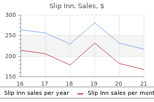
Slip Inn dosages: 1pack
Slip Inn packs: 10 caps, 20 caps, 30 caps, 60 caps, 90 caps, 120 caps, 180 caps

Buy slip inn 1pack online
The sigmoid colon can bear volvulus in either course herbals for erectile dysfunction 1pack slip inn order mastercard, clockwise or counterclockwise himalaya herbals nourishing skin cream cheap slip inn 1pack, and upon completion of the 360� flip a closed loop obstruction happens inside the affected segment. The hyperperistalsis and fluid secretion that follows further contribute to colonic distention and elevated rigidity throughout the colonic wall, which in flip leads to hypoperfusion, ischemia, and, ultimately, colonic wall necrosis. The central location of the gland in the upper retroperitoneum complicates the medical and surgical management of pancreatic illness. The dorsal pancreatic bud, destined to kind a portion of the pancreatic head and the entire body and tail of the pancreas, enlarges more quickly and extends into the dorsal mesentery. The ventral pancreatic bud, the source of the uncinate process and a portion of the pancreatic head, develops in affiliation with the hepatic rudiment and biliary ductal structures. Rotation of the ventral pancreatic bud to the left of the duodenum brings it under the dorsal bud. The pancreas is an elongated organ (12�20 cm in size in adults) that lies transversely in the upper retroperitoneum. The head of the pancreas lies on the best in the concavity of the duodenal sweep. The distal frequent bile duct traverses the top of the pancreas earlier than coming into the duodenum. The neck of the pancreas is bordered inferiorly by each the transverse mesocolon and the basis of the mesentery of the small intestine. The transverse mesocolon is attached to the inferior border of the tail of the gland; the abdomen contacts the anterior surface. The tail of the pancreas extends to the left within the leaves of the splenorenal ligament to the hilum of the spleen. The arterial blood provide of the pancreas is derived from each the celiac axis and the superior mesenteric artery. The exocrine pancreas is organized in lobular items composed of ductules and acini. Numerous zymogen granules are visualized via electron microscopic examination of the mobile apex. Centroacinar cells (which express the floor marker Hes-1, a Notch pathway signaling molecule) and ductal cells (which categorical cytokeratin-19) are extra columnar. The acini rest on a thin basal lamina penetrated by quite a few blood vessels and nerve fibers. Centroacinar cells have been recently implicated as a attainable cell of origin in pancreatic ductal carcinoma. The endocrine pancreas consists of roughly 1 million islets of Langerhans. The islets include endocrine cells that stain positively for insulin (75%�80%), glucagon (10%� 20%), and somatostatin (5%). Pancreatic polypeptide and various other different enteric peptides are also expressed within cells of the pancreatic islets. At the decrease proper is an islet of Langerhans, the endocrine portion of the pancreas, which is principally concerned in regulating glucose homeostasis. The asterisk is positioned amongst acini, which are involved in secreting varied digestive enzymes (zymogens) into the ducts (indicated by the stable arrow). Also depicted are centroacinar cells (arrow), which sit on the junction of the ducts and acini. These could additionally be broadly divided into developmental and adult patterns of anatomical variation. The boundaries of the stomach cavity, intra- and retroperitoneal divisions, and the peritoneal cavity subdivisions determine the location and appearances of the varied structural anomalies encountered since these are benign processes which are inclined to respect intact anatomy. The abdominopelvic cavity is bounded superiorly by the diaphragm, inferiorly by the pelvic floor, and circumferentially by the belly wall. The abdominopelvic cavity is split into intraperitoneal and retroperitoneal elements by the peritoneum. The peritoneal cavity is subdivided into a number of spaces and recesses via peritoneal reflections. Most notably, the peritoneal cavity is partitioned into supra and infra mesocolic compartments by the transverse mesocolon. Developmental and childhood structural anomalies of the abdominal cavity Any dysfunction of the physiological developmental rotation of the small bowel is termed malrotation. The key concern is the propensity for midgut volvulus with resultant obstruction and bowel ischaemia. Congenital intraabdominal cystic lesions embody lymphangiomata, mesenteric cysts, enteric cysts, and enteric duplication cysts. Exomphalos (omphalocoele) is a persistence of the physiological umbilical small bowel herniation and has coverings of amnion and peritoneum. Gastroschisis involves an anterior abdominal wall hernia that lies lateral to the umbilicus and lacks coverings. Congenital paediatric groin hernias are seen in 3%�5% of births and up to 30% of premature (male) infants. The vast majority are oblique inguinal hernias on account of incomplete closure of the processus vaginalis (Canal of Nuck in females). Adult stomach hernias Hernia is: "The protrusion of an organ or tissue out of the body cavity by which it usually lies. Any reason for increased intraabdominal strain predisposes to stomach wall hernia along with conditions that lead to weakening of the belly wall. Clinical analysis is sufficient to diagnose an abdominal wall hernia typically. Irreducible (incarcerated) hernias may be related to bowel obstruction, or bowel ischemia because of strangulation. The inguinal canal is an oblique channel, approximately four cm long, through the inferomedial anterior belly wall, and a web site of potential weak point. The adjacent femoral canal represents the medial compartment of the femoral sheath. Inguinal hernias account for over 70% of belly wall hernias with femoral hernias responsible for 5%�15% of instances. Lateral view exhibits a "corkscrew" appearance of the duodenum (arrow) that has torted around itself in a neonate with malrotation. These are described primarily based on location, appearances, or typically eponymously: � Pantaloon hernia occurs when direct and oblique ipsilateral inguinal hernias concurrently exist. Paraduodenal hernias represent approximately 53% of inner hernias; left-sided hernias are more widespread. Intermittent stomach ache or acute bowel obstruction (often closed-loop and strangulated) are the most generally noticed symptoms. Adult diaphragmatic hernias are mostly posttraumatic in origin with the left hemidiaphragm being extra frequently affected.
Diseases
- Krasnow Qazi syndrome
- Ventricular extrasystoles perodactyly Robin sequence
- Adenosine triphosphatase deficiency, anemia due to
- Primary agammaglobulinemia
- Amelogenesis imperfecta local hypoplastic form
- Thyrocerebrorenal syndrome
Buy slip inn 1pack fast delivery
There is usually palpable tenderness of the puborectalis/ levator muscle tissue because the examining finger moves posteriorly to anteriorly herbals 24 1pack slip inn proven. The key to the prognosis is the reproduction of the pain when the coccyx is manipulated between the examining finger and the thumb quality herbals 1pack slip inn generic with amex. Seventy to eighty % of acute pancreatitis sufferers develop gentle and uncomplicated acute pancreatitis, while 20%�30% will develop more extreme symptoms with concomitant multiple organ failure. The two most typical risk factors for acute pancreatitis are gallstone disease and extreme alcohol use. Indeed, inherited mutations in genes encoding for digestive enzymes are current in no much less than some patients with hereditary pancreatitis. In an try to establish predictors of difficult acute pancreatitis, several association research linking cytokines and chemokines with acute pancreatitis severity have been performed. Agitation, confusion, hypoxemia, and lack of enchancment within forty eight h are signs suggesting deterioration. Patients older than 55 years are also at larger threat of present process a extreme irritation of the pancreas. The affiliation between elevated haematocrit and poor outcome is of explicit interest. Strong infiltration and accumulation of immune cells are detectable round pancreatic lobules in acute pancreatitis (*). After excessive stimulation of the immune system, a paralysis of the immune system happens during the course of acute pancreatitis. The most typical risk factors for acute pancreatitis in adults are gallbladder disease (usually due to concomitant choledocholithiasis) and chronic or excessive alcohol consumption. In about 15% of patients the trigger stays unknown after thorough investigation, although this could turn out to be much less widespread as elements of genetic predisposition, environmental susceptibility, and autoimmunity are elucidated. The regular pancreas develops from the fusion of dorsal and ventral pancreatic buds throughout fetal improvement. In as a lot as 90% of individuals the ducts of both the dorsal and ventral buds fuse along with the parenchymal fusion, leading to the principle pancreatic duct draining the whole pancreas via the major papilla. Source: Courtesy of Professor Kl�ppel and Professor Esposito, Technical University of Munich. This leads on to high dorsal ductal stress during lively secretion, resulting in insufficient drainage and ductal distension. The cardinal symptom of belly ache is current in about 95% of patients and sometimes radiates in a band-like method to the lower thoracic region of the back. With biliary pancreatitis, the pain could additionally be more localized to the proper upper quadrant and extra variable in intensity over time because of the contribution of biliary colic. Surgical decompression by midline laparotomy provides the best therapy of this complication. Serum amylase typically rises within a number of hours after the onset of signs and returns to normal within 3�5 days. Conversely, amylase concentrations might be excessive in the absence of acute pancreatitis in individuals with macroamylasemia (a syndrome characterised by the formation of enormous molecular complexes between amylase and abnormal immunoglobulins), in sufferers with decreased glomerular filtration, in illnesses of salivary glands, and in extrapancreatic belly diseases related to inflammation, together with acute appendicitis, cholecystitis, intestinal obstruction or ischemia, peptic ulcer, and gynecological diseases. In patients with acute pancreatitis serum lipase remains elevated for an extended time frame than amylase, which can be helpful in patients with a delayed presentation. The biochemical markers of a biliary etiology of acute pancreatitis embrace an alanine aminotransferase elevation of more than 3 times the upper range of regular and a serum whole bilirubin greater than 3 mg%. Imaging is frequently beneficial to verify the clinical analysis, confirm the trigger, and grade the extent and severity of acute pancreatitis Table 35. Radiography, higher gastrointestinal collection, and ultrasound are of restricted value within the prognosis of acute pancreatitis. On stomach ultrasound, bowel gases typically masks focal hypoechoic areas within the pancreas. Abdominal ultrasound is useful in detecting cholelithiasis and biliary obstruction. Cholecystectomy for gallstone-induced acute pancreatitis must be carried out using a laparoscopic procedure after decision of acute pancreatitis. After the first week, distinction between these two kinds of collections is usually attainable. Cholelithiasis (*) and edematous (arrow) pancreas tissue with peripancreatic fluid areas are proven in anterior pararenal space at (a) arterial and (b) portal venous phases. Fine needle aspiration is helpful for ascertain prognosis and to acquire a specimen for culture. Classification of acute pancreatitis�2012: revision of the Atlanta classification and definitions by worldwide consensus. Complications such as bleeding, inadvertent puncture of adjacent viscera, secondary an infection, and extended durations of drainage with resultant pancreaticocutaneous fistulae could also be averted. The revised Atlanta classification of acute pancreatitis: its significance for the radiologist and its impact on treatment. Small-diameter plastic stents simply occlude, particularly with fluid collections containing debris. Stent occlusion could subsequently end in an infection and delayed decision of necrotic tissue. Moreover, surgical necrosectomy inside the first weeks is associated with excessive mortality rates (up to 65%). Pancreas is replaced by low-attenuation assortment with a well-defined rim and multiple pockets of gas. Pancreatic trauma is uncommon and associated with damage to different higher stomach viscera. Empiric antibiotics ought to embrace each cardio and anaerobic Gram-negative and Grampositive microorganisms. All images present peripancreatic secretion accumulation (arrow) and rupture in the neck of the pancreas. In the previous, parenteral vitamin seemed to be perfect for eliminating the stimulation of pancreatic secretion while stopping deterioration of dietary status and development to protein vitality malnutrition. Complications embody formation of pseudocysts, pancreatic duct stenosis, duodenal stenosis, vascular complications, biliary obstruction, malnutrition, and a persistent ache syndrome. Chronic pancreatitis is also a danger issue for the development of pancreatic cancer. The most typical explanation for an acute exacerbation of pancreatitis is continued alcohol abuse or dietary transgressions. Acute exacerbation of persistent pancreatitis manifests in two types, irrespective of underlying etiology: acute interstitial edematous pancreatitis (75%�85%) with a mortality of under 1%, and acute hemorrhagic necrotizing pancreatitis (15%�25%) with a mortality between 10% and 24%. Complications are frequent in patients with persistent pancreatitis, and pseudocysts are among the commonest findings.
Cheap 1pack slip inn fast delivery
As a handy approximation herbals for cholesterol slip inn 1pack online, the conventional clearance has typically been assumed by many clinicians to be approximately 100 mL/min herbs de provence recipes buy 1pack slip inn with mastercard. Several methods can be found for the calculation of creatinine clearance from the serum creatinine focus. These strategies ought to be used only for sufferers with intact liver operate and no abnormal muscle disease, similar to hypertrophy or dystrophy. Cockcroft and Gault (1976) compared their method with the nomogram methodology in adult males of various ages. Both strategies additionally demonstrated an age-related linear decline in creatinine excretion, which can be as a end result of the lower in muscle mass with age. Calculation of Creatinine Clearance from Serum Creatinine Concentration the issues of acquiring a whole 24-hour urine assortment from a patient, the time essential for urine collection, and the analysis time preclude a direct estimation of creatinine clearance. Serum creatinine concentration, Ccr, is related to creatinine clearance and is measured routinely within the medical laboratory. Children There are a quantity of methods for calculation of creatinine clearance in children, primarily based on body size and serum creatinine concentration. Turn the proper part of the ruler to the suitable serum creatinine value and the left side will indicate the clearance in mL/min. What is the creatinine clearance for a 25-yearold male patient with a Ccr of 1 mg/dL What is the creatinine clearance for a 25-yearold male affected person with Ccr of 1 mg/dL and a physique weight of 80 kg Connect the intersection point at Solution the patient is overweight and the Clcr calculation should be based mostly on ideal body weight. In acute renal failure and in other conditions in which kidney perform is changing, the serum creatinine might not represent steady-state conditions. If Ccr is measured day by day and the Ccr value is constant, then the serum creatinine concentration is probably at steady state. Although the Cockcroft�Gault technique for estimating Clcr has some biases, this method has gained basic acceptance for the determination of renal impairment (Schneider et al, 2003; Hailmeskel et al, 1999; Spinler et al, 1998). A suggested illustration of sufferers with various degrees of renal impairment primarily based on creatinine clearance is shown in Table 24-3. The practice issues present that, relying on the formula used, the calculated Clcr can vary significantly. Consequently, until a clinically vital change within the creatinine clearance happens, dosage adjustment will not be wanted. For aminoglycosides and vancomycin, dose adjustment is individualized in accordance with the wide range of Clcr. Each equation for the calculation of renal function from serum creatinine concentrations gives considerably completely different results. The Cockcroft�Gault methodology for estimating Clcr has been used most incessantly and tends to be the preferred approach at this time. These estimation strategies are referred to as creatinine-based strategies in the clinical literature (Stevens et al, 2006; Levey et al, 2009). The Cockcroft�Gault has a longer historical past of use but the original equation was based on fewer topics. The Cockcroft�Gault formula was developed initially with the information from 249 men with Clcr ranging from 30 to one hundred thirty mL/min. Typically, regular values for creatinine clearance are normalized by a physique floor area of 1. For many new medicine, drug dosing information for renal-impaired sufferers is now available and should be consulted within the package insert. In sufferers with continual kidney disease, the following recommendations are good practices that physicians and pharmacists ought to be conscious of (Munar and Singh, 2007): 1. Use warning for medication with lively metabolites that can exaggerate pharmacologic results in patients with renal impairment. Whether renal impairment will alter the pharmacokinetics of the drug sufficient to justify dosage adjustment is an important consideration. For many drugs that are eliminated primarily by metabolism or biliary secretion, uremia could not alter pharmacokinetics sufficiently to warrant dosage adjustment. Active metabolites of the drug may be fashioned and must be considered for additional pharmacologic results when adjusting dose. For some 786 Chapter 24 medication, the free drug concentrations may need to be thought of as a outcome of decreased or altered protein binding in uremia. Combination products that comprise two or more energetic drugs in a fixed-dose combination may be differentially affected by decreased renal operate and thus, the utilization of combination drug merchandise in uremic sufferers should be discouraged. The following strategies may be used to estimate preliminary and maintenance dose regimens. After initiating the dosage, the clinician ought to continue to monitor the pharmacodynamics and pharmacokinetics of the drug. As mentioned by Chennavasin and Brater (1981), every nomogram has errors in its assumptions and drug database. In the uremic affected person, the rate of renal drug excretion has decreased, leading to a decrease in whole body clearance. The fraction of regular renal function remaining in the uremic affected person is estimated from Clcr. After the remaining total physique clearance in the uremic affected person is estimated, a dosage regimen could also be developed by (1) decreasing the upkeep dose, (2) rising the dosage interval, or (3) altering each upkeep dose and dosage interval. Although whole body clearance is a more accurate index for drug dosing, the elimination half-life of the drug is more generally used for dose adjustment due to its convenience. Clearance permits for the prediction of steady-state drug concentrations, while elimination half-life yields information on the time it takes to attain steady-state concentration. Nomograms Nomograms are charts obtainable to be used in estimating dosage regimens in uremic patients (Bjornsson, 1986; Chennavasin and Craig Brater, 1981; Tozer, 1974). The nomograms may be based on serum creatinine where knr is the nonrenal elimination fee fixed and a is a constant. The fractions of drug excreted unchanged within the urine (fe) for medicine A, B, C, and D are 5%, 50%, 75%, and 90%, respectively. The uremic elimination fee fixed (ku) is the sum of the nonrenal elimination price fixed and the renal elimination price constant, which is decreased as a result of renal impairment. If the affected person has full renal shutdown (ie, Clcr = zero mL/min), then the intercept on the y axis represents the percent of drug elimination as a outcome of nonrenal drug elimination routes. Dose Adjustment in Renal and Hepatic Disease 787 Drug D, which is excreted 90% unchanged in the urine, has the steepest slope (equivalent to a in Equation 24. On the other hand, drug A, which is excreted solely 5% unchanged in the urine (ie, 95% eliminated by nonrenal routes), is least affected by a decrease in creatinine clearance.
Purchase slip inn 1pack with amex
Typically krishna herbals cheap slip inn 1pack without prescription, sufferers are asymptomatic between discrete episodes of rectal ache which final less than 60 min before disappearing utterly herbals scappoose oregon generic slip inn 1pack free shipping. Obstruction of the outflow of exocrine secretions both by an inflammatory mass, calcified protein plaques in pancreatic ducts, or narrowing of ducts by inflammatory scars normally causes continual pain. Gastric varices, especially within the fundus, could develop due to thrombosis of the portal vein or splenic vein. The inflammatory processes might lead to a fistula, which may hook up with the small or massive bowel and trigger quick bowel syndrome. Transabdominal ultrasound: dilated major pancreatic duct and protein plug throughout the duct (arrow). Transabdominal ultrasound: spherical, echo-poor liquid structure divided by a septum (arrow). Pancreatic pseudocysts are a frequent complication of acute and chronic pancreatitis. Transabdominal ultrasound: acoustic reflections as a result of pancreatic parenchymal calcifications (arrows). Radial endoscopic ultrasound: acoustic parenchymal reflexes because of calcifications. Improvement of major pancreatic duct visualization after secretin stimulation (d, e, f). Morphology of pancreatic ducts corresponds to grade 4 based on the Cambridge classification. Esophagogastroduodenoscopy: impression of the gastric lumen due to a pancreatic pseudocyst. Esophagogastroduodenoscopy: the endoscope is retroflexed with view to the gastric fundus. Angiography: (a) small pseudoaneurysm triggered severe bleeding (arrow); (b) successful treatment by coiling (arrow). Angiography: (a) proteolytic destruction of a big pseudoaneurysm (arrow) of the gastroduodenal artery brought on life-threatening bleeding. Duodenoscopy: (a) spontaneous perforation of pancreatic necrosis into the distal duodenal bulb; evacuation of pus and necrotic materials. This gene encodes cationic trypsinogen, the principal trypsin proenzyme in human pancreatic juice. Together, the 12 helices (h1�h12) surround an aqueous pore, which varieties a channel through the lipid bilayer and permits anions to passively move by way of the cell membrane. Common additional findings include neutropenia, and development or skeletal abnormalities. Localization of the cystic fibrosis transmembrane conductance regulator in pancreas. Localization of the cystic fibrosis transmembrane conductance regulator in human bile duct epithelial cells. As pancreatic juice flows through the intralobular duct, the protein-rich acinar secretions are diluted and alkalinized by the duct epithelial cells. This atrophic gland is fibrotic and reveals prominent lobulation with cystic modifications. Typical findings embrace focal portal and periportal fibrosis, ductular proliferation, and periductular irritation. Proximal to this obstruction, the small bowel is distended (note thickened bowel wall and gas bubbles in fecal material). Much of the mucosal surface is roofed with concretions consisting of inspissate mucofecal materials. If lively trypsin accumulates in the pancreatic parenchyma, this probably could cause an uncontrolled proteolytic chain reaction as a result of extra activated trypsin could be produced by the motion of trypsin on trypsinogen. To stop this, trypsinogen normally contains an inactivating cleavage website at an accessible site on its floor (R122). In hereditary pancreatitis, the R122H mutation prevents digestion of trypsinogen at R122 and this allows lively trypsin to accumulate in an uncontrolled method. Thus, excessive activation of trypsin is believed to be the first event inflicting pancreatic damage in many patients with hereditary pancreatitis. The pancreas shows changes consistent with chronic pancreatitis together with delicate atrophy and a number of punctate parenchymal calcifications (especially in the pancreatic head). Typical exocrine gland options embody fatty substitute of acini with sparing of ducts. Steatorrhea and diabetes mellitus typically occur two to four many years after the onset of pancreatitis, whereas pancreatic cancer is usually delayed by an additional two to three many years. Although the overall risk of malignancy could be very low, the presence of those pancreas cysts is related to a large diploma of anxiety and further medical investigation, as a outcome of issues about malignancy. They can current with dramatic hormonal symptoms due to dysregulated manufacturing and launch of endogenous and ectopic hormones. Clinical administration focuses on managing the signs of hormone extra as properly as localization and ultimately resection of the tumor mass. Necrolytic migratory erythema is a common presentation for glucagonoma and happens at analysis in 70% of circumstances. Central superficial bullae develop and rupture, forming erosions that crust and heal with hyperpigmentation. Relatively homogeneous cells are present in nesting, trabecular, or gyriform preparations. Tumor cells are small to medium sized with eosinophilic cytoplasm and uniform, round to oval, stippled nuclei. A 61-year-old girl introduced with 2 weeks of gastroesophageal reflux symptoms, stomach ache, nausea, and diarrhea. This lesion encases the splenic artery, invades the portal vein and results in a partial dilatation of the principle pancreatic duct. Wang2 1 2 University of Bari Medical School, Policlinico Hospital, Bari, Italy Saint Louis University School of Medicine, St. The majority (75%) of gallstones within the United States and Europe are cholesterol stones, which are often subclassified as both pure ldl cholesterol or combined stones, which include no much less than 50% cholesterol by weight. Pigment gallstones include mostly calcium bilirubinate and are subclassified into two teams: black (20%) and brown pigment stones (4. The three courses of biliary lipids are unesterified cholesterol (>95%), phospholipids (>95% lecithins), and bile acids, that are composed of main (cholic and chenodeoxycholic acids) and secondary bile acids (derived from 7-dehydroxylation of the first bile acids within the liver and by intestinal micro organism within the ileum and colon: deoxycholic, lithocholic, ursodeoxycholic, sulfolithocholic, and 7-oxo-lithocholic acids). Major risk elements for ldl cholesterol gallstones embrace rising age, female gender, being pregnant, metabolic syndrome, insulin resistance, speedy weight loss, bodily inactivity, high cholesterol food regimen, gallbladder stasis, estrogen and oral contraceptives, diabetes mellitus, and low serum magnesium. Liver and small gut present the most important sources of cholesterol leading to lithogenic bile.
Alchornea castaneifolia (Iporuru). Slip Inn.
- Are there safety concerns?
- What is Iporuru?
- Dosing considerations for Iporuru.
- How does Iporuru work?
- Coughs, problems with erections (impotence), diabetes, diarrhea, headache, toothache, snakebite, bronchitis, chancre sores, chills, eye inflammation (conjunctivitis), severe diarrhea (dysentery), painful or abnormal menstrual periods, arthritis, colds, and many other uses.
Source: http://www.rxlist.com/script/main/art.asp?articlekey=96112
Slip inn 1pack order line
It accommodates structural and useful proteins arranged in a tissue-specific orientation for direct healing and tissue reworking phoenix herbals 50x slip inn 1pack cheap visa. Surgisis Biodesign has been used as an alternative alternative to herbals forum purchase 1pack slip inn with visa splitthickness pores and skin grafting in human patients with full-thickness chronic leg ulcers and granulating open dermal wounds. The skin surrounding the groin harvest websites was undermined to assist in a tension-free closure. An ice pack was applied to help restrict swelling, and a Foley catheter was left in place for several days to assist in bladder drainage. Abdominal Flaps When other, more conventional choices have failed or circumstances dictate that a new tissue source should be used, stomach flaps present an alternative. Abdominal flaps are often utilized in other surgical procedures, such as breast reconstruction, and they could additionally be applicable in gynecologic reconstruction as properly. Disadvantages embody the potential for partial graft skin slough, which may require additional surgical intervention and grafting. This patient has complete vaginal obliteration despite 5 previous procedures for vaginal constriction, because of undiagnosed lichen planus. After a number of months of therapy with steroids and methotrexate the tissues look considerably improved. A giant defect along the distal posterior vaginal wall and perineal physique after excision of vaginal mesh secondary to dyspareunia. A painful vertical scar at the vaginal apex excised, and the defect bridged with the graft. The scar is incised and utterly excised, and the surrounding tissues are adequately mobilized in preparation for a perineal flap. A ruler and curved tissue forceps used to outline the world of stricture and to plan the size of graft needed for harvest. A narrowed midvaginal contracture in a longitudinal view, emphasizing a thick scar with a narrow passage connecting the upper and decrease vagina. A hinged perineal graft is then created instantly lateral to the labium majus on the facet of the contracture. Making the distal end of the flap round somewhat than pointed is suggested to reduce the danger of slough of the distal aspect of the graft. The flap is rotated into the defect and is secured to the adjoining tissue with interrupted sutures. The initial sutures safe the flap near the vaginal apex; subsequent sutures are positioned towards the introitus, with care taken to keep away from asymmetry. The tissue lateral to the labium majus is circumferentially mobilized to allow the incision to be closed in a tension-free method. Adequate control of her underlying illness course of, the will for a practical vagina, and the failure of previous vaginal dissections led to the recommendation to use bilateral perineal flaps for reconstruction. The vaginal dissection has been completed, and measurements have been taken to decide the scale of graft wanted. A broad base on the hinge preserved the blood supply, and rounding of the distal tip was carried out. The left perineal flap was rotated into the defect and was secured to surrounding tissue with interrupted sutures. When the left perineal flap was secured, vaginal dissection was repeated on the proper aspect, measurements have been taken, and the right perineal flap was mobilized. Interrupted sutures, beginning at the apex and dealing toward the introitus, were placed to secure the grafts in a symmetrical and tension-free method. After the flaps had been secured in place, the tissue surrounding the incisions lateral to the labia majora was mobilized. Often, the lateral facet of the incision can be mobilized to a larger degree than the medial facet, to keep away from a tethering impact on the remaining labial and periclitoral tissues. An preliminary layer of interrupted sutures was placed to reapproximate the subcutaneous tissues in the lateral incisions. The pores and skin was then reapproximated with a operating subcuticular, delayed absorbable suture. A Foley catheter was left in place to drain the bladder, and an ice pack was applied to restrict edema. Tissue edema within the perineal flaps was not unusual, and the grafts had been monitored for proof of vascular compromise. She eventually underwent hysterectomy, bilateral perineal flap, bilateral Singapore flap building, and, lastly, construction of a sigmoid neovagina, by which stenosis ultimately developed. The perineal space showed extensive scarring from earlier operations and pelvic irradiation. The sigmoid neovagina with a Lucite dilator in place (thick arrow) underwent stenosis and was mobilized from the left pelvic sidewall and adjacent rectum (thin arrow). After mobilization abdominally, the sigmoid neovagina was everted via the vagina and excised. With sharp dissection and cautery, the sigmoid neovagina was dissected free from the overlying bladder and urethra and underlying rectum. A preoperative left exterior ureteral stent was positioned to aid the identification and dissection of the left ureter. After the fascial edges had been reapproximated, the skin was closed, leaving a protracted, vertical midline scar. A de facto vaginectomy could additionally be performed as a result of therapy for in depth condyloma acuminata. The aim of vaginectomy is twofold: (1) to remove the illness and (2) to retain a functioning structure. The latter interprets into maintaining the vagina as a supple, nonconstricted, and suitably prolonged structure. The factor most frequently liable for vaginal deformity and accompanying dyspareunia is scar formation. As was noted in Chapter 50, neighboring organs are exceedingly shut (2-4 mm) to the vaginal mucosa. The vagina itself is a somewhat easy structure-essentially a possible area with its anterior and posterior walls in light contact in vivo. The vagina is attached at its decrease margin to the vulva and at its higher margin to the uterus, along with the uterine helps. The vagina is connected laterally to the levator ani and a mass of surrounding connective tissue (endopelvic fascia). The unfastened peripheral attachments enable movement, in addition to flexibility between the factors of relative fixation. Anteriorly, the vaginal wall and the bladder and urethral walls are in apposition. Similarly, an identical set of circumstances exists between the rectal and vaginal partitions posteriorly. When decreased to its lowest common denominator, the vagina is a pleated, flippantly muscled, highly vascularized skin tube. Intraepithelial neoplasia in the absence of glands occupies less than 1 mm of a vaginal wall cross-section. Treating the vagina more deeply to eradicate the illness provides nothing to the remedy however could adversely influence the functional outcome.
Slip inn 1pack buy mastercard
A 1: 100 diluted vasopressin resolution is injected beneath the cervical and vaginal mucosa with a 25-gauge needle and a triple-ring 10-mL syringe herbs mill 1pack slip inn otc. The cervix is kept taut by downward traction of the tenaculum and is completely freed from the rectum posteriorly quality herbals products pvt ltd slip inn 1pack discount without a prescription. The Metzenbaum scissors are directed away from the bladder and towards the stump in a rigorously executed spreadand-cut method. The cardinal and uterosacral stumps are sutured into every vaginal angle, and the vagina is closed transversely with interrupted zero Vicryl sutures. If prolapse is present, a culdoplasty or a vaginal vault suspension must be performed (see Chapters 53 and 55). As a cautionary observe, it should be understood that in the course of the supracervical hysterectomy, the bladder peritoneum could additionally be superior over the top of the cervix and sutured down posteriorly as a means of overlaying and peritonizing the stump. Conversely, the peritoneum of the sigmoid colon could additionally be advanced and sutured anteriorly for the same function. The patient subsequently desired elimination of the cervix because of a persistent foul discharge and postcoital bleeding. A scalpel is used to make a circumscribing incision into the cervix roughly 5 to 10 mm again from the exterior os. The bladder is sharply dissected from the cervix together with the anterior vagina; similarly, the posterior vagina and cul-de-sac are dissected free of the cervix. The cervical stump, after having its ligamentous and vascular pedicles secured, is minimize free via a pointy scalpel or scissors. The bladder has been dissected free from the anterior aspect of the cervix via Metzenbaum scissors. Again, notice the upward traction on the cervix, which facilitates the posterior dissection. Note that the rectum has been sufficiently mobilized off the posterior facet of the cervix. If intraepithelial neoplasia had been current or suspected, the cervix could be reduce up analogous to conization and serially sectioned. For the sake of organization, the vagina could additionally be divided into thirds: upper, center, and lower. Throughout its length, the vagina is intimately applied to the bladder and urethra anteriorly and is similarly applied to the rectum posteriorly. This particular transitional area can be thought-about the entry portal to or the exit portal from the vagina. When one is dissecting on this space, consideration should be given to the pronounced vascularity along the lateral and anterolateral walls and the necessity for vasoconstrictive agents. Middle Third the center third begins slightly below the urethrovesical junction and crosses beneath the lower margin of the symphysis pubis (posterior-inferior margin) (2. This portion, together with the cranial portion of the decrease third, has the best diploma of mobility in contrast with the remainder of the vagina. However, as one dissects caudally, the wall shared among bladder, urethra, and vagina allows no simple airplane of separation. The vagina terminates around the cervix, and the vaginal vault is divided into fornices by the protruding portio vaginalis of the cervix. Between the latter is a cold entry level between the posterior fornix of the vagina and the cul-de-sac. The relationships of the upper vagina to the bladder, urethra, and cervix require precise anatomic knowledge of the retroischial and retropubic (extraperitoneal) areas. Many gynecologists refer to the lateral areas as paravaginal, but in reality these areas represent the perivesical spaces in their entirety. The anterior boundary of the retropubic area is the symphysis pubis and the pubic bone. The perivesical areas lengthen on both facet of the bladder and end above at the pubic bone and the obturator internus muscle and beneath on the obturator internus muscle and the ischial bone. Large venous sinuses and cavernous sinuses account for this vascularity, which is most plentiful on the level of the bulb of the vestibule. Much controversy has existed as to which constructions help and maintain the place and integrity of not only the vagina but additionally its immediate neighbors: the bladder, the urethra, and the rectum. Common partitions are shared by the urethra, bladder, and vagina anteriorly and with the rectum and vagina posteriorly. The major support to the higher vagina consists of the cardinal ligaments, in addition to shared partitions amongst bladder, rectum, and, to a lesser extent, uterosacral ligaments. Also among the many cervix, higher vagina, and bladder exists a well-defined fascial layer, silvery white in colour. The deep cardinal ligaments prolong into the perivesical areas to the pelvic side wall. The upper vagina is provided by way of the pelvic plexus with input from the hypogastric plexus, prevertebral ganglia, and sacral nerves. The blood supply emanates from the descending branch of the uterine artery, the vaginal artery, and the internal pudendal artery. The rugous vaginal mucosa can be seen to merge with the smooth cervical mucosa on the far periphery of the portio vaginalis of the cervix. The decrease third of the vagina forms a unit with the labia minora, vestibule, urethra, and rectum. The bladder, the urethra, and a portion of the vestibule have been dissected freed from the anterior wall of the vagina and have been eliminated. The bladder covers the retroverted uterus, and the sigmoid colon (C) covers the uterus, which lies in the cul-de-sac. The bladder-urethra advanced has been eliminated, exposing the anterior (outside) wall of the vagina (V). The tip of the scissors is pointing to the pubocervical fascia of the vaginal wall. The blades of the scissors lie on that fascia and over the anterior vaginal fornix (F). In this case of vestibulitis, the boundary between the vagina and the vestibule is even more apparent. The Skene ducts (small arrow), paraurethral ducts (large arrow), and Bartholin ducts (white arrow) all are intimate with the outer wall of the vagina. The relationship of the Bartholin gland to the posterolateral wall of the vagina (V) is proven right here. Clamps are positioned across the upper and decrease margins of the Bartholin gland (arrow factors to the gland). Allis clamps stretch the lateral wall of the vagina over the positioning where the Bartholin gland was previously situated. A proctoscopic swab has been positioned in the defect created by extirpation of the gland.
1pack slip inn with visa
While the combination therapy is extremely promising by means of potential for durable responses herbals himalaya 1pack slip inn order otc, it carries significantly elevated toxicity herbs for weight loss 1pack slip inn cheap with amex, with 93% of sufferers experiencing treatment-related opposed occasions, and thus candidates for mixture immunotherapy have to be rigorously selected. The combination group additionally demonstrated larger rates of rash (55%), pruritis (47%), fatigue (38%), and diarrhea (34%) compared to single-agent immunotherapy alone (Wolchok et al. Furthermore, a significant proportion of patients (21�36%) on combination ipilimumab/nivolumab require discontinuation of therapy due to opposed events (Wolchok et al. Chronologically, dermatologic manifestations are often the primary to come up, followed by diarrhea or colitis, which usually present after the primary 1�3 cycles of immunotherapy. Grade three or four treatment-related antagonistic occasions happen with highest incidence in patients treated with ipilimumab/nivolumab mixture therapy (55%), and fewer regularly for single agent ipilimumab (27%), nivolumab (16%), or pembrolizumab (10%) (Larkin et al. Pruritis and rash happen in as a lot as 24% and 19% of ipilimumabtreated sufferers, respectively (Hodi et al. Pruritis might happen to a slightly lesser diploma with pembrolizumab compared to ipilimumab (Robert et al. Rarely, severe or life threatening grade 3�5 immune-mediated dermatitis can occur, as represented by StevensJohnson syndrome, poisonous epidermal necrolysis, or look of a bullous, ulcerated, or hemorrhagic rash which occurs in less than 1% of sufferers (Fecher et al. Patients on mixture ipilimumab/nivolumab do expertise larger rates of diarrhea of any grade (34�44%) (Wolchok et al. Onset of diarrhea normally occurs after the second cycle of immunotherapy, at a median time of seven. Grade 1 diarrhea consisting of less than 4 diarrheal bowel actions over baseline in 24 h ought to be managed with the antimotility agent loperamide. Patients should follow the American Dietary Association colitis food plan, increase fluid hydration, and receive electrolyte repletion as wanted (Weber et al. For grade 3�4 diarrhea outlined as a minimum of 7 diarrheal bowel movements over baseline in 24 h, immunotherapy should be completely discontinued. Oral prednisone 1�2 mg/kg every day or dexamethasone four mg every four h ought to observe as a gradual taper over no less than four weeks (Weber et al. Average time to resolution of grade 2 or greater diarrhea handled with a median dose of prednisone 80 mg daily was 2. Infliximab could be administered once each 2 weeks as essential for persistent symptoms, however is contraindicated in patients with bowel perforation or sepsis. If possible, tuberculosis testing is recommended prior to infliximab use (Fecher et al. If diarrheal signs worsen again while on reducing doses of a steroid taper, the steroids ought to be increased again to a dose of 80�100 mg per day and then more slowly tapered and inflimximab may be reinitiated if essential (Weber et al. Abdominal ache and presence of stool blood or mucous could be a signal of colitis, which may progress to bowel obstruction and barely perforation (<1%) if not promptly addressed (Hodi et al. Surgical seek the guidance of should be requested if the patient develops an ileus with severe diarrhea (Fecher et al. Hepatitis Immune-mediated hepatic impairment is uncommon and occurs in roughly 4% of ipilimumab treated sufferers (Hodi et al. Incidence of hepatic toxicity is 15�23% in sufferers treated with the ipilimumab/ nivolumab mixture, and 15% of instances are grade 3�4 (Wolchok et al. The hepatitis usually presents as an asymptomatic rise in serum liver transaminases or bilirubin or can involve fatigue and fever (Weber et al. Baseline liver function tests ought to be carried out previous to every cycle of immunotherapy and clinicians ought to contemplate screening for hepatitis B or C previous to immunotherapy initiation, particularly provided that immunotherapy trials sometimes exclude patients with energetic hepatitis B or C (Fecher et al. Clinicians ought to counsel patients in opposition to active alcohol use and ingestion of hepatotoxic medication whereas on immunotherapy (Chmiel et al. Upon any noted rise in liver function checks, clinicians should suspect illness progression, medication toxicity, or viral hepatitis, all of which must be ruled out earlier than diagnosing immunotherapy-mediated hepatitis. Once serum ranges rise to higher than or equal to 2 occasions the higher limit of normal, repeat testing is recommended every 1�3 days till stabilization or a decrease is famous, and autoimmune hepatitis evaluation together with antinuclear, antismooth muscle, antimitochondrial, and antiliver-kidney microsomal antibodies must be checked (Fecher et al. Liver function checks ought to be monitored a minimum of weekly, and day by day if the levels are greater than eight instances the conventional. In a rare cases refractory to these therapies, antithymocyte globulin has been effectively used (Chmiel et al. Grade 3 events included one case of hypopituitarism and two instances of hypophysitis. Grade 4 occasions consisted of hypopituitarism and decrease in serum corticotropin level (Hodi et al. In patients on combination ipilimumab/nivolumab in the phase I trial, 13% developed immune-mediated endocrinopathies (Wolchok et al. The most common immunotherapy-related endocrinopathy is hypophysitis and usually presents after the third cycle of ipilimumab (Fecher et al. Symptoms of hypophysitis can typically be imprecise and embody headaches, visible adjustments, fatigue, nausea, stomach ache, psychological standing modifications, and altered bowel habits. Treatment of hypophysitis first entails recognition, which may typically be troublesome as the signs are nonspecific and may mimic other common melanoma points such as brain metastases, that are essential to rule out. Immunotherapy must be held for any grade 3�4 endocrinopathy or in sufferers with symptomatic pan-hypopituitarism (Weber et al. Importantly, evidence of extreme dehydration or hypotension ought to make the clinician suspect an adrenal crisis, which is also managed with rapid initiation of excessive dose methylprednisolone. Infectious causes ought to be ruled out and an endocrinology consult is recommended. Patients with secondary adrenal insufficiency require physiologic hydrocortisone alternative and provided that pituitary dysfunction could be long-lasting or everlasting, sufferers ought to be informed that steroid upkeep could also be life-long (Weber et al. Hypothyroidism occurs at a rate of 6�10% in sufferers on ipilimumab, pembrolizumab, or nivolumab (Topalian et al. Time onset is variable, ranging from months to years after the initiation of immunotherapy (Ryder et al. Pembrolizumab has a slightly increased fee of both hypothyroidism (9% vs 2%) and hyperthyroidism (3% vs 2%) in comparability with ipilimumab (Robert et al. Most patients are both asymptomatic or present with fatigue and this often resolves with initiation of hormone supplementation (Ryder et al. Reported immune-mediated neurologic deficits include neuropathy, continual inflammatory demyelinating polyneuropathy, transverse myelitis, myasthenia gravis, and Guillane-Barre syndrome (Gaudy-Marqueste et al. Peripheral neuropathies may be only barely symptomatic and may resolve spontaneously, though persistent grade 2 neuropathy is treated by withholding the following dose of remedy and administering a prednisone or dexamethasone taper over 4 weeks (Weber et al. Pancreatitis Immune-mediated pancreatitis is rare, with an incidence of 1% or less in ipilimumab and pembrolizumab handled patients (Robert et al. Pancreatitis normally presents as asymptomatic elevations in amylase and/or lipase and sometimes contains fevers, malaise, or belly discomfort (Weber et al. Grade 1 or 2 pancreatitis might slowly resolve without intervention (Tirumani et al. Pneumonitis Incidence of pneumonitis is also very rare in ipilimumab-treated patients (<1%) and could also be slightly larger in sufferers handled with pembrolizumab (2%) (Robert et al. While grade three or 4 pneumonitis is rare, it might be extreme and analysis could additionally be delayed by initially attributing affected person signs to other causes. Symptoms and radiographic findings normally resolve with high-dose steroid therapy and discontinuation of the immunotherapeutic agent, but this could take a quantity of months and sufferers may symptomatically enhance forward of normalization of imaging findings (Barjaktarevic et al.
Effective slip inn 1pack
The saphenous vein is uncovered (scissors are beneath the vein) as it sweeps via the fats herbs de provence slip inn 1pack buy visa, extending from a medial location in the thigh and vectoring towards the midpoint below the inguinal ligament yam herbals mysore 1pack slip inn best. Close-up view of the saphenous vein penetrating the cribriform fascia and draining into the femoral vein (arrow). Several small veins may be seen to be a part of the junction of the femoral and saphenous veins. These small tributaries embrace the superficial epigastric, superficial exterior pudendal, and superficial circumflex iliac veins. The scissors are lateral to the pubic bone and the lacunar ligament, beneath the terminal portion of the inguinal ligament, and medial to the femoral vein. The Kocher clamp points to the exterior iliac artery just cranial to the purpose the place it crosses beneath the inguinal ligament. This vessel lies in its own fascial compartment and is separated from the femoral vein by powerful connective tissue (fascia) (arrows). Pressure on the nerve by the inguinal ligament when the inferior extremities are severely flexed may end up in femoral nerve palsy. The tip of the forceps points to the femoral nerve (*), which was embedded inside the substance of the psoas muscle. The exterior iliac artery (small arrow) and the exterior iliac vein (outlined small arrow) beneath the artery are located medial to the retracted muscle. Deep throughout the pelvis, above the sacrum, the femoral and obturator nerves be part of the lumbosacral trunk. The finger of the surgeon factors to the medial thigh and site of the gracilis muscle. The muscle arises from the lower portion of the symphysis pubis and pubic bone and inserts onto the medial surface of the tibia. The vulva is an integral factor of female sexual anatomy and physiology, and its loss significantly compromises an necessary day-to-day function. A modification to simple vulvectomy is "skinning vulvectomy," which is a shallower excision. Logically, the need for deep excision for intraepithelial illness is troublesome to justify as a end result of the average thickness of involved mucosa (hair-bearing areas) ranges from 0. Thus an excision of 2 to three mm will remove in excess of 95% of concerned pores and skin and appendages, predictably eradicating the disease. No justification is understood for excising the vulva to a depth greater than 5 mm unless the operation is being performed for invasive carcinoma. The incision is carried down from the lower mons to the lateral facet of the labium majus with a 3-mm peripheral margin (from the lateral crease of the labium). This is sustained to the bottom border of the labium majus and then throughout the perineum to the opposite facet. A vasopressin 1: a hundred resolution is injected alongside the shallow reduce edges of the incision. The defect created by excision of the labia majora and perineum is covered by a split-thickness skin graft, and a stress dressing is applied. If the labia minora, vestibule, and clitoris are involved in the intraepithelial neoplasia, then excision ought to include these buildings. If the hood and glans are concerned and have biopsy-proven carcinoma in situ, then the glans clitoris, sheath, and frenulum should be excised with the labia minora. Electrosurgical coagulation and dissection should be avoided on this space as a outcome of it devitalizes tissue and will increase the risk of necrotizing fasciitis. It is apparent that the surgeon should dissect superficial to the exterior anal sphincter, perineal muscle tissue, and levator ani muscles during the perineal portion of the vulvectomy. Exposure of muscle signifies that the surgeon has dissected unnecessarily too deep. The operative website should be coated with Silvadene cream three times per day and at bedtime when main closure has been applied. The rules of this operation are to deeply resect the tumor with extensive peripheral margins and to extend the zone of resection to the vaginal and anal mucosa. This is coupled with an en bloc resection of the superficial inguinal and deep femoral lymph nodes. The incisions are reduce transversely throughout the lower stomach simply above the symphysis and curving upward to the anterior superior iliac backbone. The incision is sustained in a style similar to that described for simple vulvectomy. The cribriform fascia overlaying the fossa ovale has been exposed and dissected away. The vein again is ligated at the decrease portion (apex) of the femoral triangle as a end result of a section of this vein is included with the lymph node and fat specimens. Whether or not deep node dissection is carried out, the lowest external iliac node must be extricated and sampled. The muscle is definitely separated via curved Mayo scissors from its origin on the anterior superior iliac spine. The incision at the superior portion of the mons is prolonged on the best and left sides. These vessels are suture-ligated with 3-0 Vicryl sutures after enough hemostasis has been obtained. Tension closures result in wound separation and have a tendency to then heal by granulation. The subcutaneous tissue is sutured with 3-0 Vicryl interrupted sutures and is approximated above the drains. The vestibule is sutured to the remaining perineal pores and skin with interrupted 3-0 Vicryl sutures. The inferior extremities ought to be saved elevated to enhance lymphatic drainage (wrapped in elastic bandages or pressure stockings) in the course of the postoperative course. Advantages include preservation of the groin skin layer, avoidance of incisional groin an infection, and lymphedema of the legs. This technique approaches easily the vulvar sentinel nodes as a result of the fossa ovalis and the junctions of the femoral and higher saphenous veins are in close proximity to the labialcrural fold. I choose to use postoperative stress dressings secured by keep sutures placed by way of the groin and anchored to the underlying fascia. The lateral incision alongside the labial-crural fold used for the unconventional vulvectomy can also be used to develop the skin flap over the femoral triangle with extension of the dissection above the inguinal ligaments and pubis. The proximity of the fossa ovalis and its vascular structures to the labial-crural fold is emphasized. The single Texas Longhorn incision, which is illustrated, is a more skin-sparing technique than the historical butterfly incision. Initially, only the upper a half of the unconventional vulvectomy incision is used to develop the groin skin flaps. These cavernous constructions (clitoris, bulb and vestibule, and corpora cavernosa) can and will bleed with out remission for lengthy durations, as manifested by a constant gradual ooze. Pressure of the blood on the vulvar pores and skin could compromise its blood provide, inflicting actual necrosis. Therefore when this situation occurs, the hematoma have to be drained to relieve the stress.
Slip inn 1pack buy on-line
Cephalad migration of the needle away from the back of the pubic bone is the most common explanation for bladder perforation herbals on express slip inn 1pack generic without prescription. External rotation of the handle will initially result in penetration of the obturator internus muscle by the needle tip herbs books 1pack slip inn generic visa, with the potential to injure aberrant vessels along the lateral pelvic sidewall. Continued exterior rotation of the deal with with cephalad migration of the needle may result in injury to the obturator neurovascular bundle or (D) the external iliac vessels. I favor to use basic anesthesia; however, some surgeons prefer intravenous sedation with local anesthesia to permit the efficiency of the cough stress check to facilitate appropriate tensioning of the sling. Because approximately 50% of cases are accomplished at the facet of a prolapse repair, all surgeons must be properly versed at tensioning methods beneath general anesthesia. The anterior vaginal wall is hydrodistended with a mix of lidocaine and epinephrine, with the goal of utterly blanching the anterior vaginal wall on the stage of the mid to distal urethra. A scalpel blade is used to make an incision from slightly below the exterior urethral meatus to the extent of the mid urethra. The vaginal wall is sharply dissected with Metzenbaum scissors off the posterior urethra, creating small tunnels to the inferior pubic ramus. Some physicians favor to hydrodissect the trocar trajectory bilaterally before passing the trocars by using a spinal needle, injecting fluid along the again of the pubic bone. A catheter information is placed within the indwelling Foley catheter in order that the urethra and bladder neck may be displaced away from the place the trocar is inserted. The trocar tip is inserted into the previously dissected tunnel on both sides lateral to the urethra and advanced to the undersurface of the pubic bone. The tip of the needle is carefully advanced via the endopelvic fascia into the retropubic house. When the resistance of the endopelvic fascia is overcome and the tip of the needle is in the retropubic space, the deal with of the trocar is dropped and the needle is superior through the retropubic house because it hugs the again of the pubic bone. Cystoscopy is performed with a 30- or 70-degree scope to consider the bladder for inadvertent trocar harm with the trocar in place. During repassing of the trocar, great care ought to be taken to hug the back of the pubic bone. In such instances, the patient should proceed with the voiding trial postoperatively without the necessity for discharge with an indwelling catheter as a end result of the bladder perforation is very small and is often in a excessive, nondependent portion of the bladder. If extreme hematuria is present or the perforation is in the base or trigone of the bladder, continuous postoperative bladder drainage ought to be undertaken. The length of drainage ought to be decided on the basis of the kind and extent of the bladder damage. As the ends of the mesh gadget are attached to the trocars on all sides, the mesh with its plastic sheath is pulled up through the suprapubic stab wound along the trocar trajectory. Some surgeons choose to perform the procedure beneath native anesthesia and use a cough stress check. In such situations the sling is tensioned to the point at which minimal leakage occurs during coughing. Regardless of tensioning method, the final word endpoint is to create a laxity within the mesh manifested by a ricochet of the mesh back towards the urethra if pulled on vaginally with a right-angle clamp whereas also avoiding direct mesh contact with the underside of the urethra. After, the plastic sheaths masking the mesh are removed, and tension of the mesh is rechecked. The vaginal wound is copiously irrigated and closed with a working 3-0 polyglycolic acid suture. The suprapubic stab wounds are closed with absorbable suture or liquid tissue adhesive. Vaginal packing may be inserted briefly on the completion of the case if the affected person is bleeding or concurrent prolapse procedures are being carried out. The catheter may be eliminated along with the vaginal packing in the recovery room, and the affected person is discharged after confirming voiding efficiency. Mayo or Metzenbaum scissors are used to create a tunnel to the inferior pubic ramus. The tip of the needle is placed within the small tunnel that has been created and may come into direct contact with the inferior pubic ramus, pointing toward the ipsilateral shoulder. With the index finger of the nondominant hand within the vagina and the thumb on the shaft of the needle, the tip is pushed via the urogenital diaphragm. Once the resistance of the urogenital diaphragm is overcome, the handle is dropped and the needle is moved in a medial and superior direction, whereas direct contact with the again of the pubic bone is maintained. The tip of the needle is then palpated suprapubically and is guided to exit through the beforehand created stab incision. Leakage of urine throughout a coughing stress test indicates the need for adjustment of the sling materials. Tunnels are created bilaterally to enable trocars to come into direct contact with the inferior pubic ramus. At the previously marked puncture sites in the suprapubic area, a stab incision is made on each side. A trocar is inserted into the first of the suprapubic incisions whereas aligning with the sagittal axis of the physique after which fastidiously puncturing via the anterior rectus sheath. Angling caudally and "strolling off" the superior posterior fringe of the pubic bone, the trocar is superior into the retropubic area maintaining close contact with the posterior floor of the pubic bone. In a controlled manner, the trocar is progressively advanced until the tip is visible in the vaginal incision. The mesh is hooked up to the trocars, and the trocars are withdrawn through the suprapubic stab wounds. These slings are handed through a gaggle of inner thigh muscle tissue, particularly the gracilis tendon, adductor brevis, and obturator externus. Both methods contain specially designed needles which would possibly be handed from the obturator area into the vagina or from the vagina into the obturator region. When passed from outside-in, the sling is directed from a small incision lateral to the clitoris at the inferior edge of the adductor longus tendon, by way of the obturator foramen, around the ischiopubic ramus, and into the anterior vagina at the level of the midurethra. It passes so as by way of the next structures: gracilis tendon, adductor brevis muscle, obturator externus muscle, obturator membrane, and beneath or through the obturator internus muscle and periurethral endopelvic connective tissue; it lastly exits into the opened vagina. In the technique used for the inside-out approach, the same buildings are passed through in the other way. The adductor brevis muscle is displaced to reveal the situation of the obturator externus muscle, the site of which lies immediately on the obturator membrane. The anatomic location of a transobturator suburethral sling is drawn on the cadaver. Average distance from the Monarch device to the obturator vessels in six fresh-frozen cadavers. Average distance from Monarch device to obturator nerves in six fresh-frozen cadavers. The clamp is pointing to the arcus tendineus fascia pelvis and the obturator internus muscle.