

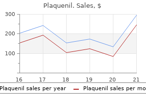
Plaquenil dosages: 400 mg, 200 mg
Plaquenil packs: 10 pills, 20 pills, 30 pills, 60 pills, 90 pills, 120 pills, 180 pills, 270 pills, 360 pills

Purchase plaquenil 400 mg otc
Intestinal microbes are documented to play an important function within the inflam matory response arthritis diet food list buy plaquenil 400 mg low price. In addition arthritis associates buy plaquenil 400 mg on-line, bacteria that may adhere to and invade the intestinal mucosa, similar to Escherichia coli, have been implicated. Constitutional symptoms similar to fatigue, fever, and weight reduction are regularly current. A thorough historical past should be obtained to exclude other causes of diarrhea (see Chapters 5 and 6). In addition, a dermatologic examination might reveal erythema nodosum, pyoderma gangrenosum, or aphthous ulcers (see Chapter 26). Related to bowel activity: peripheral arthritis, erythema nodosum, episcleri tis, aphthous stomatitis, pyoderma gangrenosum (see Chapter 26). Independent course: ankylosing spondylitis, sacroileitis, uveitis, primary sclerosing cholangitis. Related to malabsorption: anemia, cholelithiasis, nephrolithiasis, metabolic bone illness. Laboratory Features Laboratory tests are useful in assessing the severity of illness. A complete metabolic profile might reveal electrolyte abnormalities related to fluid loss, and hypoalbuminemia related to intestinal malabsorption. Markers of inflammation: � Serum Creactive protein and erythrocyte sedimentation price levels. Levels correlate positively with scientific, endoscopic, and radiologic measures of disease activity. Stool culture for bacterial pathogens and stool examination for ova and parasites should be performed in patients during their preliminary presentation. Imaging (see Chapter 27) Smallbowel followthrough and enteroclysis: � these exams are not used as firstline diagnostic tests. Microscopic examination of the mucosa (see Chapter 28) typically present crypt abscess, crypt distortion, and elevated cellularity within the lamina propria. Fourquadrant mucosal biopsies should be obtained every 10 cm, starting in the terminal ileum as the colonoscope is withdrawn for a minimum 33 biopsies; as properly as, all suspicious lesions should be biopsied. The yield of surveillance could be increased by utilizing techniques such as chromoendos copy and slim band imaging to establish areas of dysplasia. The alternative of medical remedy is determined by the extent, location, and severity of the disease and the presence or absence of fistulas. The therapeutic effect of aminosalicylates is decided by the native concentration at the inflamed mucosa. They can be found in oral (prednisone, prednisolone, budesonide), intrave nous (hydrocortisone, methylprednisolone), and enema (hydrocortisone) formulations. Budesonide is a glucocorticoid with high (>90%) firstpass liver metabolism; as a result, budesonide has considerably fewer unwanted effects than typical glucocorticoids; it has a gradual onset of action. Side results of glucocorticoids embody zits, hypertension, hirsutism, cataracts, striae, hyperglycemia, hyperlipidemia, insomnia, hyperactivity, acute psychotic episodes, adrenal suppression, and weight acquire. Idiosyncratic (doseindependent): acute pancreatitis, nausea, vomiting, diarrhea, flulike symptoms. Although the exact mechanism of motion is unknown, they inhibit Tcell proliferation and induce apoptosis. Ustekinumab: � Ustekinumab is a monoclonal antibody to the p40 subunit of interleukin12 and interleukin23. Surgery Surgery is indicated for the remedy of medicationrefractory, worsening illness and issues, including severe medication unwanted effects, fistulas, poisonous megacolon, and obstruction. It is outlined as acute colonic dilatation with a transverse colon diameter of >6 cm (on imaging examination) in a patient with a severe assault of colitis. Other danger factors include main sclerosing cholangitis, backwash ileitis, a family history of colon most cancers, youthful age at diagnosis of disease, and more extreme inflammation. C Stool culture and examinations for ova and parasites and Clostridium difficile toxin. Physical examination reveals nor mal important indicators and proper decrease quadrant tenderness without rebound tenderness. Besides a barely elevated white blood cell depend, the blood work is unremarkable. He is positioned on remedy with prednisone forty mg daily, which is tapered over the course of two months, however he becomes symp tomatic again at a dose of 20 mg per day. Magnetic resonance enterography reveals a fibrotic stric ture approximately eight inches (20 cm) in size, with proximal dilatation of the small bowel. Due to workrelated activities over the previous 3 weeks, he has not been taking his prescribed drugs frequently. On admission, the patient is pale and diaphoretic, with a pulse rate of one hundred twenty per minute, blood stress 90/55 mmHg, and temperature one hundred and one �F (38. The white blood cell depend is 18 000 mm�3, hematocrit worth 15%, and hemoglobin level 6 g dl�1. An intravenous line is started, and fluids and packed pink blood cells are administered. Colonoscopy is an affordable option to 130 Luminal Gastrointestinal Tract confirm the prognosis. Vital indicators, bodily examination, laboratory check results, and up to date endoscopy are all in keeping with mildto average disease. The subsequent step is to prescribe a glucocorticoidsparing drug, specifically azathioprine or 6mercaptopurine. Therefore, surveillance colonoscopy should be obtained annually or bien nially after eight years of disease. A full blood depend also needs to be obtained annually to consider the affected person for anemia. Surgical resection of the strictured terminal ileum with primary anastomosis of the small bowel to the colon is the greatest choice for this patient. A colonic diameter of >6 cm on a plain abdominal Xray (or computed tomography) is suggestive of toxic megacolon. All the other checks listed above are contraindicated in a affected person suspected of having poisonous megacolon. She has no nocturnal pain, early satiety, stomach cramps, diarrhea, rectal bleeding, or weight reduction. Physical examination reveals a blood stress of 130/70 mmHg, pulse price 70 per minute, and physique mass index 25; she is afebrile. Routine laboratory checks present a traditional complete blood count and normal ranges of blood glucose, serum electrolytes, creatinine, and calcium. General Constipation impacts roughly 15% of the final population, however can have an effect on as much as 50% of individuals over sixty five years of age. Physiology Colonic Motility Coordinated contraction of the colonic muscle is important to propel colonic contents in the path of the anus. Peristalsis includes contraction of the colon proximal to an area of distention and relaxation of the colon distal to the world of distention.
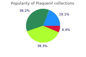
Plaquenil 200 mg purchase with mastercard
A massive arthritis x ray 400 mg plaquenil purchase fast delivery, heavy needle driver was then used to manipulate the rod and again it out through its authentic entry website arthritis foundation generic plaquenil 400 mg free shipping. If implant retrieval is important, each the previous presacral corridor and commonplace anterior retroperitoneal approached have been described. Access associated problems in anterior lumbar surgery carried out by spinal surgeons. Retrograde ejaculation after anterior lumbar interbody fusion with and with out bone morphogenetic protein-2 augmentation: a 10-year cohort controlled examine. Urological issues following use of recombinant human bone morphogenetic protein-2 in anterior lumbar interbody fusion: offered at the 2012 Joint Spine Section Meeting: clinical article. Avoiding longterm disturbance to bladder and sexual operate in pelvic surgery, significantly with rectal cancer. The infra-aortic arteries of the backbone: their variability and clinical significance. Minimally invasive presacral retroperitoneal approach for lumbosacral axial instrumentation. Particular consideration is warranted to the relatively greater failure price of two-level procedures, and additional research is critical to clarify its ongoing position as a legitimate treatment choice. The approach utilizes the retrorectal or presacral space, the plane between the visceral fascia of the mesorectum and the parietal fascia overlaying the anterior facet of the coccyx and sacrum 212 Kim et al. This allowed for maintenance or restoration of the intervertebral house for decompression of the neural parts after earlier discectomy or decompression as well as anterior help and fusion floor. The system is inserted into the intervertebral physique space of the lumbosacral spine, and is meant for intervertebral body fusion. It is meant to stabilize the spinal segment to promote fusion to limit motion and decrease pain using bone graft. The authentic indication was for L4�S1 single-level anterior fusions in sufferers who had failed a minimal of 6 months of nonoperative therapy. As evident in the name of the procedure, interbody fusion between the focused contiguous vertebrae is a necessity to the optimal outcome. The advantages of interbody fusion embody decreased instability and motion of degenerative, pathologic, or pain-generating motion segments. Diagnosis of pseudoarthrosis stays one of the functions of follow-up, although some have questioned its significance on situation that direct correlation between pseudoarthrosis and worse scientific outcomes has been troublesome to show in lumbar fusions. Other radiographic findings utilized in evaluation of pseudoarthrosis are radiolucent clear zones around pedicle screws and endplate cyst formation. Conservative remedy remains an choice, much like previous to the index process. Careful consideration must be paid to surgical affected person choice with exact prognosis of pain-generating segments, adjacent-level pathology, world spine steadiness and or deformity, and total affected person targets and expectations. Choice of instrumentation, approach or approaches, and fusion ranges must be planned. Early revision methods typically concerned explantation of the system, at occasions for migration of the interbody gadget. The common precept of revision backbone surgical procedure should be saved in mind-if you wish to change the outcome, you have to do something totally different than you did the first time. Barrier of fibrin glue or hydrogel sealant posterior to interbody device to seal annulotomy. Spurred on by constantly wonderful fusion charges regardless of early reports of problems, bigger series had been reported including one by which a polyethylene glycol hydrogel sealant (Duraseal, Confluent Surgical Inc. Furthermore, "bone resorption within the vertebral body led to graft subsidence and lack of radiographic evidence of progression towards fusion in multiple cases. The question raised was whether or not osteolysis could also be important within the early postoperative period in sufferers with new or continued pain. Histopathology of one of many patients revised for symptomatic posterior instrumentation showed granulation tissue subsequent to trabecular bone with suggestion of irritation on the site of osteolysis. In addition, Balseiro and Nottheimer36 reported two cases of postoperative pain that showed evidence of osteolysis seemingly originating from their preexisting subchondral endplate cysts, citing their preoperative existence as a potential threat factor for subsequent osteolysis. These early stories seemed to counsel affiliation of osteolysis with early unfavorable outcomes with variable longer term implications. They reported minimal associated cage migration or subsidence, though not quantified, and instructed the posterior instrumentation stabilized and negated any potential resultant instability. They calculated mean subsidence as 24% (13�40%) versus 12% (11�14%) within the two groups, respectively. The resultant lack of intrinsic strength of the graft and endplates was followed by subsidence of the graft and lack of intervertebral height. Eight of the 9 (88%) patients with cage migration required revision secondary to neurological symptoms. Later revisions discovered the cages fused of their posteriorly migrated place with both cage and heterotopic bone impingement on neural buildings. Placement of cages/spacers at peripheral places of interbody house is presumably much less susceptible to subsidence if osteolysis happens. Preexistence of subchondral endplate cysts may be a threat factor for developing adjoining osteolysis. Osteolysis with associated cage migration may be evident at or before 6 weeks postoperative on plain radiographs. Maintaining an increased awareness of those potential complications when osteolysis is present is important. Cage migration with ensuing radicular pain typically, but not always, requires revision surgical procedure with worse clinical outcomes. Complications of Posterior and Transforaminal Lumbar Interbody Fusion One problem in comparing results of osteolysis is the differing postoperative imaging protocols. No patients required revisions for osteolysis, subsidence, or migration, although no scientific outcomes were described. Subsequently, reappearance of the ache and incomplete enchancment of the numbness occur. He felt strongly the cause was a traumatic results of overzealous retraction of the nerve roots because of insufficient surgical publicity and advocated a wider exposure with partial facetectomy during nerve root decompression. Pheasant and Dyck54 of their 1982 article on failed lumbar disc surgery, attribute the "battered root" and the resulting arachnoiditis as one cause of failure. Soon thereafter in 1985, a German group reported successful remedy of the "battered root syndrome" with indwelling spinal cord stimulators. Battered root syndrome warrants additional investigation into the incidence, mechanism, prevention, and treatment. On the contrary, one potential series equally reported very excessive rates of osteolysis, however differed in that cage migration with resultant radiculopathy was also discovered at a high fee. Both revisions discovered a discrete inflammatory mass at time of decompression that exhibited histopathology of "diffuse osteoid and woven bone amidst a fibrovascular stroma densely populated by lymphocytes and eosinophils. They famous a ensuing lower in the price of radiculitis from 20 to 5% with use of the sealant. Because of its hydrophilic properties, Duraseal is understood to have expansile properties, but has been comparatively protected when used in this "off-label" fashion within the spine with solely isolated case reviews of such dramatic issues. Neidre and Macnab64 developed a classification system for lumbosacral nerve root anomalies.
200 mg plaquenil buy otc
T10 to T12 � Visceral afferent fibres travel with the sympathetic nerves and enter the cord through corresponding dorsal roots (T12 �2) arthritis pain and sweating cheap 200 mg plaquenil with visa. Vagina Vagina is a fibromuscular tube that extends from the cervix to the vestibule of the vagina rheumatoid arthritis walk 200 mg plaquenil order otc. Vaginal fornix is the recess between the cervix and the wall of the vagina and is split into three areas. Anterior fornix is located anterior to the cervix and is said to the vesicouterine pouch. The urinary bladder is palpable through the anterior fornix during a digital examination. Posterior fornix is located posterior to the cervix and is related to the rectouterine pouch (of Douglas). The rectum, sacral promontory (S1 vertebral body), and coccyx are palpable through the posterior fornix throughout digital examination. It opens into the vestibule and is partially closed by a membranous crescentic fold, the hymen. It is supported by the levator ani; the transverse cervical, pubocervical, and sacrocervical ligaments (upper part); the urogenital diaphragm (middle part); and the perineal body (lower part). Epithelium the epithelium of vagina is non-keratinized, stratified, squamous much like, and continuous with, that of the ectocervix. Arterial Supply Superior portion of the vagina is provided by the vaginal branches of uterine artery (branch of inside iliac artery). Middle and lower portions of the vagina are supplied by the interior pudendal artery, which arises from the internal iliac artery. Lower one-fourth (below the hymen) belong to perineal area and drains into the superficial inguinal nodes. Nerve Supply Upper three-fourths of vagina is equipped by the pelvic splanchnic nerves (S2, S3 and, sometimes, S4). Lower one-fourth is equipped by the deep perineal branch of the pudendal nerve (S2, S3 and S4). External Genitalia Female exterior genitalia (or vulva/pudendum) consists of a vestibule of vagina and its surrounding constructions such as mons pubis, labia majora, labia minora, clitoris, vestibular bulb and pair of larger vestibular glands. These longitudinal folds run downward and backward from the mons pubis and are joined anteriorly by the anterior labial commissure. Labia Minora are two folds of hairless pores and skin (with no fat) located medial to the labia majora that enclose the vestibule of the vagina. Labia minora are divided into higher (lateral) parts, which, above the clitoris, fuse to kind the prepuce of the clitoris, and decrease (medial) components, which fuse under the clitoris to form the frenulum of the clitoris. Posteriorly every labium minus is continuous with the fourchette, which connects the labia with the vaginal introitus. The physique of the clitoris is fashioned by two corpora cavernosa, that are steady with the crura of the clitoris. It is the space between the labia minora, which contains the urethral orifice, paraurethral glands (of Skene), vaginal introitus (incompletely covered by the hymen), greater vestibular glands (of Bartholin), and lesser vestibular glands. Bulbs of the Vestibule are a paired mass of erectile tissue on both sides of the vaginal orifice and are the homologues of the bulb of the penis of the corpus spongiosum. Each bulb is joined to one another and to the undersurface of the glans clitoris by a slim band of erectile tissue. They consist of two small oval our bodies that flank the vaginal orifice, involved with, and often overlapped by, the posterior finish of the vestibular bulb. They are situated barely posterior and on each side of the opening of the vagina. Each opens into the vestibule by a 2 cm duct, located within the groove between the hymen and the labium minora. The epithelium of the Bartholin duct is cuboidal near the gland, however becomes transitional and finally stratified squamous close to the opening of the duct. Perineal physique � the female external genitalia (or vulva/pudendum) consists of a vestibule of vagina and its surrounding structures such as mons pubis, labia majora, labia minora, clitoris, vestibular bulb and pair of larger vestibular glands. Located at the junction of anterior 1/3 and middle 1/3 of labia majora � Bartholin gland is situated on the junction of center 1/3 and posterior 1/3 of labia majora. Perineum Perineum is the diamond-shaped area between the thighs, which corresponds to the outlet of the pelvis and presents It contains perineal pouches (superficial and deep); ischiorectal fossa; pudendal canal and anal canal. Boundaries: Anterior: Pubic symphysis, arch and the arcuate ligament Anterolateral: Ischiopubic rami Lateral: Ischial tuberosities Postero-lateral: Sacrotuberous ligaments Posterior: Tip of the coccyx Floor: Skin and fascia Roof: Pelvic diaphragm and associated fascia It is divided into an anterior urogenital triangle and a posterior anal triangle by a line drawn across the surface connecting the ischial tuberosities. Pelvis Perineal Pouches Urogenital triangle accommodates the superficial and deep perineal pouches (spaces): Superficial Perineal Pouch It lies between the perineal membrane (inferior fascia of the urogenital diaphragm) and the Colles fascia (membranous layer of superficial perineal fascia). It is an open compartment, as a end result of the truth that anteriorly, the house communicates freely with the potential area lying between the superficial fascia of the anterior stomach wall and the anterior abdominal muscle tissue. The superficial perineal muscular tissues are eliminated in the left of the diagram to show crus and bulb of the penis. The superficial perneal muscular tissues have been eliminated within the left half of the diagram to show bulb of the vestibule and higher vestibular gland. Deep perineal pouch is enclosed partly by the perineum, and situated superior to the perineal membrane (inferior fascia It lies between the superior and inferior fasciae of the urogenital diaphragm. Recently the deep pouch is being described as the area between the perineal membrane and pelvic diaphragm. It incorporates muscles like external urethral sphincter and deep transverse perinei, attaching to the perineal body. The ducts cross by way of the perineal membrane to reach superficial perineal pouch and open into the bulbous portion of the spongy (penile) urethra. Clinical Correlations � Episiotomy is a surgical incision of the perineum (and the posterior vaginal wall) to enlarge the vaginal opening during childbirth. Perineal Fascia Perineal fascia has two components (superficial and deep) and each of these could be subdivided into superficial and deep elements. Posteriorly, it curves across the superficial transverse perineal muscle to join the lower margin of the perineal membrane. It emerges from the inferior side of the perineal membrane and continues alongside the ventral (inferior) penis with out covering the scrotum. It separates the pores and skin and subcutaneous fat from the superficial perineal pouch and covers the muscle tissue within the pouch. It becomes continuous with the dartos tunic of the scrotum, with the superficial fascia of the penis, and with the Scarpa fascia of the anterior abdominal wall. Straddle accidents may rupture of the bulbous spongy urethra beneath the perineal membrane, leading to extravasation of urine into the superficial perineal pouch, which can unfold inferiorly into the scrotum, anteriorly across the penis, and superiorly into the decrease a half of the belly wall. Location: It is the roof (superior boundary) of the superficial perineal pouch, and the ground (inferior boundary) of the deep perineal pouch.
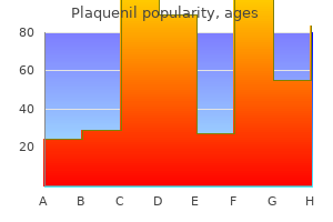
Cheap 200 mg plaquenil
Upper border of the guts is slightly oblique and atrial (mainly the left atrium) arthritis diet johns hopkins plaquenil 200 mg buy mastercard. Ascending aorta and the pulmonary trunk cross anterior to it (obscuring it) and at its right extremity arthritis medication enbrel plaquenil 400 mg low price, the superior vena cava enters the right atrium. Right border of the guts corresponds to the best atrium and is barely convex to the best. The inferior border separates the sternocostal(anterior) floor from the diaphragmatic (inferior) surface. Left border of the guts (obtuse margin) separates the sternocostal and left surfaces. It descends obliquely, convex to the left, from the left atrial appendage to the cardiac apex. It is round and mainly shaped by the left ventricle and partly the left atrial appendage. The coronary heart has the following three surfaces: Sternocostal (anterior), diaphragmatic (inferior) and left floor. Anterior (sternocostal) surface is shaped largely by right ventricle and right auricle and partly by left ventricle and left auricle. Anterior interventricular groove is evident on this floor which separates proper and left ventricle. The left atrium is hidden on the entrance by the ascending aorta and pulmonary trunk. It lies in entrance of the center 4 thoracic vertebrae (T5�T8) within the lying-down place and descends one vertebra in the erect posture (T6�T9). The base is separated from vertebral column by the oblique pericardial sinus, esophagus, and aorta. Some authors consider the base of the heart because the higher border of the guts where great blood vessels (superior vena cava, ascending aorta and pulmonary trunk) are attached. Cardiac apex is the blunt rounded extremity of the center shaped by the left ventricle, which is directed anteroinferiorly and to the left. The apex is situated mostly behind the fifth left intercostal house, near or a little medial to the midclavicular line. It is shaped primarily by the left ventricle (2/3) and partly proper ventricle (1/3) that are separated from one another by the posterior interventricular groove. Left floor is shaped primarily by the left ventricle and partly by the left atrium and auricle. The wall of the center consists of three layers: inside endocardium, center myocardium, and outer epicardium. Grooves/sulci Coronary (atrioventricular) sulcus is current on the external floor of the center, in a circumferential method across the heart, marks the division between the atria and the ventricles. The crux is the purpose at which the interventricular and interatrial sulci cross the coronary sulcus. Left half of anterior half is small and lodges circumflex branch of left coronary artery. Base of the guts is the posterior surface of coronary heart and extends from T 5 to T eight thoracic vertebra ranges in supine posture. Left and proper atrium Base of the center is the posterior floor of coronary heart and is principally contributed by left atrium and partly right atrium. Diaphragmatic floor of heart is majorly contributed by left ventricle and partly proper ventricle. Contains left anterior descending coronary artery Atrioventricular groove (coronary sulcus) separates atria from ventricles. Right coronary artery lodges in right a half of coronary sulcus and left coronary artery gives circumflex branch in left a part of coronary sulcus. Left anterior descending coronary artery runs within the anterior interventricular groove. Heart Chambers Right atrium has an anterior rough-walled portion (atrium proper and the auricle) lined with pectinate muscles and a posteriorly situated smooth-walled (sinus venarum) into which the two venae cavae open. Sulcus terminalis is a groove on the exterior surface of the right atrium (embryologic junction of the sinus venosus and primitive atrium) comparable to crista terminalis on inner floor. Pectinate muscles are the outstanding ridges of atrial myocardium located in the inside of each auricles and the right atrium. The inside of minimi) auricle presents reticular sponge-like network of the muscular ridges 608 Thorax Right atrium is bigger however thinner than the left atrium. Right auricle is the conical muscular pouch of the upper anterior portion of the proper atrium, it covers the proximal a half of the best coronary artery. Fossa ovalis is an oval-shaped despair within the interatrial septum and represents the location of the foramen ovale, via which blood runs from the right atrium to the left atrium in fetal circulation. Four valveless pulmonary veins from lungs (oxygenated blood) open into the left atrium. Right ventricle is essentially evident anteriorly and contributes to the main portion of the sternocostal floor of the center. Trabeculae carneae are irregular anastomosing muscular ridges, which form the trabeculated a part of the ventricles (inflow tract) and develop embryologically from the primitive ventricle. Supraventricular crest (a C-shaped inside muscular ridge), marks the junction between the trabeculated part and clean part of the proper ventricle. Papillary muscles are cone-shaped muscle tissue enveloped by endocardium, prolong from the anterior and posterior ventricular walls and the septum, and their apices are hooked up to the chordae tendineae. These contract to tighten the chordae tendineae, stopping the cusps of the tricuspid valve from being everted into the atrium, preventing regurgitation of ventricular blood into the proper atrium. Chordae tendineae lengthen from one papillary muscle to more than one cusp of the tricuspid valve. It is called the moderator band for its ability to stop overdistention of the ventricle and carries the best limb (Purkinje fibers) of the atrioventricular bundle from the septum to the sternocostal wall of the ventricle. Table 29: Differences of inflowing and outflowing parts of the best ventricle Inflowing decrease half It develops from primitive ventricle It is large in measurement and lies below the supraventricular crest Outflowing upper part It develops from bulbus cordis It is small in size and lies above the supraventricular crest It is tough as a end result of presence of the muscular ridges-the trabeculae It is easy and varieties higher 1 inch conical a part of the right ventricular carneae. It types a lot of the proper ventricular chamber chamber-the infundibulum, which supplies rise to pulmonary trunk Left ventricle is especially evident at the posterior view of the heart, its apex is directed downward, forward, and in course of the left. Crista terminalis divides right atrium into easy posterior part and tough anterior part. Superior vena cava Inferior vena cava opening is guarded by eustachian valve (rudimentary), coronary sinus by Thebesian valve and atrioventricular opening by tricuspid valve. Aortic sinus bulging into proper atrium Torus aorticus (aortic mound) is the outstanding area of the right atrial septum, which marks the projection of the noncoronary aortic sinus into the right atrial wall. Because the esophagus is so near the upper chambers of the guts, clear photographs of these coronary heart buildings and valves can be obtained. Valves Heart valves are located across the fibrous rings of the cardiac skeleton and are lined with endocardium. They incorporate leaflets or cusps, which close collectively to seal and forestall backflow. There are two pairs of valves in the coronary heart: (a) a pair of atrioventricular valves, and (b) a pair of semilunar valves. Pulmonary valve is the semilunar valve that lies between the best ventricle and the pulmonary artery and has three cusps (anterior, proper, and left).
Purchase 200 mg plaquenil with amex
Abducent Nerve Abducent nerve nucleus is in the pons arthritis in your back treatment plaquenil 400 mg purchase without prescription, axons leave the mind on the pontomedullary junction anteriorly after which pierces It passes via the cavernous sinus mendacity inferolateral to inner carotid artery and enters the orbit via the Abducent nerve pierces dura mater relatively early in its intracranial course and has received the longest intradural course arthritis pain or bone cancer generic plaquenil 200 mg with mastercard, It is the earliest nerve to get involved in raised intracranial rigidity. The patient will present with a medial deviation of the affected eye (internal strabismus) or diplopia on lateral eye motion. [newline]Lesion of the abducens nerve might result from a sepsis or thrombosis in the cavernous sinus. Sympathetic provide from cervical plexus nerve from cervical sympathetic chain � � � � Dilator pupillae has the radially organized fibres on iris and is provided by the T-1 sympathetic fibres, which start from the lateral horn cells of the spinal twine. These fibres synapse in superior cervical ganglion on the cervical sympathetic chain. The post-ganglionic fibres move through around the branches of inside carotid arteries to reach the eyeball muscle tissue like dilator pupillae through long ciliary nerves. It is innervated by the sympathetic system, which acts by releasing noradrenaline, which acts on 1-receptors. Threatening stimuli that activates the fight-or-flight response, this innervation contracts the muscle and dilates the iris, thus quickly letting more light/information reach the retina. There seems to be misprint as cornea is provided by branches of nasociliary (not nasolacrimal) nerve. The ophthalmic division of the trigeminal nerve offers nasociliary nerve which further sends lengthy ciliary nerves and brief ciliary nerves to provide cornea. Cornea is equipped by lengthy and quick ciliary nerves branches of nasociliary nerve, which itself is a department of ophthalmic division of the trigeminal nerve. Abducent (not trochlear) nerve has received the longest intradural course, because it pierces dura mater relatively early in its intracranial course. Has longest intradural course � Light and Accommodation Reflex Light reflex controls the diameter of the pupil in response to the depth of sunshine that falls on the retina. Pathway: In response to gentle shown into the pupil, the ganglion cells in retina project the information bilaterally to the pretectal nuclei, which further sends (through the posterior commissure) crossed and uncrossed fibers to the accent oculomotor (Edinger-Westphal) nucleus. Edinger-Westphal nucleus of oculomotor nerve offers preganglionic parasympathetic fibers, which synapse in ciliary ganglion. Ciliary ganglion offers postganglionic fibers to innervate the sphincter pupillae muscle of the iris. The frontal cortex sends corticotectal tract to the superior colliculus and pretectal nucleus. The superior colliculus and pretectal nucleus project to the oculomotor complex of the midbrain. This advanced includes the following buildings: the rostral accessory oculomotor (Edinger-Westphal) nucleus, which mediates pupillary constriction through the ciliary ganglion. The caudal accessory oculomotor (Edinger-Westphal) nucleus, which mediates contraction of the ciliary muscle. Self Assessment and Review of Anatomy It receives enter from the contralateral frontal eye area and initiatives to the ipsilateral lateral rectus muscle. Activation of the frontal eye fields transfer both the eyes in contralateral path. For example, activation of left frontal eye fields moves both (right and left eyes) in the course of right facet. Subcortical center for vertical conjugate gaze is located in the midbrain on the degree of the posterior commissure. On attempted lateral conjugate gaze, the only muscle that features is the intact lateral rectus. Argyll Robertson pupil is pupillary light�near dissociation, leading to absence of a miotic response to light, each direct and consensual, with the preservation of a miotic reaction to close to stimulus (accommodation�convergence). Horner syndrome is caused by transection of the oculo-sympathetic pathway leading to miosis, ptosis, enophthalmos and hemianhidrosis. Relative afferent (Marcus Gunn) pupil outcomes from a lesion of the optic nerve, the afferent limb of the pupillary light reflex. The analysis could be made with the swinging flashlight take a look at Transtentorial (uncal) herniation could occur because of a brain tumor or hematoma (subdural or epidural). Ipsilateral hemiparesis occurs on account of strain on the corticospinal tract, which is situated within the contralateral crus cerebri. Compression of the posterior cerebral artery, results in ischaemia in visual cortex, resulting in contralateral homonymous hemianopia. Papilloedema is noninflammatory congestion of the optic disk on account of elevated intracranial pressure. It causes medial rectus palsy on attempted lateral conjugate gaze and monocular horizontal nystagmus within the abducting eye. When an attempt is made to gaze contralaterally (relative to the affected eye), the affected eye shows no adduction. For example, if the right eye is affected the patient develops diplopia on looking to the left. Infratemporal region Infratemporal Fossa is the irregularly shaped cavity, situated below and medial to the zygomatic arch, not totally enclosed by Its boundaries are: Anteriorly is the maxilla bone (posterior surface) and posterior is the temporal bone (styloid and mastoid processes). At the medial boundary is the sphenoid bone (lateral pterygoid plate) and lateral is the mandible bone (ramus and coronoid process). At the roof is bigger wing of the sphenoid and infratemporal crest and the ground is open below. Temporal fossa is the shallow depression on the side of the skull bounded by the temporal traces and terminating under the extent of the zygomatic arch. Its boundaries are: Anteriorly is the frontal bone (zygomatic process) and zygomatic bone (the frontal process). The floor is contributed by components of the frontal, parietal, temporal, and larger wing of the sphenoid bone. Contents are: Temporalis muscle, the deep temporal nerves and vessels, the auriculotemporal nerve and the superficial temporal vessels. Pterygopalatine Fossa and Ganglion Pterygopalatine fossa is a cone-shaped paired depression deep to the infratemporal fossa and posterior to the maxilla on each side of the cranium, located between the pterygoid course of and the maxillary tuberosity, close to the apex of the orbit. Head and Neck the boundaries and relations of pterygopalatine fossa: Anterior Wall: Posterior surface of the maxilla bone. The openings and their contents in posterior wall are: Foramen rotundum to middle cranial cavity (contains maxillary nerve); pterygoid canal to foramen lacerum (contains nerve of the pterygoid canal); palatovaginal canal to choana (containing pharyngeal branch of the maxillary artery and pharyngeal nerve from the pterygopalatine ganglion). The opening is the sphenopalatine foramen to the nasal cavity, which transmits the sphenopalatine artery and nasopalatine nerve. The opening is the inferior orbital fissure to the orbit which transmits the maxillary nerve. The opening is the larger palatine foramen to the palate, which transmits the greater palatine nerve and vessels. Contents: Maxillary nerve passes via foramen rotundum to enter the pterygopalatine fossa, pterygopalatine ganglion, maxillary artery (third part).
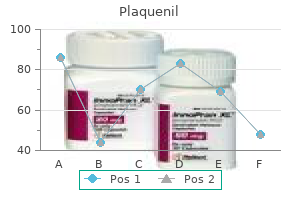
Plaquenil 400 mg buy discount
It runs obliquely downward by the facet of the nostril degenerative arthritis in neck and back generic plaquenil 200 mg online, is linked with the cavernous sinus by the superior and inferior ophthalmic veins which are devoid of valves arthritis knee yoga exercises buy plaquenil 400 mg on line. It begins as angular vein, joins anterior division of retromandibular vein to type common facial vein, which in turn drains into internal jugular vein. Cavernous sinuses receive blood from the facial vein through the tributaries superior and inferior ophthalmic veins. Danger space of the face is the area of the face near the nostril drained by the facial veins. Pustules (pimples) or boils or different skin infections, notably on the aspect of the nostril and higher lip, might spread to the cavernous venous sinus through the facial vein, pterygoid venous plexus, and ophthalmic veins. Bacteria in the sphenoid and ethmoid sinuses can unfold to the cavernous sinuses by way of the small emissary veins and are the most common websites of major infection leading to septic cavernous sinus thrombosis. Surface marking for inside jugular vein is drawn as a line joining some extent on the neck, medial to the ear lobule to a point at the medial end of the clavicle. Line passing from ear lobule to medial finish of clavicle � Lymphatic Drainage Lymph nodes in the head and neck are organized in two horizontal rings and two vertical chains on both aspect. The outer(superficial) ring consists of the occipital, preauricular (parotid), submandibular and submental nodes. All lymph vessels of the top and neck drain into the deep cervical nodes, both instantly from the tissues or indirectly via Lymph reaches the systemic venous circulation by way of both the right lymphatic duct or the thoracic duct. Superficial lymph nodes of the pinnacle Lymphatics from the face, scalp, and ear drain into the occipital, retroauricular, parotid, buccal, submandibular, submental, and superficial cervical nodes, which themselves drain into the deep cervical nodes (including the jugulodigastric and juguloomohyoid nodes). Deep lymph nodes of the top Nasal cavity & paranasal sinuses drain into the submandibular, retropharyngeal, and upper deep cervical. Larynx drains into the higher and decrease deep cervical; Pharynx drains into the retropharyngeal and higher and decrease deep cervical. Thyroid gland drains into the lower deep cervical, prelaryngeal, pretracheal, and paratracheal nodes. Superficial cervical lymph nodes lie alongside the exterior jugular vein in the posterior triangle and alongside the anterior jugular vein within the anterior triangle. Superior deep cervical nodes lie alongside the inner jugular vein in the carotid triangle of the neck. They receive afferent lymphatics from the again of the top and neck, tongue, palate, nasal cavity, larynx, pharynx, trachea, thyroid gland, and esophagus. The efferent vessels be a part of those of the inferior deep cervical nodes to form the jugular trunk, which empties into the thoracic duct on the left and into the junction of the internal jugular and subclavian veins on the right. Inferior deep cervical nodes lie on the interior jugular vein near the subclavian vein. They obtain afferent lymphatics from the anterior jugular, transverse cervical, and apical axillary nodes. The vertical chain consists of superior and inferior groups of nodes related to the carotid sheath. Anterior palate � � � � � � Submental (suprahyoid) lymph node are situated between the anterior bellies of the digastric muscle. They drain the central parts of the lower lip and ground of the mouth and the tip of the tongue. Their efferents cross to the submandibular lymph nodes and partly to a gland of the deep cervical group. Anterior half of nasal cavity (including anterior part of nasal septum) drains into submandibular nodes. Postauricular nodes Development of Face, Palate and Nose Face is fashioned by three swellings: the frontonasal prominence, maxillary prominence (pharyngeal arch 1), and mandibular Nasal placodes are the bilateral ectodermal thickenings which develop on the ventrolateral aspects of the frontonasal Nasal placodes invaginate into the underlying mesoderm to type the nasal pits, thereby producing a ridge of tissue that Nasolacrimal groove varieties between the maxillary prominence and the lateral nasal prominence and finally types the nasolacrimal duct and lacrimal sac. Structures contributing to formation of the Face Prominence Frontonasala Maxillary Medial nasal Lateral nasal Mandibular a prominence (pharyngeal arch 1). Structures formed Forehead, bridge of nostril, and medial and lateral nasal prominences Cheeks, lateral portion of higher lip Philtrum of upper lip, crest, and tip of nose Alae of nose Lower lip the frontonasal prominence is a single unpaired structure; the other prominences are paired. Age 4th week (28th day) fifth week (31 to 35 days) 6th week seventh week 8th week tenth week Events � Buccopharyngeal membrane breaks down, stomodaeum communicates with foregut � Frontonasal, maxillary and mandibular processes are distinct � Lens and nasal placodes are distinguished � Nasal placodes invaginate, nasal pits are formed � Lateral and medial nasal prominences are evident � Tubercles (for pinna) appear � Palatal course of appear from the maxillary course of � Eyelids established � Maxillary course of fused with medial nasal process � Eyes shifted from a lateral to a frontal location � Palatal processes and nasal septum fused with each other Primary palate is fashioned by the medial nasal prominences on the midline. Posterior to the primary palate, the maxillary process on each side sends a horizontal plate (palatal process); these plates Secondary palate is fashioned by fusion of the lateral palatine processes (palatal shelves) that develop from the maxillary Definitive palate is formed by fusion of the first and secondary palates at the incisive foramen. Palatine shelves from maxillary prominence of pharyngeal arch 1 = orange, nasal septum from the medial nasal prominences = green, major palate from the medial nasal prominences = green 421 Self Assessment and Review of Anatomy Development of varied components of the adult palate Component Source of improvement. In entrance of incisive fossa (premaxilla) carrying four incisor teeth Fused medial nasal processes of frontonasal course of Fusion of palatine cabinets (palatal process) of maxillary processes b. Soft palate Unossified a part of fused palatine shelves (palatal processes) of two maxillary processes, which lengthen posteriorly past the nasal septum Clinical Correlations � Cleft palate is assessed as anterior or posterior. The anatomical landmark that separates anterior from posterior cleft palate defects is the incisive foramen. Midline cleft lip (hare lip) is due to non-fusion of the 2 medial nasal processes within the midline. Development of Nasal Cavity Nasal pits are ectoderm lined depressions that outcome from proliferation of mesenchyme in lateral and medial nasal swellings. Oronasal membrane initially separates nasal cavities from the oral cavity, however its rupture permits communication between nasal and oral cavities by way of the primitive choanae. Floor of the nasal cavity is shaped by fusion of the medial nasal process (nasal septum) with the palatine processes of the maxilla. Paranasal sinuses develop as diverticula of the lateral nasal wall and lengthen into the maxilla, ethmoid, frontal, and sphenoid bones. Maxillary processes Medial nasal processes Medial and lateral nasal process Medial nasal and maxillary course of. Medial nasal processes � � � � Midline cleft lip (hare lip) is due to non-fusion of the two medial nasal processes in the midline. Failure of fusion between medial nasal and maxillary process leads to unilateral cleft lip. Facial development begins at week 4 and the final phases of facial improvement occur in the interval from 7�8th week. Connective tissue types a thick, vascularized subcutaneous layer which is dense, contains quite a few blood vessels and nerves, sweat glands, and hair follicles. The arteries anastomose freely and are held by the dense connective tissue around them, and thus, they have a tendency to remain open when minimize, inflicting profuse bleeding. Aponeurosis (galea aponeurotica) is a tendinous sheet that covers the vault of the cranium and continues as the frontal muscle anteriorly and the occipital muscle posteriorly. Arterial provide: Scalp is supplied by the supratrochlear and supraorbital branches of the internal carotid and by the superficial temporal, posterior auricular, and occipital branches of the exterior carotid arteries. Nerve provide: Scalp is innervated by the supratrochlear, supraorbital, zygomaticotemporal, auriculotemporal, lesser occipital, larger occipital, and third occipital nerves. Arteries Facial artery arises from the external carotid artery just superior to the lingual artery (above the upper border of the hyoid bone).
Discount 400 mg plaquenil with amex
Lymphatics Lymph Drainage from the Lateral Quadrant Majority of the lymph (>75%) drains as follows: Axillary nodes (humeral rheumatoid arthritis united states generic 400 mg plaquenil overnight delivery, subscapular sarcoid arthritis definition plaquenil 200 mg on line, pectoral, central, and apical) infraclavicular and supraclavicular nodes proper subclavian lymph trunk (for the right breast) or left subclavian lymph trunk (for the left breast). Remaining (25%) of lymph drainage occurs by way of the interpectoral, deltopectoral, supraclavicular, and inferior deep cervical nodes. Lymph Drainage From the Medial Quadrant Parasternal nodes proper bronchomediastinal lymph trunk (for the proper breast) or left bronchomediastinal lymph trunk (for the left breast). Nipple is provided by sixth intercostal nerve � Nipple lies in the fourth intercostal area and is provided by fourth intercostal nerve. Anterior axillary � Lymphatics from higher outer quadrant of breast drain into anterior (pectoral) axillary lymph nodes. Lateral group lies along lateral thoracic vessels � Lateral group of axillary lymph nodes lie alongside the axillary vein. Apical � Anterior (pectoral), posterior (subscapular) and lateral (humeral) teams drain the lymph into the central group of lymph nodes, which finally drain into the terminal apical lymph nodes (near the apex of axilla). Superior thoracic artery � Mammary gland receives blood from the axillary artery branches (lateral thoracic artery, thoracoacromial artery); the posterior intercostal arteries and the internal thoracic (mammary) artery branches. Anatomical Snuff Box the anatomical snuff box is an elongated triangular depression seen on lateral aspect of the wrist immediately distal to the radial styloid course of, gets extra distinguished when the thumb is absolutely extended. Boundaries Borders � Anterolaterally (Radial) � Tendons of abductor pollicis longus � Extensor pollicis brevis � Posteromedially (ulnar) � Tendon of extensor pollicis longus � Proximal border � Styloid means of the radius � Distal border � Apex of the schematic snuffbox isosceles triangle Scaphoid, trapezium and base of first metacarpal bone* � Skin and superficial fascia � Cephalic vein (subcutaneous) � Radial nerve branches (subcutaneous) Contents Radial artery (pulse) * At the ground, two joints are partly evident - the wrist joint and the first carpometacarpal joint. Floor Roof Note: the superficial radial nerve, may be rolled from side to aspect on the tendon of extensor pollicis brevis. The tendons concerned are abductor pollicis longus and extensor pollicis brevis and Finkelstein check becomes constructive. Radial Artery � Anatomic snuff field is a triangular interval bounded anterolaterally by the abductor pollicis longus (and extensor pollicis brevis) and posteromedially by the tendon of the extensor pollicis longus. Floor is shaped by extensor carpi longus and brevis tendons � Abductor pollicis longus and extensor pollicis brevis kind the anterolateral wall of anatomical snuff box. It extends from the lateral margin of the radius to the styloid process of the ulna, the pisiform, and the triquetrum and is crossed superficially by the superficial department of the radial nerve. Posterior interosseous nerve runs beneath which compartment of extensor retinaculum: a. Flexor carpi radialis � the tendon of flexor carpi radialis passes by way of a tunnel created by a fascial slip on the roof of flexor retinaculum. Abductor pollicis longus � First extensor compartment of wrist has two tendons: Abductor pollicis longus and extensor pollicis brevis. The aim is to distinguish infectious tenosynovitis from superficial or localized abscess. Midpalmar longitudinal crease signifies the lateral restrict of the hypothenar eminence. Proximal row (lateral to medial): Scaphoid, Lunate, Triquetral, Pisiform Distal row: Trapezium, Trapezoid, Capitate, Hamate Metacarpals are miniature lengthy bones consisting of bases (proximal ends), shafts (bodies), and heads (form knuckles). Capitate begins to ossify within the second month; the hamate on the finish of the third month; the triquetrum in the third yr; and the lunate, scaphoid, trapezium and trapezoid within the fourth year in females (fifth year in males). The pisiform begins to ossify within the ninth or tenth 12 months in females, and the twelfth in males. Some authors mention: Lunate-fourth yr, Scaphoid and trapezoid-fifth yr and trapezium-sixth yr 711 Self Assessment and Review of Anatomy Clinical Correlations � Scaphoid is the most generally fractured carpal bone. A fracture leads to osteonecrosis of the scaphoid bone (proximal fragment) � There is tenderness on the ground of the anatomical snuff field. Major supply from dorsal surface � Major blood supply (~80%) of scaphoid comes by way of dorsal surface via dorsal branches of radial artery. Trapezium � the capitate bone is the biggest of the carpal bones in the human hand, and occupies the middle of the wrist. Lies in the proximal row of carpal bones � the carpal bones within the proximal row are scaphoid, lunate, triquetral and pisiform. Epiphysis is on the head � Epiphysis is present on the heads of all metacarpals, besides first metacarpal (epiphysis is on the base). Joints Midcarpal Joint is a plane synovial aircraft joint between the proximal and distal rows of carpal bones and allows gliding First Carpometacarpal Joint is a saddle synovial joint between trapezium and the primary metacarpal bone, permitting flexion Plane synovial joints are present between the carpal bones and the medial four metacarpal bones, allowing a easy Metacarpophalangeal Joints are ellipsoid (> condyloid) joints that enable flexion and extension, and abduction and Interphalangeal Joints are hinge synovial joints that permit flexion and extension. It has 120� of flexion and extension, about 40� of abduction and adduction, and some rotation possible. Fascia Palmar aponeurosis is a triangular fibrous layer overlying the tendons within the palm and protects the superficial palmar arterial arch and palmar digital nerves. Three subgroups of radial (abductor pollicis longus and extensor pollicis brevis), central (abductor pollicis brevis and opponens pollicis) and ulnar (flexor pollicis brevis) muscle tissue are concerned. Lumbricals 1 and a pair of arise from lateral aspect of lateral two tendons of the flexor digitorum profundus. Lumbricals 3 and 4 take origin from adjoining sides of medial three tendons of the flexor digitorum profundus. The tendons of lumbricals cross the lateral facet of metacarpophalangeal joints to be inserted into the lateral aspect of dorsal digital growth of the corresponding digit from second to fifth. Patient is requested to tightly hold a card between the fingers whereas the examiner tries to pull it out. On the index finger and little finger, the growth is strengthened by extensor indicis and extensor digiti minimi, respectively, which blends with it. Self Assessment and Review of Anatomy Clinical Correlations � Mallet finger (hammer or baseball finger) is a finger with permanent flexion of the distal phalanx as a result of an avulsion of the lateral bands of the extensor tendon to the distal phalanx. It occurs because of A forceful blow on the tip of the finger causing sudden and powerful flexion of the phalanx. Adductor pollicis � Dorsal digital enlargement is a tendinous modification of extensor digitorum and receives attachment of lumbricals, interossei but not adductor pollicis. The lumbricals of the hand, the palmar and dorsal interossei of the hand, additionally insert on these bands. High Yield Points 716 � the tip of the hook gives attachment to the flexor retinaculum. Claw hand is hyperextension at metacarpophalangeal joint and flexion at the interphalangeal(s). Abductor pollicis brevis � Pen check is to check anterior abduction of thumb, carried out by abductor pollicis brevis (median nerve supply). Palmar interossei � Card check - A card is positioned between the 2 fingers of the patient to grasp. Supplied by median nerve � Adductor pollicis muscle has two heads, both supplied by the ulnar nerve. Interosseous muscle � Clumsiness of hand happens due to paralysis of intrinsic muscle tissue of hand like lumbricals and interossei.
Buy plaquenil 200 mg
Chronic illnesses of the exocrine pancreas rheumatoid arthritis dogs cheap plaquenil 400 mg on-line, corresponding to cystic fibrosis and persistent pancreatitis ginger for arthritis in dogs plaquenil 200 mg cheap without a prescription, result in a lack of pancreatic mass and deficiency of pancreatic enzymes, and may result in maldigestion. Endocrine Function the first function of the endocrine pancreas is to secrete insulin and different hormones to control serum levels of glucose and triglycerides and amino acid balance. The endocrine portion of the pancreas constitutes roughly 10% of the gland and is made up of the islets of Langerhans. Autonomic nerves, metabolites such as glucose, circulating hormones, and local paracrine hormones regulate the operate of the islets. Gallbladder dysfunction is outlined as an ejection fraction less than 35% Secretin is concerned in alkalinizing the duodenum; pancreatic enzymes are inactivated by an acidic pH An inhibitory hormone. Inhibits gastric acid secretion Stimulates pancreatic secretion Inhibits gastric acid secretion Stimulated by acid and fat in the duodenum Secretin Somatostatin D cells (in the pancreatic islets and gastric and intestinal mucosa) Inhibits gastric acid and Stimulated by acid. Pancreatic Anatomy and Function 279 Pearls the splenic vein and artery course alongside the size of the pancreas. Splenic vein thrombosis might lead to isolated gastric varices (gastric varices within the absence of esophageal varices). A traumatic injury would most probably be localized to which of the following regions The blood stress is 92/54 mmHg, pulse fee a hundred and fifteen per minute, and temperature 102 �F (38. Abdominal ultrasonography reveals dilatation of the bile duct to 12 mm, multiple gallstones within the gallbladder, but no gallbladder wall thickening or pericholecystic fluid. Laboratory exams show a normal full blood depend, whole bilirubin level of 5 mg dl�1, aspartate aminotransferase a hundred forty five U l�1, alanine aminotransferase 79 U l�1, amylase 196 U l�1, and lipase 246 U l�1. The primary care doctor obtains an abdominal Xray, which reveals air�fluid levels within the abdomen and first portion of the duodenum. Esophagogastroduodenoscopy is performed and reveals narrowing in the second portion of the duodenum with normalappearing duodenal mucosa. Answers 1 2 D D this affected person has gentle acute pancreatitis as evidenced by epigastric pain and mildly elevated serum amylase and lipase ranges. Ascending cholangitis is unlikely within the absence of fever, jaundice, or elevated liver enzyme ranges. Peptic ulcer typically presents with epigastric ache and presumably with bleeding, but not usually with elevated serum amylase and lipase levels. Primary sclerosing cholangitis presents with elevated liver biochemical check levels in a cholestatic pattern (elevated serum bilirubin and alkaline phosphatase levels). Amylase isoforms could be obtained to determine if the salivary amylase stage is elevated in serum and can be useful in a patient with isolated hyperamylasemia. The spleen can be injured in motor vehicle collisions; nonetheless, in this case ultrasonography revealed fluid across the pancreas and no damage to the spleen. The bile duct is dilated, and the serum aminotransferase and pancreatic enzyme levels are elevated. Together with the presence of gallstones, this pattern is most suggestive of bile duct obstruction secondary to choledocholithiasis. Gastrointestinal manifestations of cystic fibrosis include pancreatic insufficiency, steatorrhea, failure to thrive, and belly pain. The ache is sharp, radiates to the back, and is associated with a decrease in appetite. She reviews that over the earlier weekend she was at a wedding and drank six glasses of wine. Her medicines include hydrochlorothiazide, niacin, and naproxen one to two tablets per week for joint pain. Her household history is outstanding for hypertension and diabetes mellitus in her father. She has consumed one to two glasses of red wine daily for many years, and admits to binge ingesting every so often. On bodily examination, the blood strain is 124/78 mmHg, pulse price 110 per minutes, and respiratory fee 12 per minute. General Acute pancreatitis is an acute inflammatory situation of the pancreas that will prolong to native and distant extrapancreatic tissues. The majority (80%) of circumstances of acute pancreatitis are brought on by gallstones or alcohol. In addition, microlithiasis (small [<3 mm] stones) and sludge (biliary debris) cause acute pancreatitis. Gallstones cause acute pancreatitis by obstructing the pancreatic duct, or through reflux of bile or particles from the bile duct into the pancreatic duct. Persons with acute pancreatitis due to alcohol may have underlying continual pancreatitis (see Chapter 19). Other Causes these account for 10�15% of instances of acute pancreatitis: � Hypertriglyceridemia (serum triglyceride stage >1000 mg dl�1): the third most common cause, accounts for 3�5% of circumstances of acute pancreatitis. Anatomic Causes Pancreas divisum: affects up to 7% of the population, however pancreatitis happens in 15�20% of persons with pancreas divisum. Genetic Causes these are associated with continual pancreatitis which will present acutely: � Cystic fibrosis. On pancreatic biopsy, granulocyte epithelial lesions that always cause destruction of the pancreatic ducts are seen. Idiopathic Acute Pancreatitis the cause of pancreatitis is unknown in 5�10% of circumstances. Pathophysiology Normal mechanisms that protect the pancreas (see additionally Chapter 17): � Activation of pancreatic proenzymes occurs exterior the pancreas throughout the duodenal lumen. Necrotizing pancreatitis: 5�10% of patients develop necrosis of the pancreatic or peripancreatic tissue, which may stay sterile or become infected; contaminated necrosis is uncommon in the course of the first week of acute pancreatitis. Phases of Acute Pancreatitis Early phase: lasts about one week, mediated by the systemic inflammatory response and associated with an increased danger of extrapancreatic organ failure. Late section: only happens in sufferers with moderatetosevere acute pancreatitis, and is characterized by the presence of native problems, an increased danger of an infection, and organ failure. Complications Local Pancreatic: ascites, fistula, peripancreatic fluid assortment, pseudocyst, abscess, necrosis (sterile and infected). Nonpancreatic: bile duct obstruction, gastric outlet obstruction, duodenal ulcer, splenic and portal vein thrombosis. Organ failure is best outlined by the modified Marshall scoring system, which is predicated on pulmonary, cardiovascular, and renal dysfunction (Table 18. Organisms most regularly seen in contaminated pancreatic necrosis embody Escherichia coli, Klebsiella spp. Cr, creatinine; PaO2, partial stress of oxygen in arterial blood; FiO2, fraction of impressed oxygen. Prognosis the bulk (75�80%) of instances of acute pancreatitis are delicate and selflimiting. Some 20�25% of circumstances are severe, and despite aggressive efforts at preliminary resuscitation, as much as 50% of these patients will develop multiorgan failure or pancreatic necrosis. Assessment of Severity Several scoring techniques have been developed to predict severity, so as to establish extreme acute pancreatitis and predict pancreatic necrosis, mortality, and the need for immediate aggressive fluid resuscitation and intensive affected person monitoring.