

Nortriptyline dosages: 25 mg
Nortriptyline packs: 60 pills, 90 pills, 120 pills, 180 pills, 270 pills, 360 pills

Cheap nortriptyline 25 mg overnight delivery
Transcutaneous magnetic stimulation44 and extracorporeal shock-wave therapy45 have also been used efficiently to reduce neuroma-related pain anxiety 3 months postpartum order 25 mg nortriptyline amex. Intraoperative photograph of a patient who developed a sensory neuroma close to her incision following a hip substitute anxiety symptoms but dont feel anxious buy generic nortriptyline 25 mg line. Her painful space was marked (hatched area) utilizing a conscious pain-mapping procedure intraoperatively. Intraoperative testing revealed good overlap of the stimulation paresthesias with her painful area. During her 1-week exterior trial, she skilled important ache relief, and he or she later underwent permanent placement of the stimulator system. Ultimately, prevention of neuroma formation by inhibiting axonal growth into the nerve scar would be best. Perhaps nerve growth inhibitors could be injected into injured nerves before neuroma formation, or maybe a way of pharmacologically silencing mechanosensitive nerves subsequent to neuroma formation would be a comparably efficient strategy. The position of conduits in prevention of primary and secondary neuroma formation stays to be additional elucidated. The most notable advancements have been in the field of neurostimulation, with novel purposes of electrical stimulation concentrating on pain relief to specific painful areas and limiting unwanted paresthesias in adjacent, nonpainful areas. Future studies will hopefully set up whether or not these much less invasive techniques, as well as the percutaneous ablative techniques such as radiofrequency ablation, are effective. Development of ongoing exercise, mechanosensitivity, and adrenaline sensitivity in severed peripheral nerve axons. Cold intolerance in surgically handled neuroma sufferers: a potential follow-up study. The efficacy of morphine, pregabalin, gabapentin, and duloxetine on mechanical allodynia is different from that on neuroma pain within the rat neuropathic ache model. Efficacy of chemical neurolysis for the therapy of interdigital nerve compression of the foot: a retrospective study. Sonographically guided cryoneurolysis: preliminary experience and clinical outcomes. Implantation of sensory nerve into muscle: preliminary clinical and experimental observations on neuroma formation. Treatment by collagen conduit of painful post-traumatic neuromas of the sensitive digital nerve: a retrospective research of 10 circumstances. Perineural fat grafting in the therapy of painful end-neuromas of the higher limb: a pilot study. Peripheral subcutaneous electrostimulation for management of intractable post-operative inguinal pain: a case report sequence. Extracorporeal shockwave therapy for interdigital neuroma: a randomized, placebo-controlled, doubleblind trial. Interest of telemicrosurgery in peripheral nerve tumors: a couple of series of seven instances. Burchiel Integration of sensory information is a multistage process leading to perception with several ranges of modulation along pathways. The function of this region was popularized in 1965 within the gate-control principle by Melzack and Wall. Later, lesions were made with the laser beam by Levy and coworkers, Powers and associates, and Young and colleagues14-18 and with the ultrasonic probe by Dreval and Kandel and associates. Lesions and deafferentation can occur on account of a variety of pathologic processes. The procedure intends to preferentially interrupt the (nociceptive) fibers grouped in the lateral bundle of the dorsal rootlet and the (excitatory) medial a part of the tract of Lissauer. The small myelinated and unmyelinated fibers carrying nociceptive impulses course to the lateral aspect, which makes them appropriately located to enter into the tract of Lissauer, where they run for one or two segments before penetrating the grey matter of the spinal twine. The giant myelinated fibers course medially to enter instantly into the dorsal column. Activity of the second neuron is influenced each by upper ranges and by peripheral afferents. Somatogenic pain as a result of most cancers invasion is actually an necessary part; nonetheless, neurogenic ache linked to compression and destruction of neural structures or iatrogenic ache (after surgery or radiation treatment) can usually be encountered. In latest years, progress in oncology has significantly extended survival of sufferers with most cancers, and the long-term use of opioids can, even when administered intraspinally, have unwanted effects infringing on the standard of life. The use of ablative procedures may be thought of, subsequently, in sufferers with refractory pain and in those requiring excessive doses of morphine. For surgical procedures performed within the cervicothoracic region, control of pain was good in 87% of patients; and for surgeries performed in the lumbosacral area, management of ache was good in 78%. Other series report a relatively small variety of sufferers, and results are somewhat heterogeneous. Principles for indications are a limited, managed cancer in a patient with good enough basic standing to stand up to main surgical procedure and wellidentified and well-characterized ache as a end result of a clearly defined lesion. An early surgical determination could be made with profit to the patient when it supplies the likelihood to avoid extraordinarily disturbing and heavy pain medicine. The follow-up Brachial plexus accidents occur after main trauma, sometimes bike crashes in young adults. Trauma results in stretching of the roots as a lot as their complete avulsion from the spinal cord. Pain happens in 30% to 90% of patients, and its incidence is related to the localization of the lesion. Both ache elements are described by two thirds of sufferers; solely the background pain is noted in 20%, and solely paroxysms appear in only 10%. Major motor deficits are usually related and extra often located on the C7 to C8 territories. Its onset can be immediately after the trauma or delayed even years after the preliminary event. In the period following the avulsion, most sufferers underwent direct nerve suturing, grafting, or neurotization, all theoretically known to prevent the installation of persistent ache. For these patients going through severe uncontrollable ache, varied surgical methods have been tried with limited effect. Spinal twine stimulation is generally ineffective because of fiber degeneration up to the brainstem. Root meningocele is usually related to root avulsion, but avulsion may be present even within the absence of meningocele. Several levels are normally affected, but all roots of the brachial plexus are avulsed in about 40% of patients. A-B, T2-weighted magnetic resonance imaging reveals full avulsion of both ventral and dorsal roots and in addition of part of the lateral twine (A), and a pseudomeningocele on the lower cervical spine on the left facet with a hyperintensity of the ipsilateral dorsal column subsequent to the trauma and the stretching with avulsion of the dorsal root (B). C, Schematic drawing of the floating microelectrode (arrow) implanted into the dorsal horn following its axis.
Nortriptyline 25 mg generic fast delivery
Cilia have become recognized as being essential in neural tube closure anxiety insomnia 25 mg nortriptyline order with amex, probably as mediators of neural tube formation and neural fold fusion anxiety 8dpo buy nortriptyline 25 mg lowest price. Before the neural tube utterly closes, the cranial portion begins to undergo speedy enlargement as a result of increased growth of the tube and enlargement of the ventricular system. The in depth proliferation of the nervous tissue causes curvature of the embryo along its lengthy axis. Progressive dorsal flexion leads to raising and isolating the embryo from its membranes. With the appearance of the vesicles (at concerning the 5-mm embryo stage), the neural tube bends ventrally to type two flexures: a cervical flexure at the junction of the spinal cord and hindbrain, and a cephalic flexure at the stage of the lengthy run midbrain. Even within the late 1800s, transient segments of the neural tube, often known as neuromeres, had been described as early morphologic evidence of neural segmentation. A continuing controversy exists in regards to the function of some neuromeres, particularly those who give rise to the forebrain (prosomere) and the midbrain (mesomere),seventy three but by comparability, the hindbrain (rhombomere) is quite well understood. These gradients set in movement the sequential expression of sets of embryonic gap genes, pair-rule genes, and section polarity genes, with each gene set leading to progressively higher segmentation. This anterior area contributes to the future forebrain and part of the midbrain. Otx2 regulates forebrain and midbrain dedication, whereas Otx1 features in a extra restricted method by determining cortical and sense organ development. The multiple Hox genes confer segmental identity, and their expression starts sequentially within the primitive streak and types overlapping time- and site-specific expression domains, generally known as the Hox code, along the physique axis. Distinct neural and glial identities are acquired by neuroepithelial progenitor cells by way of progressive restriction of histogenetic potential beneath the influence of local environmental signals. Evidence for morphogenetic regulatory processes at particular locations of the creating neural primordium has led to the idea of secondary organizers, which regulate the identity and regional polarity of neighboring neuroepithelial areas. Their subsequent exercise refines native neural identities alongside the anterior-posterior or dorsal-ventral axes, and regionalizes the anterior neural plate and neural tube. Furthermore, this experiment exhibits that evenand odd-numbered rhombomeres display distinct properties. Indeed, most of the rhombomere genes seem to have unique rhombomeric expression profiles. Evidence for this is seen most clearly in r4, which is patterned by the intrinsic expression of Hoxb1 and the r4-specific expression of a number of downstream genes that promote or suppress r4 id. The regulation of rhombomeric interactions that drive cell migration, expression of proteins, and apoptosis promotes neuronal maturation. This area is laid out in a zone between the early anterior domains specified by Otx99 and the posterior domain specified by Gbx and Hox. It is now thought that the isthmus constriction colocalizes with the possible isthmic organizer (IsO) at rhombomere r0, as defined by expression of the fibroblast progress issue eight gene (Fgf8). Fgf8 signaling works cooperatively in suggestions loops with En1/2,103 Pax2/5,78 Pax3/7,seventy seven and Otx2104 to drive both the diencephalon tegmentum or tectal differentiation. The boundary between the metencephalon and myelencephalon is marked by the third or pontine flexure, fashioned after the cervical and cephalic flexures. Segmentation is quickly apparent in the rhombencephalon with repetitive units, known as rhombomeres. Initially four rhombomeres, designated A by way of D, served as very helpful anatomic landmarks in the human embryo. For example, rhombomere B was defined because the neuroectoderm reverse the otic plates. The boundaries between rhombomeres are established progressively throughout improvement and permit consideration of the rhombomeres as self-contained, comparatively cell lineage�restricted compartments. Each rhombomere expresses a novel combination of transcription elements and establishes specific patterns of neuronal differentiation93 expressed in its personal set of ganglia and nerves. In distinction, if an odd-numbered rhombomere is grafted adjoining to one other odd-numbered rhombomere, the cells of the two rhombomeres intensively mix. Barkovich proposed that defects could be expected because of both displaced rostral or caudal activity of the IsO. The resulting defect would manifest as a shortened midbrain and elongated or enlarged pons and cerebellum, r0 and r1 derivatives. Up to 5 forms of pontocerebellar hypoplasia have been reported that may symbolize caudal IsO perform. Imaging findings include absent/hypoplastic cerebellar vermis, cerebellar hemisphere hypoplasia, cystic fourth ventricular dilation, elevated tentorium, and associated hydrocephalus. Cerebellar foliation has been thought to be a consequence of mechanical forces that induce fissure formation. The Engrailed household of homeobox genes, En1 and En2, are expressed early and are concerned within the formation of the tectum and cerebellum. En1 mutants lack the tectum and a lot of the cerebellum, whereas En2 mutants are discovered to have a hypoplastic cerebellum with defects of foliation. This type of medulloblastoma tends to come up in very younger youngsters, displays a large cell/anaplastic or desmoplastic histology, and has a comparatively poor prognosis. The first is altered anterior and ventral alerts from irregular functioning of the prechordal plate. These problems comprise either anomalies of telencephalic commissures (agenesis of the corpus callosum, anterior commissure, hippocampal commissure, and posterior commissure) or midline septal defects (agenesis of septum pellucidum; cysts of cavum septum pellucidum, cavum vergae, and cavum velum interpositum). The forebrain arises from the anterior neural plate during gastrulation, and by the end of somitogenesis it contains the dorsally positioned telencephalon, the ventrally positioned hypothalamus, and the caudally located diencephalon. Fate-mapping research have instructed that the dorsal and ventral forebrain arise from totally different areas of the neuroepithelium. Unlike the rhombencephalon, no prosomeric subdivisions have been reported in the prosencephalon, and only distinct histogenetic protosegments have been recognized. Therefore, Shh mutants have rostral midline cortical defects however are capable of forming a dorsal telencephalic midline and two cortical hemispheres. The two hemispheres of the telencephalon communicate by way of axonal commissures, all of which develop in a reasonably comparable time-frame. Axons destined to turn into the anterior commissure begin to cross in the lamina reuniens at 10 weeks of gestation, and this is full by week 11. The diencephalic prosomere has been divided into three elements, named p1, p2, and p3. The dorsal roof and alar plates of each element correspond to each diencephalic primordia: p1 becoming primordia of the pretectum and epithalamus; p2 changing into primordia of the thalamus; and p3 becoming primordia of the ventral or prethalamus. The diencephalic prosomere basal plate accommodates the rostral a part of the ventral tegmental area of the Tsai�substantia nigra complicated and different nuclei of the prerubral tegmentum collectively. The diencephalic zone requires Emx2 and Otx2 to balance the affect from the IsO to ensure diencephalon improvement. The pallium is additional subdivided into the hippocampal formation (archicortex), neocortex, olfactory/piriform cortex (paleocortex), and claustrum/pallial amygdala. Factors similar to Numb promote neuronal differentiation and could additionally be expressed in the apical half or basal a half of the ventricular zone.
Purchase nortriptyline 25 mg with mastercard
An interval time period between the 2 required mind death evaluations is optionally available in sufferers 18 years of age and older anxiety early pregnancy generic 25 mg nortriptyline visa. Because of the difficulties in performing a mind dying examination in youthful patients anxiety symptoms peeing discount 25 mg nortriptyline overnight delivery, nevertheless, two examinations-including apnea testing with every examination, separated by an remark period-are required (Table 185-3). When ancillary research are used, a second clinical examination and apnea check ought to nonetheless be carried out, and elements that may be accomplished must yield results which are according to mind demise. Permission from the household may be requested for analysis of the patient by an organ and tissue procurement company. Pediatric neurocritical care groups can make meaningful contributions via a multidisciplinary method to brain damage in critically unwell children and thru the event of greatest clinical follow pathways and efficient quality improvement efforts. As new know-how becomes available, the contribution to consequence of poorly characterized physiologic variables, such as autoregulation of cerebral blood circulate, shall be better understood. New methods to optimize affected person management through implementation of evidence-based finest medical practices will pave the best way for future scientific trials of pharmacological neuroprotective and rehabilitation interventions. Resuscitation of blood strain and oxygenation and prehospital brain-specific therapies for the severe pediatric traumatic brain damage patient. Brain accidents and neurological system failure are the most typical proximate causes of dying in children admitted to a pediatric intensive care unit. Variation in intracranial strain monitoring and outcomes in pediatric traumatic mind harm. Craniocervical arterial dissection in kids: clinical and radiographic presentation and outcome. Pediatric neurocritical care: a neurology session mannequin and implication for training and coaching. Emerging subspecialties in neurology: building a career and a subject: pediatric neurocritical care. Length of stay and mortality in neurocritically ill patients: impression of a specialized neurocritical care staff. Effect of implementation of a paediatric neurocritical care programme on outcomes after extreme traumatic brain harm: a retrospective cohort study. Multimodal monitoring in traumatic mind harm: present status and future instructions. Brain tissue oxygen monitoring after extreme traumatic mind injury in children: relationship to end result and association with different clinical parameters. Transcranial Doppler-based assessment of cerebral autoregulation in critically ill kids during diabetic ketoacidosis therapy. Cerebral hyperemia measured with near infrared spectroscopy throughout therapy of diabetic ketoacidosis in youngsters. The epidemiology of vasospasm in children with moderate-to-severe traumatic brain injury. Optic nerve sheath diameter as a marker for analysis and prognostication of intracranial pressure in Indian sufferers: an observational examine. Tissue oxygen index: thresholds for cerebral ischemia utilizing near-infrared spectroscopy. Acute care scientific indicators related to discharge outcomes in children with severe traumatic mind harm. Incidence of hypo- and hypercarbia in severe traumatic mind damage before and after 2003 pediatric pointers. Cerebrovascular response in infants and younger youngsters following severe traumatic mind injury: a preliminary report. Part 1: relation to age, Glasgow coma rating, consequence, intracranial pressure, and time after injury. Childhood arterial ischaemic stroke incidence, presenting options, and threat elements: a potential population-based research. Intelligence after stroke in childhood: evaluate of the literature and suggestions for future research. Neurologic end result in survivors of childhood arterial ischemic stroke and sinovenous thrombosis. Report of the National Institute of Neurological Disorders and Stroke workshop on perinatal and childhood stroke. A multispecialty pediatric neurovascular convention: a mannequin for interdisciplinary administration of advanced illness. Use of alteplase in childhood arterial ischaemic stroke: a multicentre, observational, cohort research. Maternal and infant traits related to perinatal arterial stroke within the toddler. Stroke in youngsters: the coexistence of a quantity of danger elements predicts poor consequence. Prospective evaluation of risk factors for recurrent stroke throughout childhood-a 5-year follow-up examine. Arteriopathy prognosis in childhood arterial ischemic stroke: results of the vascular effects of infection in pediatric stroke research. Arterial ischemic stroke in neonates, infants and youngsters: an outline of underlying conditions, imaging methods, and remedy modalities. Risk of recurrent childhood arterial ischemic stroke in a population-based cohort: the significance of cerbrovascular imaging. Ischaemic stroke from dissection of the craniocervical arteries in childhood: report of 12 sufferers. Prognosis of occlusive disease of the circle of Willis (moyamoya disease) in youngsters. Lipoprotein (a) and genetic polymorphisms of clotting issue V, prothrombin, and methylenetetrahydrofolate reductase are danger components of spontaneous ischemic stroke in childhood. Outcome following decompressive craniectomy for malignant center cerebral artery infarction in kids. Cerebral venous sinus thrombosis in children: risk components, presentation, analysis and consequence. Cerebral venous sinus thrombosis presenting with excessive subarachnoid hemorrhage in a 14-yearold boy. Management of sickle cell disease: summary of the 2014 evidence-based report by professional panel members. Therapy insight: stroke risk and its administration in sufferers with sickle cell illness. Pathophysiology and treatment of stroke in sickle-cell illness: current and future. Primary hemorrhagic stroke in youngsters with sickle cell disease is related to recent transfusion and use of corticosteroids. Moyamoya syndrome in childhood sickle cell illness: a predictive issue for recurrent cerebrovascular events. Reversible posterior leukoencephalopathy syndrome and silent cerebral infarcts are related to extreme acute chest syndrome in kids with sickle cell disease. Parent training and biologic components affect on cognition in sickle cell anemia. Prevention of a first stroke by transfusion in children with sickle cell anemia and abnormal outcomes on transcranial Doppler ultrasonography.
Cheap nortriptyline 25 mg visa
This method is significantly less efficient if the affected person has preoperative hydrocephalus anxiety symptoms for hours cheap 25 mg nortriptyline overnight delivery. CystoperitonealShunt Some advocate placement of a cystoperitoneal shunt as a first-line treatment to achieve early cyst obliteration whereas avoiding massive fluid shifts associated with microsurgery anxiety symptoms aspergers nortriptyline 25 mg discount fast delivery. Cystoperitoneal shunting could provide the benefit over microsurgical fenestration for improved discount in postoperative cyst quantity (74% versus 58%). Strategies to avoid these issues include placement of an adjustable stress valve, attempted elimination when cyst obliteration is demonstrated, and avoidance of low-pressure or valveless shunts. Finally, for patients presenting with quadrigeminal cistern or pineal region arachnoid cysts, Torkildsen shunt variation95 offers the ability for cyst drainage with out the danger for overdrainage. Ventriculocystocisternostomy could produce extra durable results compared with ventriculocystostomy. Distortion created by the cyst could make the standard landmarks troublesome to acknowledge, and frameless navigation allows the surgeon to guide the tip of the endoscope to the desired location without the dangers related to head pinning. They are probably to be positioned within the thoracic backbone, present with refined symptoms, and may mimic wire compression. The targets of surgical intervention are similar to those of intracranial cysts, and extensive fenestration of the cyst alone could additionally be sufficient to keep away from recurrence. EndoscopicFenestration Neuroendoscopy has emerged as an effective different to microsurgical fenestration and excision. The surgical technique for cyst fenestration stays the identical, however endoscopy presents a quantity of benefits over microsurgery. For midline and intraventricular cysts, endoscopy provides the ability to visualize the boundaries of the cyst in addition to crucial adjoining structures however reduces the need for in depth dissection. Although a big proportion of cysts are asymptomatic, the signs they produce can range from hydrocephalus or focal deficits to epilepsy, cognitive deficits, and psychiatric issues. Surgical intervention could additionally be helpful for sufferers who exhibit symptoms associated to the cyst. However, treatment paradigms are operator dependent and produce comparable outcomes. Transoccipital endoscopic fenestration of atrial cysts causing ventricular entrapment. Endoscopy versus microsurgical cyst excision and shunting for treating intracranial arachnoid cysts. Clinical characteristics and remedy methods for idiopathic spinal extradural arachnoid cysts: a single-center experience. Language impairment associated with arachnoid cysts: recovery after surgical treatment. Intracranial arachnoid cysts: impairment of upper cognitive capabilities and postoperative improvement. A schizophrenia-like psychotic disorder secondary to an arachnoid cyst remitted with neurosurgical remedy of the cyst. Quantitative proteomics comparability of arachnoid cyst fluid and cerebrospinal fluid collected perioperatively from arachnoid cyst patients. Bilateral arachnoid cysts of the temporal fossa in four kids with glutaric aciduria sort I. Intracranial arachnoid cyst household with autosomal recessive trait mapped to chromosome 6q22. The fetal subarachnoid cisterns: an ultrasound research with report of a case of congenital communicating hydrocephalus. Arachnoid cysts: entrapped collections of cerebrospinal fluid variably communicating with the subarachnoid house. Endoscopic statement of a slit-valve mechanism in a suprasellar prepontine arachnoid cyst: case report. Teaching neuroimages: head banging with out head trauma: subdural hemorrhage in association with arachnoid cysts. Analysis of a bleeding mechanism in patients with the sylvian arachnoid cyst using a finite element model. Risk elements for pediatric arachnoid cyst rupture/hemorrhage: a case-control examine. How often do persistent extra-cerebral haematomas occur in sufferers with intracranial arachnoid cysts Arachnoid cysts of the middle cranial fossa: experience with 77 patients who had been treated with cystoperitoneal shunting. Epilepsy related to a cerebellar arachnoid cyst: seizure management following fenestration of the cyst. Complete decision of medically refractory temporal lobe epilepsy after arachnoid cyst fenestration. Bobble-head doll syndrome: some atypical options with a new lesion and evaluate of the literature. Suprasellar arachnoid cyst presenting with bobble-head doll movements: a report of three circumstances. Bobble-head doll syndrome because of a suprasellar arachnoid cyst: endoscopic treatment in two circumstances. Bobble-head doll syndrome: a surgically treatable situation manifested as a rare motion dysfunction. Bobble-head doll syndrome efficiently handled with an endoscopic ventriculocystocisternostomy. Growth, puberty and hypothalamic-pituitary function in kids with suprasellar arachnoid cyst. Suprasellar arachnoid cyst presenting with bobble-head doll syndrome: Report of three circumstances. Suprasellar arachnoidal cyst as a explanation for precocious puberty�report of three sufferers and literature overview. Supratentorial interhemispheric cysts associated with callosal agenesis: surgical therapy and end result in 16 youngsters. Quadrigeminal cistern arachnoid cyst: A sequence of 18 patients and a evaluation of literature. Arachnoid cyst of the cerebellopontine angle manifesting as contralateral trigeminal neuralgia: case report. Hemifacial spasm associated with a cerebellopontine angle arachnoid cyst in a younger adult. Facial spasm and paroxysmal tinnitus related to an arachnoid cyst of the cerebellopontine angle�case report. Percutaneous endoscopic therapy of suprasellar arachnoid cysts: ventriculocystostomy or ventriculocystocisternostomy Cine-magnetic resonance imaging evaluation of communication between center cranial fossa arachnoid cysts and cisterns. Proton magnetic resonance spectroscopy and diffusion-weighted imaging in intracranial cystic mass lesions.
25 mg nortriptyline buy visa
Spinal wire stimulation in therapy of persistent benign pain: challenges in therapy planning and current standing anxiety symptoms for 2 weeks 25 mg nortriptyline purchase otc, a 22-year experience anxiety symptoms confusion nortriptyline 25 mg discount otc. Computer modelling of spinal cord stimulation and its contribution to therapeutic efficacy. Optimum electrode geometry for spinal twine stimulation: the narrow bipole and tripole. Spinal wire stimulation electrode design: a prospective, randomized, managed trial evaluating percutaneous with laminectomy electrodes. Rechargeable spinal cord stimulation versus non-rechargeable system for patients with failed again surgery syndrome: a cost-consequences evaluation. Technical efficiency of percutaneous leads for spinal cord stimulation: a modeling research. Electrical stimulation of excitable tissue: design of efficacious and secure protocols. Is constant present or fixed voltage spinal twine stimulation superior for the suppression of nociceptive visceral and somatic stimuli Patient-perceived differences between fixed current and constant voltage spinal twine stimulation techniques. Conventional and kilohertzfrequency spinal wire stimulation produces intensity- and frequencydependent inhibition of mechanical hypersensitivity in a rat mannequin of neuropathic ache. Spinal cord stimulation reduces mechanical hyperalgesia and glial cell activation in animals with neuropathic pain. Analgesic efficacy of high-frequency spinal cord stimulation: a randomized doubleblind placebo-controlled study. Topographical distribution of paresthesiae- a preliminary analysis of 266 mixtures with contacts implanted 178 1445. Minimally invasive placement of epidural plate electrodes underneath local anaesthesia in spinal cord stimulation. Minimally invasive implantation of epidural spinal twine neurostimulator electrodes by utilizing a tubular retractor system: technical notice. Implantation of surgical electrodes for spinal twine stimulation: classical midline laminotomy method versus minimal invasive unilateral approach mixed with spinal anaesthesia. The use of intraoperative electrophysiology for the placement of spinal cord stimulator paddle leads under basic anesthesia. Complications of spinal cord stimulation, suggestions to improve consequence, and monetary impact. Over the ensuing years as anatomic and physiologic data of pain methods grew, new methods aimed at new targets were developed for an array of pain circumstances. Careful detailed descriptions of these techniques and their purported efficacy from pioneers in the field established the significance of surgery for the remedy of ache. The invasive nature of these methods and associated comorbidities, in addition to the excessive degree of technical ability required for his or her use, led to the development and widespread adoption of newer prescribed drugs and nondestructive treatments. As a result, the utilization of destructive strategies in the surgical therapy of ache has dramatically declined over the earlier couple of many years. For instance, the long-term administration of opioids is associated with dependancy, opioid-induced hyperalgesia, cognitive disorders, high prices, and the suppression of the immune and reproductive techniques. In this text we summarize the printed information on destructive procedures for both malignant and nonmalignant causes of ache to be able to decide the extent of proof supporting continued use and help define areas that warrant additional study. Therefore we reviewed human scientific research that reported outcomes for harmful strategies used in the therapy of nonmalignant ache conditions. Reviewed studies had been grouped based on the surgical target, beginning with peripheral or "first-order neuron" targets. Preventive Services Task Force,14 as follows: I: Evidence obtained from no much less than one correctly randomized controlled trial. Dramatic ends in uncontrolled experiments (such as the results of remedy with penicillin within the 1940s) is also included on this class. In this edition, we additionally review later peer-reviewed English literature from January 2009 to December 2014 for both malignant and nonmalignant ache etiologies. Rhizotomy Our database search and secondary review of references located fifty six rhizotomy studies that generally concerned spinal or facet ache and facial pain syndromes (eTables 179-1A via F), and eleven papers that addressed cancer pain (eTable 179-1G). The majority of the research (43/56) reported data relevant to either trigeminal neuralgia (eTable 179-1A, 33 studies15-46) or lumbar side syndrome (eTable 179-1B, 15 studies47-61), whereas the remainder evaluated the consequences of rhizotomy on a big selection of truncal and extremity neuralgias (eTable 179-1C, 1 study62), cervical ache (eTable 179-1D, 9 studies63-71), cluster headaches (eTable 179-1E, three studies72-74), vagoglossopharyngeal neuralgia (eTable 179-1F, 1study75), or most cancers pain of various etiologies (eTable 179-1G76-84). Despite the variability in handled pain syndromes, number of sectioned roots, outcome measures, and follow-up, the outcomes have been discouraging enough that the process was relatively abandoned and replaced with modified rhizotomies directed at side denervation for the therapy of lumbar facet syndrome. Outcome measures and patient selection differed between the teams sufficiently to preclude a meta-analysis. In the final research, intrafacetal rhizotomy was found to be superior to extrafacetal rhizotomy. Proportions of patients with "good outcomes" varied from 41% to 75% in long-term follow-up. In the possible research,73 83% of sufferers had quick ache aid but also demonstrated a 39% recurrence rate at long-term follow-up. The open nature of the process, the fading effect, and the presence of more practical procedures all contributed to the abandonment of this process. One case report was available for the management of facial pain secondary to squamous cell carcinoma of the tongue. Despite sufficient power, this examine found no statistical distinction between the groups. In another research, a case sequence of sixty one patients with sciatica, 59% of sufferers had "markedly reduced" postoperative pain at follow-up ranging between 1 month and 15 years. Follow-up Outcome Dubuisson92 Fedder93 Geurts et al89 1995 1990 2003 Hosobuchi94 1980 Case collection Case report Multicenter, randomized, double-blind, managed trial Case series Total anesthesia, including loss of dysesthesia, hyperesthesia, and unique pain, occurred in 2 of the 3 sufferers 80% of sufferers experienced enchancment 1 affected person had complete pain relief, 2 partial ache reduction 11 4 eighty three: forty five sufferers had radiofrequency lesioning, 38 patients had control therapy (local anesthetics) three Jansen95 Kato et al96 Unclear 1 mo-2 yr, eight mo 19-48 mo 2000 1990 Case sequence Case report Lozano et al97 1998 Case collection Murphy98 Occipital neuralgia of varied etiologies Failed again surgery syndrome 18 66 Mean 7 mo thirteen Mean 5. Unfortunately, long-term follow-up knowledge had been unclear, rendering any conclusions troublesome. Other printed series on ganglionectomy performed for occipital neuralgia have been small and without standardized follow-up knowledge. One retrospective paper evaluated the effects of occipital neurectomy for the remedy of occipital neuralgia of various causes. Interestingly, all case collection reported good to excellent results even in patients with postherpetic neuralgia. In one series, for instance, deep ache was eliminated or considerably decreased in all of six sufferers with postherpetic craniofacial dysesthesia. Two small sequence, one with 4 patients93 and one other with 10 patients,102 reported on sufferers treated with thoracic ganglionectomies for a big selection of symptoms. The number of patients in these case sequence ranged from 1 to 72, and follow-up was between 1 month and 41 years.
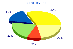
Generic nortriptyline 25 mg line
The filum terminale is classically believed to fixate the distal end of the cord and thereby decrease its movement inside the vertebral canal anxiety 1 week before period purchase nortriptyline 25 mg otc. This viscoelastic band often allows the distal part of the wire to move slightly i have anxiety symptoms 247 buy generic nortriptyline 25 mg online, but when its nature is compromised by both fatty infiltration22 or irregular thickening, undue caudal tension may outcome. However, in uncommon cases, caudal Symptoms the symptoms of a spinal wire displaced caudally as a result of a fat-infiltrated filum terminale embrace pain and orthopedic, urologic, and neurological issues. Commonly, the initial scientific symptom in patients with a tethered cord secondary to a fat-infiltrated filum is gradual and progressive lack of coordinated bladder activity. This may manifest as repeated bouts of urinary tract an infection or primary or secondary urinary incontinence. Axial T1-weighted magnetic resonance picture demonstrating a fat-infiltrated filum terminale. The mixture of upper and lower motoneuron disturbances in the decrease extremities is the signature of this pathologic situation. Nonradicular pain within the again and legs may be the primary manifestation in adults with fatty filum terminale. Additionally, patients with the Currarino triad and cloacal exstrophy usually tend to have such malformations. In general, three standards are essential to verify the medical impression of a tethered spinal wire on imaging: caudal descent of the conus, fatty infiltration and thickening of the filum terminale, and a drawn-out appearance of the distal conus. Occasional adult patients have many a long time of symptom-free life only to come to medical consideration because of irreversible bladder dysfunction. Operative picture demonstrating a fatty filum terminale (centrally positioned and working over the suture). The affected person had had recurrent bouts of urinary tract an infection and episodes of urinary incontinence. Results of preoperative urodynamics had been irregular however, at 1 yr after surgical procedure, had been discovered to be within regular limits. Electrocautery is performed at two points, and the filum transected at every level. The dura mater and overlying delicate tissues are then closed in routine fashion (Video 232-1). They regularly penetrate the dura of an related bony median septum or may simply exit dorsally. This tethering ends in neurological deficits referable to the caudal spinal wire. Tubbs and associates46 beforehand reported 19 patients with meningocele manqu� confirmed at surgery. An related cut up twine malformation was present in 74% (4 of whom had bony septa, and three fibrous septa). At surgery, 63% of the sufferers have been discovered to have intradural bands from the cord to the inside side of the dorsal dura, whereas 37% had intradural bands that pierced the dura and Treatment There is evident consensus within the neurosurgical community that the symptomatic patient with a low-lying conus (conus positioned under the L1-L2 interspace) and a fatty filum should undergo surgical untethering. Sectioning of the filum is carried out with an strategy by way of the L5-S1 interspace with laminotomies of L5 and S1 to help in publicity. Twenty-one p.c of the bands occurred at one vertebral level, 42% at two vertebral levels, and 21. Symptoms In the Tubbs sequence,46 84% of sufferers had irregular neurological findings at the initial evaluation. The commonest symptom was decrease motoneuron disturbance similar to atrophy of the decrease extremity muscle tissue. In 37% of the sufferers, the initial findings on neurological examination had improved on the time of follow-up. In 47% they had been unchanged, and in 16%, the findings on examination had worsened at the time of follow-up. In the sequence reported by Tubbs and associates,46 just one patient had two nonadjacent vertebral levels of involvement. In the appropriate clinical setting, exploration of the intradural contents to search for these points of fixation is justified. A small congenital opening within the skin may be overlooked on physical examination and may not be thought-about until a patient has suffered a neurological insult. These entities are seen most commonly in the lumbosacral space and could additionally be apparent only with assistance from magnification. Cranial dermal sinus tracts usually travel inferiorly towards deeper neural structures, whereas lumbosacral tracts ascend from their origin to their destination. Inclusion tumors (dermoid, epidermoid, teratoma) could also be found wherever along the dermal sinus tract in up to 60% of sufferers. Approximately 60% of all dermal sinuses enter the subarachnoid space, with approximately 25% being connected to neural components. The bands sometimes pierce the dura and terminate on the undersurface of the laminae. One affected person described by Tubbs and associates46 had a band that left the dura and shaped a tense connection with a midline cutaneous defect that was exquisitely delicate to touch. Tethering bands have additionally been described as touring and attaching ventrally and may be discovered within the cervical area. The congenital dorsal bands extend from the right posterolateral aspect of the spinal cord. This band was adherent to the underside of the dura and tented the spinal twine dorsally. If the spinal twine is involved, the attachment or infiltration is nearly all the time dorsal. Sacrococcygeal dimples, which are typically innocuous, are found inside to the natal cleft, are seen in 2% to 5% of youngsters, and are generally confused with true dermal sinus tracts. They reported that not one of the a hundred and eighty simple dimples (<5 mm in diameter, midline, inside 2. Symptoms Despite the presence of cutaneous markers, dermal sinuses typically come to medical attention solely after an infectious or neurological complication develops. Intramedullary spinal cord abscesses most likely represent essentially the most serious problems of dermal sinus tracts. Reflecting a common ontogenic dysfunction, dermal sinus tracts are seen, for example, in 15% to 40% of sufferers with cut up twine malformations. The literature helps a small male preponderance, although Jindal and Mahapatra,66 in a evaluation of 23 sufferers with dermal sinus tracts, discovered that females outnumbered males by a ratio of 16: 9. These connections between the skin and underlying structures often happen within the midline however may be present in a paravertebral location. Interestingly, this assortment extended for a quantity of vertebral ranges and had a component that rested within the posterior mediastinum. The dura is usually tented posteriorly at the point the place the dermal sinus penetrates the thecal sac. Arachnoiditis from earlier cyst rupture or infection could distort the course of the nerve roots by clumping them and create a ring-like configuration across the isointense mass. Intraoperative photograph of a dermal sinus tract (over the neurosurgical cotton patty). Pathologic specimen of a dermal sinus tract with a terminal dermoid (dilated tissue on the far proper of the tract).
Nortriptyline 25 mg effective
The capacity to predict and perceive the stereotypical deformity from statement of the vault suture suggests its major role anxiety genetic 25 mg nortriptyline cheap overnight delivery. Distraction devices anxiety videos 25 mg nortriptyline mastercard, such as those developed by Ilizarov for the distraction of extremity bones and by McCarthy for mandibular hypoplasia, are effective in elongating bone in animals. Using spring-like expanders, Persing and associates20 internalized the distraction of skull bone experimentally, and subsequently Maltese and colleagues21 employed this method within the management of human craniosynostosis. The advantage of the distraction and spring techniques is that bone elongation is a gradual process, with minimal surgical intervention and operative time. Virchow described fusion of vault sutures with local development restriction and extra distant cranial vault compression. Moss described a useful matrix theory whereby skull base and mind development dictate form. In addition, the removal of spring and bone distraction gadgets requires an additional operative process. Similar to implanted steel fixation plates and screws within the cranium, if left in place too lengthy, these metal gadgets can migrate intracranially, with progressive resorption of the endocranial floor of the cranium bone and positioning of bone on the ectocranial floor because the skull and brain enlarge. Concern about transcranial migration has been the main impetus for the avoidance of metallic fixation plates and screws in young children. Resorbable plates and screws, in addition to suture materials, have largely supplanted titanium gadgets in patients youthful than three years. Additional research are being carried out to document skull progress changes by surgical manipulation, particularly by means of mechanical devices to control or improve growth influences. Syndromic craniosynostosis is way less common and appears to be a extra generalized dysfunction of mesenchymal improvement. However, the cause of craniosynostosis remains to be unknown, although in some instances, a genetic affect is plain. Except for Saethre-Chotzen syndrome, these syndromes are the outcome of fibroblastic development factor receptor gain-of-function mutations. We have modified the Virchow hypothesis of skull deformity to clarify the cranium shapes associated with individual types of craniosynostosis. A, Fused left coronal suture (dashed line) joining the frontal and parietal bone, which act as a single progress middle (bone plate) with reduced development potential. B, the margin of the abutting sutures peripheral to the "fused bone plate" deposits a higher amount of bone on the suture (plus signs) than on the margin of the suture adjacent to the fused bone plate (dots). C, Compensation for restricted cranium progress occurs symmetrically at sutures which are "in line" with the fused suture. D, the best diploma of compensation occurs in sutures closest to the fused suture (curved arrows), and the least diploma occurs in sutures distant from the affected suture (straight arrow). This exact definition is essential as a end result of cranium deformity associated with craniosynostosis must be distinguished from that related to uterine molding; circumstances similar to plagiocephaly with out craniosynostosis will enhance with time and require no surgical procedure, whereas craniosynostosis deformities either persist or improve with time, indicating the need for surgery. Experimental evidence demonstrates that improved skull kind is achieved with an operation earlier than the human equal of 6 months of age. Correction of the cranial base deformities established in unilateral coronal synostosis was additionally more successful if surgical procedure was carried out early. Heller and associates28 confirmed a normal cephalic index in one hundred pc of sufferers undergoing whole-vault cranioplasty; however, the cranium shape returned to normal solely in patients who underwent the extra extensive cranioplasty process. As a end result, surgeons advocating this strategy are performing progressively more in depth operative procedures. Brain development as it pertains to the surgical fixation method for particular person components of bone during skull reconstruction. Note that nonrestrictive shorter appearing fixation methods are most popular throughout fast phases of brain progress. This method, especially when applied to the posterior calvarium, has gained widespread reputation for the therapy of turribrachycephaly seen in bilateral coronal synostosis. Spring expanders, that are comparable in idea however extra self-contained, have been first described in rabbits and subsequently in people. However, there have been reports that posterior cranium distraction has resulted in improved anterior cranium morphology even when compensatory modifications are present. Many luminaries of the early years of neurosurgery have been caught up in a world wave of unscientific enthusiasm for linear craniectomy as a therapy to "unlock" the brain of severely impaired, microcephalic youngsters. A passionate marketing campaign led by Abraham Jacobi, patriarch of the specialty of pediatrics and founding father of the American Academy of Pediatrics, and supported by Harvey Cushing, amongst others, ultimately drove the surgical therapy of mental retardation into deserved obscurity (see Feinsod and Davis36 for the complete story). When craniosynostosis later emerged as a distinct diagnostic entity, and as surgical interventions became safer and simpler from a beauty standpoint, the question of the relationship between cranium deformity and mind development arose as quickly as again, and it has proved proof against simple answers. None of these mechanisms seems satisfactory alone, nor has surgery proved absolutely effective in stopping neurocognitive disabilities. Evidence would additionally indicate that sellar erosion and suture diastasis have excessive specificity in all ages. Measurements above 15 torr had been thought-about abnormal, and measurements between 10 and 15 torr had been thought-about borderline. It was more prevalent in sufferers with multiple suture involvement and among syndromic instances. Patients who presented after 1 12 months of age had larger rates of intracranial hypertension. The generalization of those knowledge in support of early surgical treatment is troublesome to accept. A few studies have reported focal areas of hypoperfusion4,fifty five,56 or hypometabolism57 subjacent to irregular sutures among small numbers of selected infants with single-suture craniosynostosis. Recent investigations using subtle strategies of image evaluation have begun to try and relate calvarial deformities to deformation of the underlying neuroanatomic buildings and to growth knowledge. The priorities in care, within the remedy of those children, relate primarily to two factors: intellectual function and skull form. Historically, strip craniectomies had been carried out to release a fused suture because, presumably, removal of the fused suture would allow the restricted brain and cranium to develop usually. It was believed that "refusion" of the sutures accounted for comparatively poor head form outcomes in plenty of sufferers. Silastic sheets have been wrapped across the reduce edges of the bone at the craniectomy web site with the assumption that this barrier would stop irregular bone growth at the cranial sutures; presumably, this is ready to ameliorate deformity of the skull and brain. This led to the event of "comprehensive" cranioplasties through the past 30 to 40 years,sixty seven,68 yielding better anatomic adjustments in cranium form, and, purportedly, good neurological outcomes as nicely. Early timing of surgery and perceived efficacy in remodeling the skull form added to the popularity of the procedures,28,70 significantly when accompanied by long-term helmet remedy after surgical procedure. Concern, nevertheless, has arisen associated to the delay in full correction of the constricted skull and mind by this strategy, as nicely as the necessity to assemble a quantity of helmets (and their costs) for extended periods (approximately 1 year). In specific, long-term longitudinal studies have been needed, utilizing applicable neurological assessment tools that predict outcomes reliably. The very existence of neurological sequelae was referred to as into question in nonsyndromic single-suture craniosynostosis. However, Kapp-Simon72 noted long-term neurological incapacity, and Magee and associates73 demonstrated that infants with sagittal synostosis present process (strip craniectomy) surgical procedure, examined at 6 to sixteen years of age, (when more dependable exams of mental achievement might be precisely used) have a excessive share of studying disabilities. With this info in hand, there was an impetus to reexamine each the timing and type of surgical procedure (comprehensive vault cranioplasty versus the endoscopic strip craniectomy) in children with nonsyndromic forms of craniosynostosis. Although whole-vault (or comprehensive) cranioplasty techniques supply the opportunity for immediate and full correction of the cranium constraint of the mind on the time of surgery, they entail longer operative instances, extra blood loss, and prolonged hospital stays.