

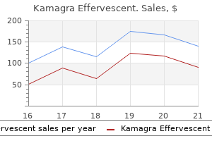
Kamagra Effervescent dosages: 100 mg
Kamagra Effervescent packs: 1 pack, 2 pack, 3 pack, 4 pack, 5 pack, 6 pack, 7 pack, 8 pack, 9 pack, 10 pack

100 mg kamagra effervescent buy with amex
B young living oils erectile dysfunction kamagra effervescent 100 mg buy without a prescription, Choroidal malignant melanoma erectile dysfunction doctors in toms river nj kamagra effervescent 100 mg sale, epithelioid cell type, with noncohesive, bigger, polygonal cells with abundant, glassy cytoplasm, distinct cell borders, and prominent nucleoli (H&E, 60�). A metastatic melanoma arising from an extraocular web site may show uveal metastatic spread. Loss of a whole chromosome three (or monosomy 3), which is an early occasion in tumorigenesis, is detected in roughly 50% of tumors. In a new tumor classification scheme molecular markers are integrated and tumors are classified as class 1 (disomy 3/6p gain) and class 2 (monosomy 3/8q gain). Treatment and Prognosis: the first objective of therapy for uveal melanomas is to stop metastasis. A inset, Melanocytoma cells exhibiting small, centrally located round to oval nuclei (H&E bleached, 10�). C, Choroidal nevus displaying spindleshaped cells with spherical to oval nuclei and vague cytoplasm (H&E, 40�). B, Eye in congenital Coats disease with retinal vasculature telangiectasia leading to repeated microhemorrhages, yellow subretinal exudates, and retinal detachment (hematoxylin and eosin, 1�). It has a yearly incidence within the United States of 1/15,000 to 18,000 stay births, with 250 to 300 new circumstances per year. The imply age at analysis for the sporadic sort is 24 months, while that for the heritable type is 12 months. Patients with the heritable form of retinoblastoma have germline mutations of the retinoblastoma gene in a single allele. Another common transpupillary thermotherapy, photocoagulation, photodynamic remedy, and native resection. Regardless of treatment, monitoring for metastasis ought to be carried out frequently and affected person compliance with follow-up must be confused. Even with early analysis, appropriate therapy, and shut follow-up, an estimated 40% to 50% of all sufferers will eventually die of metastatic disease. Metastasis generally occurs via local extension or though infiltrating into the circulatory system. Systemic metastasis is most commonly discovered within the liver, with 80% to 90% of preliminary extrascleral tumors identified at this site. B, Retinoblastoma tumor cells with hyperchromatic blue small cells surrounded by areas of tumor necrosis and microcalcifications (hematoxylin and eosin [H&E], 4�). C, Flexner-Wintersteiner rosettes exhibiting a wreath-like pattern with a central lumen formation recapitulating the retina, and Homer Wright rosettes exhibiting a wreath-like pattern with central processes, but no lumen formation (H&E, 40�). Less frequent displays are a red, painful, infected eye, orbital cellulitis, and infrequently a cataract or a fungating mass on the skin of the eye. The mutation of one irregular allele is transferred as an autosomal dominant situation with 90% penetrance; nonetheless, the disease solely manifests with inactivation of each alleles. The heritable form of the disease can also be associated with a predisposition to pineoblastoma. In Coats disease, congenital telangiectasia of the retinal vasculature ends in repeated microhemorrhages, yellow subretinal exudates, and retinal detachment. Toxocariasis, an ocular infection of the canine parasite ascarid Toxocara canis, causes the formation of a retinal inflammatory mass that might be mistaken for a tumor. B, Cut part of optic nerve margin exhibiting involvement by retinoblastoma (H&E, 4�). Retinoblastoma cells are immunoreactive to neuron-specific enolase and focally to glial fibrillary acidic protein. In areas of differentiation, photoreceptor proteins may be discovered (cone opsin, rod opsin, interphotoreceptor retinol-binding protein, and S-antigen). Tumor cells are primitive but bigger, and organized in cords and sheets that may resemble embryonic retina. B, Cystic areas lined by primitive massive cells arranged in cords and sheets resembling embryonic retina (H&E, 10�). Treatment and Prognosis: Enucleation is not the only mortality-reducing modality, and globe-sparing therapy options at the second are generally employed when appropriate. Squamous cell papilloma of the conjunctiva as a outcome of human papillomavirus agulation, and plaque radiotherapy, typically with the addition of adjuvant chemotherapy. In bigger tumors, pretreatment with chemoreduction is adopted by an ablative process. Squamous carcinoma and dysplasia of the conjunctiva and cornea: an evaluation of 101 instances. Histochemical analysis and immunohistochemical profile of mucoepidermoid carcinoma of the conjunctiva. Molecular heterogeneity in mucoepidermoid carcinoma: conceptual and sensible implications. Incidence of cutaneous sebaceous carcinoma and risk of associated neoplasms: insight into Muir-Torr� syndrome. Non-caseating conjunctival granulomas in patients with multifocal choroiditis and panuveitis. Subconjunctival herniated orbital fat: a benign adipocytic lesion that may mimic pleomorphic lipoma and atypical lipomatous tumor. Granular cell tumor (myoblastoma) of the palpebral conjunctiva inflicting pseudoepitheliomatous hyperplasia of the conjunctival epithelium. Carney complex, Peutz-Jeghers syndrome, Cowden illness, and BannayanZonana syndrome share cutaneous and endocrine manifestations, however not genetic loci. Concurrent Ki-67 and p53 immunolabeling in cutaneous melanocytic neoplasms: an adjunct for recognition of the vertical progress part in malignant melanomas Distinction of conjunctival melanocytic nevi from melanomas by fluorescence in situ hybridization. Pathology of the Conjunctiva, Orbit, Lacrimal Gland, and Intraocular Tumors 1149 fifty three. Conjunctival melanoma and melanosis: a reappraisal of terminology, classification and staging. Histologic features of conjunctival melanoma predictive of metastasis and death (an American Ophthalmological thesis). Conjunctival lymphoid tumors: medical evaluation of 117 circumstances and relationship to systemic lymphoma. Prospective trial of targeted radioimmunotherapy with Y-90 ibritumomab tiuxetan (Zevalin) for front-line remedy of early-stage extranodal indolent ocular adnexal lymphoma. Lesions of the caruncle: an outline of forty two instances and a evaluate of the literature. Survey of 1264 patients with orbital tumors and simulating lesions: the 2002 Montgomery Lecture half I.
Kamagra effervescent 100 mg cheap mastercard
Significant organ disfunction could develop with progressive inflammatory and fibrotic adjustments erectile dysfunction treatment levitra 100 mg kamagra effervescent best. Up to 20% of patients with identified systemic sarcoidosis current with ocular involvement erectile dysfunction causes stress buy 100 mg kamagra effervescent overnight delivery. B, Predominance of IgG4-positive plasma cells (IgG4 immunohistochemical stain, 20�). Other causes of granulomatous irritation must be ruled out, in particular tuberculosis, fungal an infection, and foreign bodies, among other causes of continual inflammation. The International Society of Amyloidosis 2012 guidelines describe 30 distinct human and 10 animal amyloid fibril proteins. Orbital amyloidosis is regularly benign, localized, and related to major illness. Signs and symptoms commonly involve tissue infiltration, ptosis, diplopia, irritation, and epiphora, and might mimic different orbital lots and diseases resulting in delayed diagnosis. Treatment options for ocular amyloidosis differ relying on patient symptoms and the visual significance of amyloid protein deposition. Excision can be tried, but complete excision of the lacrimal gland may be surgically difficult. The immature stem cells current throughout the tumor growth could have originated from the placenta. Capillary hemangioma is now referred to as childish hemangioma; cavernous hemangioma is now considered a venous malformation. Low-flow malformations contain mixtures of capillary, venous, and lymphatic components. High-flow malformations comprise arterial components together with other vascular buildings. This benign, noninfiltrative tumor tends to happen in adults during the third to fifth many years of life, is slowly progressive, and is mostly intraconal (within the cone of extraocular muscles). It produces mass impact with proptosis and can lead to restriction of ocular motility. The Masson trichrome histologic stain highlights the fibrotic vascular partitions and small foci of intramural easy muscle. Sections stained with Verhoeff-Van Gieson show the disorganized elastic fiber components of the lesion. Differential analysis includes capillary hemangioma, lymphangioma, and combined venous lymphatic malformations. It normally manifests as a subcutaneous eyelid lesion with progressive proptosis and swelling. B inset, Congo purple stain exhibiting apple-green birefringence under polarized mild (Congo purple stain, 4�). Orbital polyangiitis with granulomatosis (Wegener granulomatosis),seventy two orbital angiolymphoid hyperplasia with eosinophilia and Kimura disease,seventy two Rosai-Dorfman disease,72 and other tumors have been reported. Orbital Vascular Lesions Vasoproliferative lesions have undergone adjustments in their nomenclature by the International Society of the Study of Vascular Abnormalities. B, Orbital lymphangioma composed of dilated ectatic vascular channels with thin delicate fibrous partitions and lymphoid follicles (H&E, 4�). Patients can current with acute proptosis after minor head trauma, as a gradual proptosis, or after an higher respiratory an infection. Dilated ectatic vascular channels crammed with clear fluid or blood with skinny delicate fibrous partitions and flattened endothelial cells are discovered on histology. Lymphangioma may bleed into itself, causing cystic spaces crammed with blood inside the tumor. Differential diagnosis features a cavernous hemangioma, a varix (low-flow vascular malformation), and a combined venous lymphatic malformation. A cavernous hemangioma reveals dilated vascular channels crammed with blood and is a well-defined lesion. A varix is a dilatation of one or more veins that show thrombosis and hyalinization within a dilated vein histopathologically. Combined venous lymphatic malformations can come up in the superficial or deep orbit and could additionally be associated with intracranial vascular malformations. Histopathology exhibits options of venous and lymphatic channels; lymphoid follicles could additionally be seen. Treatment of orbital lymphangioma could be difficult because complete surgical excision is usually tough as a end result of poor definition of the lesion and bleeding. Clinical remark, aspiration of the hemorrhage, and/or surgery are all issues in the administration of orbital lymphangiomas. It is a congenital lesion shaped from entrapped epithelial cells throughout embryogenesis beneath the surface epithelium, often near bones. Orbital dermoid cysts are mostly positioned superotemporally on the zygomaticofrontal suture (70%) and superonasally on the maxillofrontal suture (20%), with few within the nasal gentle tissues (5%) and different areas. They sometimes present in a young youngster as firm, fixed subcutaneous lesions close to the orbital rim superotemporally. The cyst might present conjunctival epithelial lining as an alternative, particularly these positioned in the orbital gentle tissues nasally. The cysts might rupture and present typical persistent overseas body�type granulomatous inflammation. The differential diagnosis includes orbital mucocele, an orbital respiratory epithelial cyst, an orbital teratoma, and a easy conjunctival cyst. Orbital mucocele arises from a chronically inflamed paranasal sinus, most frequently the frontal or ethmoid sinuses, which secondarily entails the orbit. Mucoceles are lined by pseudostratified columnar epithelium with variable quantities of inflammation. B, Axial computed tomography scan displaying a superior medial orbital lesion molding to the globe and bones protruding into the subcutaneous tissues. Most dermoid cysts are surgically eliminated, especially if they rupture and show secondary inflammatory adjustments. It usually happens within the first two decades of life, with a mean age of eight years and a slight male predilection. The great majority of patients current with proptosis and downward and lateral displacement of the globe due to a superior or superonasal location of the mass. B, Orbital alveolar rhabdomyosarcoma appearing as loosely dispersed cells mimicking a pulmonary alveolar pattern (H&E, 40�). B, Focal positivity with desmin stain in orbital embryonal rhabdomyosarcoma (desmin immunohistochemical stain, 10�). Orbital rhabdomyosarcoma has been described to occur many years after orbital irradiation for retinoblastoma. The histologic variants of rhabdomyosarcoma that happen mostly within the orbit embody embryonal, alveolar, and anaplastic. Large, polygonal-shaped cells with eosinophilic cytoplasm and cross-striations could also be present. Alveolar rhabdomyosarcoma reveals a uniform population of cells with a excessive nuclear-to-cytoplasmic ratio.
Order 100 mg kamagra effervescent with amex
The neoplastic cells are small erectile dysfunction at age of 20 kamagra effervescent 100 mg order visa, with barely to moderately irregular nuclear contours and plentiful pale cytoplasm erectile dysfunction protocol scam or real buy generic kamagra effervescent 100 mg line. Other lymphomas could uncommonly current as localized illness involving the thyroid gland, together with follicular lymphoma, other small B-cell lymphomas, Burkitt lymphoma, Hodgkin lymphoma, and T-cell lymphomas. Patients can present with signs of nasal obstruction, nasal tumor mass, facial swelling and discharge, epistaxis, visual disturbances, or headaches. Overall, low-grade tumors tend to form plenty within the concerned nasal cavity or paranasal sinus and trigger obstruction, whereas high-grade lymphomas cause more aggressive symptoms, similar to facial edema, epistaxis, and facial ache. High-grade lymphomas can destroy bone and instantly prolong into the central nervous system. A and B, Diffuse infiltrate of huge plasmablasts and immunoblasts with vesicular chromatin, eccentric nuclei, and prominent central nucleoli. Primary circumstances occur, but most instances characterize extension from the upper respiratory tract. Radiologically, the lesion produced by lymphoma is commonly homogeneous with well-defined margins, in contrast to carcinoma, which is more infiltrative and less bulky. Lymphomas may come up within the external auditory canal or temporal bone, but more often these sites are involved by systemic lymphomas as a manifestation of dissemination. Follicle-center cell lymphoma arising in the skin usually presents as a solitary lesion, or as grouped plaques and tumors, preferentially positioned on the scalp or forehead. A clear-cut follicular growth sample is extra commonly observed in lesions arising on the scalp than those presenting on the trunk. It is important to rule out systemic disease before the analysis of major cutaneous follicular middle cell lymphoma is established. B, the neoplastic cells are a mixture of small lymphocytes, lymphoplasmacytoid cells, and peripheral plasma cells. The infiltrates are composed of small lymphocytes, centrocyte-like cells, lymphoplasmacytoid cells, and plasma cells, admixed with small number of massive cells resembling centroblasts or immunoblasts. Rare cases of main cutaneous T-cell/histiocyte-rich B-cell lymphoma have been reported. This distinction requires medical assessment; a history of lesions that come and go supports lymphomatoid papulosis. These lesions are clinically indolent however have been shown to carry a monoclonal T-cell receptor gene rearrangement supporting the diagnosis of lymphoma. B, the neoplasm is composed of small lymphoid cells with blastic nuclear chromatin and inconspicuous nucleoli. In many neoplasms, these cells have barely irregular nuclear contours and comparatively abundant clear cytoplasm resembling monocytoid B cells. In different circumstances, these cells are small and carefully resemble small lymphocytes, or can exhibit plasmacytoid differentiation. In some neoplasms, the tumor cells appear biphasic, with one component being small lymphoid cells with minimal cytoplasm, while the other component displays intensive plasmacytoid differentiation, with many cells resembling mature plasma cells. In yet different tumors, the neoplastic small cells have markedly irregular nuclear contours and resemble centrocytes. However, in neoplasms with in depth involvement, lymphoepithelial lesions may be troublesome to recognize as a result of glands are destroyed. A keratin immunostain can be helpful in figuring out residual epithelium invaded by tumor cells. When these cells turn out to be numerous and type sheets, the neoplasm has reworked to massive B-cell lymphoma, with a correspondingly poorer prognosis and requiring aggressive therapy. In common, the tumor is labeled a large-cell transformation when sheets of huge cells are present. The tumor cells also may accumulate within the follicle centers (referred to as follicular colonization) with the tumor then acquiring a nodular low-power look. However, these neoplasms additionally may exhibit true plasmacytoid differentiation in as much as a third of cases. Instead, extranodal infiltrates composed of small lymphoid cells typically admixed with plasma cells, histiocytes, and lymphoid follicles had been categorized as "pseudolymphomas," since scientific research confirmed that patients with these lesions had an indolent clinical course. Immunophenotypic and gene rearrangement research have since shown that nearly all "pseudolymphomas" specific monotypic Ig gentle chain and comprise monoclonal immunoglobulin gene rearrangements. Histologically, evidence of cytologic atypia and Dutcher bodies, if quite a few, help lymphoma. As acknowledged earlier, the presence of a monotypic B-cell population helps lymphoma. The presence of monoclonal immunoglobulin gene rearrangements without immunophenotypic evidence of monoclonality is extra controversial. Particularly when using polymerase chain reaction-based methods, small monoclonal B-cell populations or chromosomal abnormalities may be present in lesions that histologically and immunohistochemically meet the factors for a benign course of. Older names for these neoplasms within the literature include polymorphic reticulosis, lethal midline granuloma, and malignant midline reticulosis. In the early stages of disease, comparatively few neoplastic cells are present, and these cells may be of small measurement. As a outcome, biopsy specimens obtained at an early stage of illness could be misinterpreted as a benign course of. Over time, these neoplasms accrue larger numbers of huge atypical cells and the prognosis is more simply established. A, Low-power magnification of one case exhibiting a diffuse infiltrate of neoplastic cells and zonal necrosis. B, High-power magnification of one other case during which the neoplastic cells are small with irregular nuclei. Most different nonHodgkin lymphomas of the sinonasal space are B-cell lymphomas or lymphomas that secondarily involve this website. Immunohistochemical studies as well as information of the clinical historical past will assist in figuring out these cases. These patients are sometimes handled with a mixture of radiation remedy and chemotherapy. The prognosis of these sufferers is comparatively good compared to sufferers with disseminated disease. Burkitt Lymphoma the late British surgeon Denis Burkitt first described a form of lymphoma that he noticed as a big jaw mass in children from Uganda. Endemic Burkitt lymphoma was first described in equatorial Africa, within the geographic area occurring 15 levels latitude north or south of the equator. The median age of patients with endemic Burkitt lymphoma is 7 years, with a male-to-female ratio of three:1. The jaw is the best-known website of illness, involving the maxilla or mandible in 60% of patients, but large belly plenty involving retroperitoneal constructions, the gastrointestinal tract, or the gonads are additionally common.
Buy discount kamagra effervescent 100 mg
Effect of sort 3 intramural fibroids on in vitro fertilization�intracytoplasmic sperm injection outcomes: a retrospective cohort study erectile dysfunction caused by vicodin kamagra effervescent 100 mg buy amex. Three-dimensional hysterosonography versus hysteroscopy for the detection of intracavitary uterine abnormalities erectile dysfunction fast treatment 100 mg kamagra effervescent cheap otc. Three-dimensional colour Doppler sonography and uterine artery arteriography of fibroids. Fibroids and reproductive outcomes: a scientific literature evaluate from conception to delivery. While a genetic disposition have to be given, as Africans have a much higher frequency of a quantity of myomas than Caucasians, sure up- and down-regulations within the genes of sufferers with or with out myomas have been described. However, as but, no clear tips for the prevention of fibroids can be found. Hereditary leiomyomatosis and renal cell carcinoma syndrome are uncommon syndromes involving fibroids. Individuals with the gene that results in both fibroids and pores and skin leiomyomas have an elevated risk of developing a uncommon case of kidney cell most cancers (papillary renal cell carcinoma). Most guesses relating to these "candidate genes" end up to be incorrect, and much research is still required to learn the way these genes result in illness. There are additionally small variations, called polymorphisms, in genes that may play a role in influencing the risk of fibroids. Both polymorphisms and mutations are adjustments in the sequence of genes, but the difference is within the degree of change. A mutation makes a serious change in the gene that results in a change within the protein the gene is coding for. For example, it could change the amino acid from alanine to glycine or trigger the protein to be prematurely cut off. Smooth muscle cells are organized in order that the organ can stretch as an alternative of being arranged in inflexible items, just like the cells in skeletal muscle in arms and legs, which are designed to "pull" in a specific path. In girls with fibroids, tissue from the endometrium usually appears regular underneath the microscope. The presence of this abnormality, referred to as aglandular functionalis (functional endometrium with no glands), in girls having bleeding problems is typically a medical clue for their doctors to look extra intently for a submucosal fibroid (PattersonKeels et al. A second pattern of endometrium, termed chronic endometritis, also can recommend that there could also be a submucosal fibroid, although this sample may also be associated with different issues, corresponding to retained products of conception and various infections of the uterus. When deciding whether or not to launch a model new idea, firms usually have a glance at the quantity at present spent for different remedies. The economics of fibroids has been discussed chiefly in terms of the healthcare costs of hysterectomy. According to a 2006 estimate, within the United States, more than $2 billion is spent every year on hospitalization costs because of uterine fibroids alone (Flynn et al. Additionally, one study estimates that the health-care costs because of uterine fibroids are greater than $4600 per woman per yr (Hartmann et al. However, whenever you incorporate all the costs of fibroids, the way of remedy becomes even more important. First, up to a sure measurement of the enlarged uterus, laparoscopic subtotal hysterectomy utterly solves the issue, and if girls need to get rid of each danger of recurrent fibroids, hysterectomy is their solely selection. Time and type of 36 remedy have to be chosen individually and are dependent on the affected person and the treating gynecologist (Table 5. Expectant Management Wait-and-see is a possibility if patients are asymptomatic, decline medical or surgical treatment, or have contraindications to any type of treatment. However, present knowledge describe the possibility that fibroids shrink considerably either by optimizing endocrinological problems, such as hypothyroidism, or during the postpartum period (Peddada et al. To pursue the idea of expectant management, the pelvic mass must undoubtedly be categorized as a fibroid and differentiated from an ovarian mass. The complete blood depend ought to be regular, particularly in sufferers with severe symptoms, corresponding to menorrhagia or hypermenorrhea. Women must additionally be informed that the danger of miscarriage, untimely labor and supply, irregular fetal place, and placental abruption is increased throughout pregnancies with uterine fibroids (Zaima and Ash 2011). Medical Therapy the advantage of medical remedy in the administration of women with symptomatic fibroids remains to be tough to show. Medical remedy can provide sufficient symptom aid, particularly in instances the place hypermenorrhea is the main problem. The good thing about symptom enchancment decreases in long-term treatment durations and so greater than 50% endure surgical procedure within 2 years (Marjoribanks, Lethaby, and Farquhar 2006). Nevertheless, there was a shift in conventional pondering that medical treatment of fibroids relies solely on the manipulation of steroid hormones. A deeper analysis and understanding of particular genes or pathways associated with leiomyomatosis might open new possibilities for prevention and medical remedy (Al-Hendy et al. Primarily as a preoperative treatment to lower heavy bleeding in sufferers with fibroids, hormonal therapy with selective progesterone modulators, corresponding to ulipristal acetate 5 to 10 mg every day, has turn out to be extensively used throughout the final 2 years (Donnez et al. A catheter is launched via the femoral artery beneath native anesthesia, and particles are injected to block the blood circulate to the fibroid. Magnetic resonance�guided centered ultrasound: it is a more recent remedy technique for uterine fibroids in premenopausal women. In this non-invasive thermal ablative approach, multiple waves of ultrasound power are converged on a small quantity of tissue, leading to maximal thermal destruction. Uterine-Preserving Surgical Treatment of Fibroids the surgical removing of fibroids continues to be the primary pillar within the remedy of leiomyomas. Hysterectomy is the one definitive solution and may be carried out as supracervical or whole hysterectomy. Myomectomies performed by hysteroscopy, typical laparoscopy, or laparoscopy with robotic assistance and by the open or vaginal strategy are various surgical methods. Abnormal uterine bleeding problems (hypermenorrhea, dysmenorrhea, menorrhagia, and metrorrhagia) 2. Primary or secondary infertility and recurrent being pregnant loss Counseling and Informed Consent Patients undergoing an operative procedure should learn of the risks and potential problems as properly as different operating methods. Counseling before surgical procedure should embrace dialogue of the entry technique and the associated dangers: harm of the bowel, urinary tract, blood vessels, omentum, and other surrounding organs and (at a later date) wound infection, adhesionassociated pain, and hernia formation. Counseling needs to combine the individual danger dependent on the body mass index of the 38 affected person. The enucleation of fibroids by any methodology is an efficient remedy for bleeding issues or displacement strain within the pelvis. Furthermore, if some other pathologies may be causative or only co-causative for the symptoms (such as adenomyosis uteri), these issues will persist (Wallach and Vlahos 2004). Complications arising at myoma enucleations and pregnancy-related problems have been investigated extensively. All working potentialities, especially laparoscopic versus laparotomic but just lately also laparoscopic versus roboticassisted myomectomy, have been evaluated.
Diseases
- Bardet Biedl syndrome
- Seafood poisoning
- Dysferlinopathy
- Epilepsy juvenile absence
- Jankovic Rivera syndrome
- Thumb stiff brachydactyly mental retardation
- Ectopia pupillae
100 mg kamagra effervescent buy fast delivery
Keratoacanthoma varieties a cup-shaped projection from the pores and skin surface with a collarette of well-differentiated proliferating squamous epithelium surrounding a central keratin core erectile dysfunction treatment in unani order kamagra effervescent 100 mg. Within the canal erectile dysfunction new treatments 100 mg kamagra effervescent order, delay in analysis and deep invasion by the tumor can necessitate partial temporal bone resection for adequate elimination. The origins, scientific manifestations, intercourse distribution, therapy, and prognosis of squamous cell carcinoma differ depending on the positioning of origin: the pinna or the exterior auditory canal. There is a female preponderance with a mean age of 60 years, a decade sooner than in sufferers with squamous cell carcinoma of the pinna. Symptoms, often present for less than a 12 months, embody drainage, ache, and listening to loss, indicating an intensive, invasive lesion. Squamous cell carcinoma is characterized by proliferation of squamous cells with abnormal cytologic manifestations of malignancy and irregular keratinization (dyskeratosis). Keratoacanthoma is a definite clinicopathologic entity simulating well-differentiated squamous cell carcinoma. The surrounding proliferating, welldifferentiated squamous epithelium has an indistinct basement membrane and might appear to be invasive. Local excision of pinna lesions normally suffices for native control, but residual or recurrent illness still occurs in 25% of sufferers. Approximately 10% of invasive squamous cell carcinomas metastasize to regional lymph nodes; patients with such metastases can be treated with neck dissection. If after 2 to 3 weeks no decision has occurred, the lesion must be excised with a border of regular tissue and a carcinoma should be excluded by histopathologic examination. Squamous cell carcinomas of the canal metastasize to regional nodes more regularly than do pinna lesions, but the 5-year tumor-related death rate of roughly 50% is as a outcome of of local invasion quite than metastases. The defect is roofed with a skin or muscle flap and is postoperatively irradiated. Malignant melanoma, though comparatively rare, is the third most common malignancy of the exterior ear, accounting for 7% to 16% of all head and neck melanomas. When introduced with a poorly differentiated epithelioid or sarcomatoid neoplasm, one must all the time think about melanoma, and immunohistochemical studies must be done to rule it out. Melanoma arises in a preexisting nevocellular lesion of the skin or mucous membrane, and junctional change ought to be recognized to differentiate primary from metastatic melanoma. A, Spindle cell (sarcomatoid) carcinoma, C, spindle cell amelanotic melanoma, and E, atypical fibroxanthoma are virtually indistinguishable with hematoxylin-eosin stain. Treatment varies depending on the placement on the ear: peripheral pinna lesions endure wide local excision or amputation; more central lesions, adjacent to or involving the exterior auditory meatus, require rather more extensive surgical procedure together with partial temporal bone resection. Seven of the sixteen instances reported by Davidsson and colleagues84 were nodular, and depth of invasion varied from zero. All patients had extensive excision, however of the four (25%) who died, three did so within 1 yr of analysis. Dermatofibromas87 are benign polymorphous cutaneous tumors to which several names have been utilized: subepidermal nodular fibrosis, dermatofibroma, fibroxanthoma, and sclerosing hemangioma, in addition to fibrous histiocytoma. These painless lesions are sometimes found in the higher dermis of the extremities in adults. They are poorly circumscribed, normally with infiltrating margins, and are composed of spindly mononuclear or multinuclear cells with amphophilic cytoplasm that are classically arranged in a storiform pattern. Birefringent collagen is surrounded by spindle cells at the periphery of the lesion. Various modifying adjectives are utilized to fibrous histiocytomas depending on the mixture of cell varieties, vascularity, and hemosiderin in the lesion: angiomatoid, epithelioid, giant cell, or cellular. There is exceptional variability in nuclear and nucleolar measurement and shape, however mitotic exercise is low at 1 per 10 high-power fields. The cellular and nuclear pleomorphism resulted in the initial pathologic diagnoses within the 21 circumstances: sarcoma in 10, carcinoma in 4, melanoma in 1, and atypical fibroxanthoma in 6. Recently, three cases of an atypical fibroxanthoma variant have been described during which osteoclast-like giant cells were dispersed within the tumor. Although 26% of those lesions recurred inside 3 years after insufficient excision, none metastasized. The atypical fibroxanthoma should be differentiated from the actinic malignancies that it mimics by its pleomorphism: spindle cell (sarcomatoid) squamous cell carcinoma and melanoma. Squamous cell carcinoma has desmosomes and intracellular keratin; melanoma has melanosomes and premelanosomes; fibrous histiocytoma has myofilaments attribute of myofibroblasts. A battery of immunohistochemical stains can help within the differentiation of these spindle cell neoplasms (Table 12. Patients are approximately equally divided between women and men, with a imply age of 27 years (range, 1�76 years), a population much like that of fibrous histiocytoma described earlier. Myxomas of the external ear contain both the pinna and the canal in a rare familial autosomal dominant syndrome generally known as the Carney complicated. The lesions happen equally in males and females, with ages ranging from birth to 41 years (mean, 19. Histologically, polypoid or sessile myxomas occurring on the pinna or in the canal are composed of a bland myxomatous stroma and a proliferating, often basaloid, surface squamous epithelium. As a consequence of the latter, differential diagnoses based mostly on superficial biopsies could vary from squamous papilloma to epidermal inclusion cyst, trichoepithelioma, and basal cell carcinoma. When myxoma is recognized, the clinician must be alerted to the chance of the Carney advanced due to the occurrence of doubtless deadly cardiac myxomas. Recognizing that the Carney complicated includes both cardiac and aural myxomas, stories of myxoma from the cardiac atrium metastasizing to the temporal bone should be regarded with some skepticism. Merkel cell carcinoma100�104 is a malignant neuroendocrine small cell neoplasm found in the skin of aged (1. Merkel cell tumors are predominantly situated in the head and neck (face, oral, and nasopharyngeal mucosa; 47%) and trunk (40%). However, only 20 cases arising on the auricle were discovered by Rinaldo and colleagues101 of their literature evaluation. The origin of the tumor in both hair follicle mechanoreceptor Merkel cells or scattered stem cells in oral mucosa, epidermis, and dermis is under debate. Ultrastructural traits of the tumor cells embody perinuclear keratin fibrils, peripheral cytoplasmic dense core granules, and zona adherens intercellular junctions. The reported 5-year survival rate ranges from 40% to 68% of sufferers, with immunosuppressed sufferers doing relatively much less nicely. The use of keratin immunohistochemical research is recommended in evaluating lymph nodes for metastases. Mucinous (Colloid) Carcinoma Primary mucinous (colloid) carcinoma of the skin104 is a rare neoplasm having a propensity for the pores and skin of the face and neck and homologous to mucinous carcinoma of the breast. Of 37 reported circumstances presenting as a solitary nodule, 3 were found in the external ear helix and lobe and 1 in the retroauricular pores and skin. There is a 2:1 female-to-male predominance, with age starting from 31 to 89 years (mean, 65 years; median, 67 years).
100 mg kamagra effervescent purchase overnight delivery
Clinical historical past and special stains and/or cultures should distinguish blastomycosis from condyloma latum latest news erectile dysfunction treatment cheap kamagra effervescent 100 mg with visa. The hyperplasia of condyloma latum can also be confused with laryngeal malignancy erectile dysfunction 30 kamagra effervescent 100 mg buy visa. A, Mixed cell inflammation of the surface epithelium, and dense irritation of the subepithelial stroma with plasma cells. B, Immunohistochemistry reveals numerous spirochetes within the surface epithelium and few within the stroma. These latter infections could be separated on the basis of culture and particular stains. Patients allergic to penicillin may be handled with erythromycin, tetracycline, or ceftriaxone. Ferdinand von Hebra and Moritz Kohn (formerly named Kaposi) coined the time period rhinoscleroma to describe those sufferers presenting with hard (sclero) noses (rhino),35 which they originally concluded should be secondary to indolent malignancy. Interestingly, aniline dye employees in San Salvador have been famous within the late nineteenth century to have an elevated prevalence of rhinoscleroma. Alvarez found a bacillus in the legume, which he known as Bacillus indigogenous, and concluded that it was identical to Frisch bacillus. Human-to-human transmission has been assumed to be the one mode of contact, and infection will result only after prolonged exposure. Increased incidence amongst members of the family and contacts has been famous, but this stays controversial. Rhinoscleroma has been referred to as the disease of the great unwashed; social conditions may vary, however it has been confused that poor hygiene and crowded environments are frequent options. In a mountainous Indonesian endemic web site, many households sleep together in giant, poorly ventilated homes, huddled along with their dogs and home fowl for heat. The term scleroma is preferred over rhinoscleroma as a result of the whole upper aerodigestive tract could also be involved, from nostril to trachea, in addition to the nasopharynx, paranasal sinuses, and Eustachian tube. The early stage is the atrophic catarrhal stage by which the mucosa is reddened and atrophic, with foul purulent discharge and crusting. The scientific differential prognosis on this early stage contains infection with Klebsiella ozaenae. The second stage or granulomatous stage occurs months to years later, with waxy, ulcerating inflammatory masses that distend and deform the mucosal surfaces. The inflammatory lots lengthen by way of the exterior nares in severe instances and should distort the delicate tissues of the midface, leading to a rhinoceroslike look. The final sclerotic stage is characterized by fibrosis and inflammation and culminates in stenosis. Common signs of laryngeal involvement embody progressive dyspnea, cough and hoarseness. Histologically, the catarrhal stage is dominated by nonspecific lymphoplasmacytic infiltrate; bacilliladen foamy macrophages (Mikulicz cells) are sparse. At low power, one sees a diffuse lymphoplasmacytic infiltrate with quite a few foamy histiocytes (Mikulicz cells). Warthin-Starry silver stain reveals bacterial rods within these histiocytes (inset). The differential diagnoses include atypical mycobacterial infection, which also appears as a histiocytic infiltrate quite than a granulomatous reaction. Lepromatous leprosy additionally appears as a diffuse histiocytic infiltrate (Virchow cells) and should be ruled out by a Fite-Faraco stain. When a lymphoplasmacytic infiltrate dominates the histology, the diagnosis of syphilis should also be considered. Long-term (3�6 months) parenteral antibiotic therapy, similar to with tetracycline, streptomycin, chloramphenicol, or ciprofloxacin (1 g/day) has been reported. The causative agent was named Actinomyces bovis (Actinomyces: strahlenpilz or ray fungus). It was initially thought to be related to the behavior, by both man and cow, of chewing straw. It is classed as a filamentous, gram-positive, nonacid quick anaerobic micro organism, quite than a fungus, as a outcome of it reproduces by fission rather than sporulation (as do good fungi) or filamentous budding (as do imperfect fungi) and it incorporates muramic acid in cell partitions and absent mitochondria, both options of bacteria. Actinomycetae are a part of the traditional oral, gastrointestinal, and urogenital tract microbiota. They are of low pathogenicity and need mucosal barrier disruption to cause illness. If the integrity of the mucosal barrier is compromised, for instance throughout dental manipulation, trauma, or surgery, entry to the soft tissue and bone is gained. These organisms turn into pathogenic and may initiate a prolonged chronic inflammatory course of, leading to tissue destruction, formation of abscesses and sinus tracts, which can occasionally be long and communicating with the gentle tissues of the again and chest, in addition to fistula, fibrosis and tumor-like plenty. The recognized danger elements embody surgery, trauma, diabetes mellitus, alcoholism, and remedy with bisphosphonates, chemotherapy, and irradiation. In the oral cavity, Actinomycetae can be found on mucosal surfaces and within the tonsillar crypts, in gingival sulci, periodontal pockets, and in carious lesions. It is, therefore, commonly misdiagnosed as a outcome of it could mimic other circumstances, such as malignancy and tuberculosis. The pale yellow sulfur granules or grains (originally referred to as drusen) noticed clinically are microcolonies of bacilli. Microscopically, Actinomyces sometimes trigger suppurative irritation with colonies of filamentous micro organism, which appear as spherical or oval basophilic lots with a radiating arrangement of eosinophilic terminal "clubs," measuring 1 to 2 mm in diameter. Special stains are helpful to affirm the analysis, as Actinomyces stains with silver impregnation methods, and Brown and Brenn staining (gram positive). The distinction between these two filamentous bacteria is necessary, as their sensitivities to antibiotics differ: penicillin is the drug of alternative for Actinomyces, whereas Nocardia are unresponsive to penicillin and could be treated with sulfa medicine. It has been claimed that culture leads to proper prognosis in only 20% to 40% of circumstances, whereas the remaining circumstances are recognized by biopsy. Biopsy is considered as the quickest and most delicate means of diagnosis actinomycosis. Actinomycosis is often handled with extended excessive doses of antibiotics, often penicillin G. Laryngeal candidiasis is usually not seen in immunocompetent hosts, however could additionally be seen in those with diabetes or sufferers immunosuppressed because of steroid remedy, chemotherapy, or extreme persistent sickness. Laryngeal candidiasis can clinically current with a mass lesion, mimicking carcinoma. Small, oval, globose yeast cells lead to a "spaghetti and meatballs" appearance. If disseminated disease is current, then systemic antifungal therapy (amphotericin B or flucytosine) is indicated. Laryngeal aspergillosis is mostly seen as secondary invasion from the lungs and tracheobronchial tree in immunocompromised hosts. Biopsy samples taken from laryngoscopy present both spores and broad septate hyphae, nearly all of which exhibit acute angle branching on histopathological examination.
Cheap kamagra effervescent 100 mg without prescription
Occasionally erectile dysfunction medication cialis kamagra effervescent 100 mg buy overnight delivery, taller columnar cells erectile dysfunction blogs forums 100 mg kamagra effervescent amex, which hardly ever could have clear cytoplasm, could also be seen proliferating within the ducts, and even small foci of early squamous metaplasia can be recognized. Therefore you will need to carefully pattern these tumors, as more than 98% of the tumor could also be oncocytic, with only very focal areas having the everyday bilayered appearance (intercalated duct-like inside layer surrounded by clear myoepithelial cells). F, Oncocytic variant (left) and double-layered clear cell variant; note that there are areas with both internal epithelial layer and outer myoepithelial layer demonstrating clear cell adjustments (right). G, Apocrine variant; note numerous secretory snouts and cells with larger nuclei and outstanding nucleoli (lower right and inset). Then, the two-layered nature of the neoplasm could also be discernible solely with immunohistochemistry. Identical areas may be seen in pleomorphic adenoma1312 and polymorphous low-grade adenocarcinoma, but these foci are restricted and never seen all through the tumor. Acinic cell carcinoma and mucoepidermoid carcinoma can have clear cell areas, which once in a while could also be extensive, but careful sampling will show scattered cells with the characteristic options. An important practical differential diagnosis for any clear cell tumor is metastatic renal cell carcinoma, which can first current as a parotid mass. In addition, renal cell carcinoma characteristically has larger nuclear pleomorphism and atypia. Other comparable research have highlighted the immmunohistochemical variations between salivary clear cell neoplasms and renal cell carcinoma. They found 26 cancer-related transcripts inside this group, as well as 16 transcripts related to mitochondrial dysfunction. In studies, including a 1992 literature evaluation of sixty seven circumstances, recurrences have been noted in 31% of sufferers, cervical lymph node metastases in 18%, distant metastases in 7%, and demise caused by tumor in 7%. A larger incidence of recurrence (50%) was reported in a welldocumented collection of twenty-two tumors from the largest oncologic referral middle in Portugal, more than likely reflecting sufferers with more advanced disease. Even with full excision, however, recurrence and distant metastasis might trigger issue. For a discussion on the latter, please check with the "Myoepithelial Carcinoma" section. It has a squamoid phenotype and lacks options of other clear cell-rich salivary gland carcinomas"1274; myoepithelial markers are uniformly negative. This carcinoma was previously considered a diagnosis of exclusion, however recent molecular knowledge has allowed its scientific and morphological spectrum to be better characterized. Tumors are normally less than three cm in diameter, but not often could also be larger (mean 2�2. Very focal ducts could be seen, though a few of these could represent entrapped salivary tissue. The predominant tumor cell population is characterized by a round to polygonal form, with abundant clear cytoplasm, though at instances, significantly in the deeper elements of the tumors, the cells might lose their clarity when their cytoplasm seems weakly eosinophilic. The nuclei are centrally located, with delicate pleomorphism and inconspicuous nucleoli; mitotic figures are uncommon. C, Tumor cells are mildly pleomorphic and often have clear cytoplasm (left), but may have areas with eosinophilic cytoplasm (right). The diagnosis on this state of affairs should be made on the idea of tumor morphology and tumor immunophenotype. A monomorphic proliferation of tumor cells with clear cytoplasm could be seen in myoepithelial carcinoma. Radiation remedy may be added in sufferers with constructive or close margins, recurrent illness or in tumors with high-grade features. No further metastases or native recurrence developed in any of the 10 patients with follow-up in the sequence of Milchgrub and colleagues,1341 despite the fact that two introduced with cervical lymph node metastases. Rinaldo and colleagues1345 reported a case with two cervical lymph node metastases separated by 5 years, before a base of the tongue main was detected 2 years later (the affected person is alive and properly 6 years after excision of the tongue mass, thirteen years after removal of the primary lymph node metastasis). Patients have to be adopted long term (>10 years), because the recurrence price will increase significantly after 5 years. Although many tumors originate in the salivary duct system, this term ought to be reserved only for tumors that histologically resemble ductal carcinomas of the breast. Occasional patients have longer scientific histories, with one report of an roughly 38-year history1376; facial paresis may be present. The parotid gland is the commonest website of origin (88% of cases1378), however tumors in the submandibular1379,1380 and sublingual gland,1381 oral minor salivary glands,1375,1381�1383 sinonasal tract,1384 and larynx have additionally been reported. Infiltration into the surrounding parenchyma is normally apparent, but occasional tumors could appear to be circumscribed. It must be noted that sometimes, the in situ lesions may be masked by extensive progress of invasive carcinoma and are morphologically delicate, although they can be highlighted by using basal-myoepithelial markers. This tumor is composed of numerous irregular nests of infiltrating tumor in a desmoplastic background. The tumor cells reveal moderate atypia, with areas of stable and cribriform progress and comedo-type necrosis, similar to ductal carcinoma of the breast. D, Micropapillary sample composed of multiple intently packed irregular tumor nests with out fibrovascular cores surrounded by a transparent area. There are several nests of typical salivary duct carcinoma with a outstanding cribriform pattern surrounded by a markedly pleomorphic, atypical spindle cell inhabitants of tumor cells. There are areas with a stable progress sample (G, left) and outstanding pools of mucin surrounding small collections of carcinoma cells (G, proper; H). Detail of 1 tumor nest with pseudostratified, hyperchromatic nuclei and minimal to average quantities of cytoplasm (G, inset). I, Rhabdoid variant composed of a sheet of enormous, pleomorphic, dyscohesive, ovoid carcinoma cells with eosinophilic cytoplasm, eccentric nuclei, and prominent nucleoli (inset). They differ from examples of the true micropapillary variant, which resemble comparable neoplasms in the breast. Typically, there are in depth lymphovascular tumor emboli (31% of cases), perineural invasion (60% of cases), plus lymph node metastases in this variant. When correlated with survival, this revised classification has proven to be predictive of tumor habits with the apocrine A subtype having a greater progression-free survival than the opposite subtypes. A excessive frequency of reworking development factor and epidermal growth issue receptor suggests a mechanism of carcinogenesis just like that of prostatic carcinoma. The histologic adjustments are just like those discovered in the breast, in which expanded ducts, which are regularly cystic, show epithelial proliferation with solid, Roman bridge formation, papillary or cribriform development, and varying degrees of nuclear atypia. This tumor was rising in a broad-based pushing fashion (not shown) and has a prominent cystic component with areas of stable and cribriform progress (A). It consists of bland tumor cells (grade 1 nuclei) arranged in papillary (B and C), cribriform (A and D), and solid (E) patterns, usually with out necrosis. An S100 protein immunohistochemical stain was diffusely positive (F), confirming the diagnosis. This well circumscribed tumor is composed of a sheet of tumor cells, with ample oncocytic cytoplasm surrounding scattered irregular ducts containing eosinophilic secretory material (J); nuclei are bland, predominantly eccentrically positioned with very mild nuclear pleomorphism. Tumor cells have plentiful eosinophilic cytoplasm, apocrine snouts, and decapitation secretions (K) and have robust nuclear staining for androgen receptor (L); p63 highlights the peripheral myoepithelial layer surrounding ducts containing tumor, confirming its intraductal nature (M). More than 80% of tumors categorized underneath this designation are purely in situ carcinomas, with the remainder demonstrating solely minimal invasion.
Purchase kamagra effervescent 100 mg free shipping
Evaluation of the positioning erectile dysfunction first time purchase kamagra effervescent 100 mg with visa, size erectile dysfunction medications cost kamagra effervescent 100 mg cheap without prescription, and spread of neoplastic lesions with refined imaging strategies is necessary. Middle ear lesions current with hearing loss, and, with an otoscope, a mass can be seen behind the conventional or bulging eardrum. Chronic otitis media is a sequel of acute otitis media in youngsters, however in adults, chronic otitis media is an indication of a systemic illness or neoplasm. Hearing loss happens early in the midst of center ear disease owing to encroachment on the conductive chain of ossicles. An enlarging lesion may cause the drum to bulge into the external canal or destroy the drum. Both neoplasms and an infection unfold from the middle ear posteriorly into the mastoid, medially into the jugular fossa, and superiorly through the tegmen into the cranial cavity and laterally via the drum. Inner ear tumors are predominantly nerve sheath tumors, meningiomas, and uncommon lipomatous tumors of the eighth cranial nerve involving the internal auditory meatus. Aggressive papillary middle ear tumors and endolymphatic sac tumors (associated with von Hippel-Lindau disease), stay uncommon and controversial lesions. Secondary tumors within the ear/temporal bone are invariably late manifestations of previously identified tumors, either as metastases or direct local invasion. A and B, Polypoid lesion containing a subcutaneous bar of mature cartilage surrounded by normal pores and skin. Congenital Developmental Anomalies Congenital anomalies of the external ear include the deformed pinna and chronic tracts, pits, cysts, duplications, and anomalous (hamartomatous) glandular tissues in and around the exterior and middle ear, exemplifying the complexities of embryologic branchial arch derivation. Gustave Davis, the most common congenital lesion within the Bridgeport Pathology information (see Table 12. Accessory tragus most frequently occurs sporadically however could be associated with the congenital Goldenhar syndrome (oculoauriculovertebral dysplasia). Preauricular cysts and fistulous tracts14,15 above the hyoid bone and associated with the parotid gland and exterior ear are derived from the first branchial arch. The fistulas, cysts, and sinuses prolong inferiorly and deeply to involve the parotid gland. The tissue wall incorporates a variable quantity of lymphoid tissue, which is covered by nonkeratinizing squamous (and typically respiratory and/or mucinous cuboidal) epithelium. Congenital anomalies attributable to incomplete closure of the first branchial cleft happen as clefts, cysts, and tracts in and concerning the ear. The anomalies have been classified as sorts 1 and a pair of relying on their location and composition. These lesions often present in infancy and childhood however could first be recognized in adults. The cysts and tracts are lined predominantly by squamous epithelium with various admixtures of respiratory and mucinous epithelium. Imaging studies are needed for the prognosis of lesions of the parotid gland and temporomandibular joint, which can present as masses anterior or posterior to the tragus (see subsequent discussion). Type I anomalies may be merely excised but occasionally require superficial parotidectomy and facial nerve dissection. Congenital dermoid cysts17 of the pores and skin are present in strains of closure close to the ears, eyes, nose, and cheeks. The dermoid cyst is lined with epidermal squamous cells containing keratohyalin granules and surrounded by normal accessory hair follicles and glands. A, Epidermal cyst is lined with squamous epithelium replicating skin with a distinct granular layer, keratohyalin granules, and hard keratin filling the cyst. B, Pilar cyst (wen) is lined with squamous epithelium derived from the hair shaft, lacking distinct keratohyalin granules and maturing to make gentle keratin, which fills the cyst. C, Pilomatricoma (Malherbe calcifying epithelioma) is a cystic tumor of the hair root matrix just like the wen, but the squamous lining contains hair root matrix cells and the soft keratin throughout the cyst accommodates outlines of "ghost" cells and foci of calcification. D and E, Trichofolliculoma, a cystic tumor of the distal hair shaft, is lined with keratinous squamous epithelium containing well-developed hairs. Simple excision is curative, however ruptured cysts can incite an extensive international body granulomatous reaction to keratin squame and hair. Cartilaginous pseudocyst, or idiopathic cystic chondromalacia, of the auricular cartilage19�22 presents as a painless diffuse mass in the concha of the upper half of the auricle. This is likely a heterogeneous group of lesions representing congenital dysplasia of the cartilage in infants and traumatic pseudocysts in kids and adults. B, the keloid is a hypertrophied scar composed of dense birefringent collagen bundles. A, Xanthogranuloma forms a nodule that tasks above the pores and skin surface and should ulcerate. B, the dermis accommodates a discrete nodular combination of macrophages, Touton big cells, lymphocytes, and eosinophils. The medical differential diagnoses of relapsing polychondritis, chondrodermatitis nodularis helicis, and traumatic perichondritis could be disregarded by the absence of pain and irritation. Histologic examination eliminates vascular or cartilaginous neoplasms, but medical presentation and location are attribute. Treatment varies from aspiration of oily serous fluid22 to oral administration of steroids. The auricle is the location of traumatic harm various from direct physical damage leading to in depth scarring (cauliflower ears)23 of the pinna to keloid formation in the earlobes. The earlobes produce a lot of the traumatic material eliminated and despatched to the pathology service. Metallic pigmentation, argyria,24 the deposition of silver, and sarcoid-like allergic contact granulomas25 also happen. Granuloma (acanthoma) fissuratum, or spectacle frame acanthoma,26 is a traumatic deformity of the pinna brought on by the pressure of tight eyeglass frames that scar and groove the posterior aspect of the pinna. Xanthogranulomas are nodules in the pores and skin of neonates, infants, and adults which are characterized histologically by giant foamy macrophages (xanthoma cells). Ear piercing and the stimulus of foreign materials are also implicated in the etiology of xanthogranuloma. The ages ranged from start to 50 years, with peaks at youthful than 1 year and between 20 and 30 years of age. Juvenile xanthogranuloma in infancy29 is more often a number of, with rare visceral involvement reported; most will involute by the top of the first yr of life. The adult kind, in adolescents and younger adults, tends to be localized to the earlobe. Epidermal pseudoepitheliomatous hyperplasia (not illustrated) can mask the underlying infiltrating granular cell tumor. The dermis is infiltrated by vague pale granular cells which have small, centrally positioned nuclei. The differential analysis consists of infections because of mycobacteria (leprosy31,32 in addition to tuberculosis and different related mycobacteria), fungi, and parasites, which would require the utilization of acid-fast and different tissue stains and cultures. Infectious ailments endemic in the Middle and Near East and Latin America are actually seen in Europe and North America because of the benefit of population movements and navy incursions. Leishmaniasis,33�36 a protozoan an infection spread by the bite of the sandfly, causes visceral and mucocutaneous syndromes relying on the species of the parasite and the immunocompetence of the contaminated individual. However, amastigotes are scarce in histologic sections from mucosal leishmaniasis (espundia), and immunofluorescent antibody or polymerase chain response testing of tissues can enhance detection.