

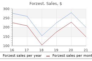
Forzest dosages: 20 mg
Forzest packs: 10 pills, 30 pills, 60 pills

20 mg forzest buy visa
A change in plasma osmolality results in a shift of total body water between internalmedicinebook erectile dysfunction hiv medications order forzest 20 mg without prescription. Renal Handling of Sodium In addition to renal water dealing with erectile dysfunction and smoking purchase forzest 20 mg with visa, sodium reabsorption and excretion are essential for maintenance of water homeostasis. The renin-angiotensin-aldosterone system is activated by lowered arterial perfusion strain sensed by the juxtaglomerular equipment of the afferent renal arteriole. Reduced arteriole effective volume (low or perceived) is sensed by the juxtaglomerular equipment, which secretes renin, activating the reninangiotensin system. Renal Handling of Water the most important osmoregulatory hormone is arginine vasopressin, also referred to as antidiuretic hormone, which is synthesized in the paraventricular and supraoptic nuclei of the hypothalamus. Secretion happens with a 1% to 2% rise in osmolality (>288 mOsm/kg), as detected by receptors in the anterolateral walls of the hypothalamus adjoining to the third ventricle. Vasopressin secretion is inhibited when the plasma osmolality is decrease than 280 mOsm/kg. The major nonosmotic stimulus is a decrease in efficient circulating quantity, which is detected by baroreceptors within the aortic arch and carotid sinuses. Although this mechanism requires a big drop (10%�15%) in blood stress, the secretory response is more robust than for will increase in osmolality. As such, acutely lowered blood pressure can override the inhibitory sign of low osmolality due to the necessity to preserve perfusion. The renal website of motion of vasopressin is the V2 receptors on the basolateral membrane of collecting duct cells within the distal nephron. The hormone-receptor interaction initiates intracellular signaling through cyclic adenosine monophosphate�dependent pathways, leading to translocation of cytoplasmic aquaporon-2 channels to the surface of the amassing duct luminal membrane. These channels enable motion of water again into the cell for later reabsorption into the circulation. The main symptoms and indicators of hyponatremia are neurologic in nature and are a scientific manifestation of swelling of the cells in the central nervous system, which ends up in cerebral edema. The most devastating consequence is herniation as a outcome of anatomic limitations on brain volume within the confines of the cranium. A main compensatory mechanism within the central nervous system is the extrusion from the cells of intracellular solutes, which prevents further water influx. In the first few hours, inorganic ions (potassium, sodium, chloride) move out of the cell. After a few days of persistent hypoosmolality, the cells additional compensate by extruding organic osmoles (glutamate, taurine, inositol). The clinician should pay attention to this protecting adaptation, as a outcome of it necessitates a slower time course of correction throughout treatment. A fast rise in plasma osmolality from aggressive therapy causes water to quickly shift out of the cells, resulting in demyelination of neurons. In the past, this was termed pontine demyelinosis, however it has additionally been reported for extrapontine neurons and is now referred to as osmotic demyelination. There is scientific development from lethargy to a change in have an effect on, to mutism and dysarthria, and at last to spastic quadriparesis and pseudobulbar palsy. On examination, special consideration ought to be paid to psychological status and neurologic abnormalities; manifestations of cardiac, hepatic, or renal illness; and indicators of adrenal insufficiency or hypothyroidism. Finally, it must be determined whether the hyponatremia is acute (<48 hours) or chronic, because this will decide the time course of remedy. It is probably going that these checks have already been performed, motivating the evaluation of hyponatremia. The immediately measured osmolality ought to be within 10 to 12 mOsm/kg of the calculated worth; an elevated osmolar hole compared with the calculated value points toward the presence of further osmoles in the plasma which are causing hypertonicity. Pseudohyponatremia can occur if the plasma lipid or protein content is tremendously increased within the plasma (usually to >6%�8% of volume), as in excessive hypertriglyceridemia and paraprotein issues. These additional components lower the aqueous portion of the plasma volume and thereby interfere with the laboratory measurement of sodium by dilutional, indirect strategies corresponding to flame photometry. Overall, the whole physique water and whole body sodium are unchanged in this scenario. An important medical example occurs with hyperglycemia, and the reported sodium value ought to be corrected for high glucose by the clinician by including 1. Because glucose is included in the calculation of osmolality, there might be no significant osmolar gap. Other osmotically energetic solutes encountered clinically are mannitol, which is used to manage increased intracranial stress, and glycine, which is used for irrigation in urologic procedures. High levels of alcohol or ethylene glycol additionally increase the osmolality, however these substances are so rapidly metabolized that an osmolar gap is in all probability not apparent by the time testing is carried out. Once pseudohyponatremia and hypertonicity have been ruled out, the analysis is narrowed to hypoosmolar hyponatremia. The mixed bodily examination and laboratory outcomes enable classification of the patient as having hypervolemic hyponatremia with extra complete body sodium, hypovolemic hyponatremia with a deficit of complete physique sodium, or euvolemic hyponatremia with near-normal whole body sodium. Signs of quantity contraction are obvious on examination, including poor skin turgor, skin tenting (forehead), decreased or undetectable jugular venous pressure, dry mucous membranes, and orthostatic adjustments in blood pressure and pulse fee. If the urine osmolality is lower than maximally concentrated (<500 mOsm/kg), an infusion of 0. With hypovolemia, the sodium will start to right and the affected person should improve clinically. Extrarenal causes of hyponatremia include gastrointestinal losses and third-space losses corresponding to in severe burns and pancreatitis. In the volume-depleted state, with intact renal function, the urine sodium is low (<10 mEq/L), reflecting a traditional response by the kidney to maximally reabsorb sodium in response to volume depletion. The renal causes of hypovolemic hyponatremia involve the inappropriate loss of sodium into the urine, and this is mirrored by a urine sodium focus of greater than 20 mEq/L. Only 5 Endocrine and Metabolic Disorders Assess medical volume status Hypovolemic Extrarenal � Gastrointestinal losses � Cerebral salt losing � Sweat losses � Third spacing losses (burns, pancreatitis, trauma, muscle, peritonitis) Renal � Thiazide diuretic remedy � Cerebral salt losing � Mineralocorticoid deficiency � Salt wasting nephropathy: � Medullary cystic illness � Chronic interstitial nephritis � Polycystic kidney illness � Analgesic nephropathy � Partial urinary tract obstruction � Chronic glomerulonephritis. These sufferers might have an abnormally sensitive thirst response to the mild hypovolemia induced by the diuretics, causing elevated water consumption. Elderly ladies are the most prone, and hyponatremia can occur within days after initiation of thiazide therapy. It is appropriate to stop the diuretic and restore potassium ranges and quantity standing. Loop diuretics, such as furosemide, are a less frequent cause of hyponatremia, which occurs solely after long-term therapy. In the absence of diuretic use, a urine sodium focus larger than 20 mEq/L with hypovolemia is evidence for underlying renal pathology. Because of the large web sodium loss, this situation is handled with salt tablets.
Forzest 20 mg order otc
In addition erectile dysfunction pills viagra forzest 20 mg generic on line, decreased right atrial pressures lead to inhibition of secretion of atrial natriuretic peptide erectile dysfunction caused by prostate surgery forzest 20 mg with mastercard, contributing to the water and sodium acquire. Edema, ascites, and orthostasis are absent, and thyroid, adrenal, and renal capabilities are normal. The major event is the discharge of vasopressin, which leads to water retention. The increased intravascular quantity stimulates a natriuresis, which really is suitable, but the resulting loss of sodium compounds the hyponatremia. A variety of medications are also known to stimulate vasopressin release or to potentiate its antidiuretic properties at the degree of the kidney (see Tables 1 and 2). A spot urine sodium degree shall be higher than 20 mEq/L, and often higher than 30 mEq/L. Vasopressin launch continues to be regulated, but at a decrease threshold of plasma osmolality. Interventions to raise the sodium often have short-lived effects, and the sodium resets at its previous worth over time. A physiologic example of reset osmostat is seen in pregnancy, when the normal sodium range is lowered by 5 mEq/L. Low solute consumption mixed with high fluid intake also can trigger hyponatremia, as with beer potomania or a low-protein "tea and toast" food regimen in elderly patients. Hyponatremia is also widespread after pituitary surgery (transsphenoidal or by craniotomy) and could also be a results of damage to the hypothalamic-pituitary tract that causes release of preformed vasopressin from broken neurons. Hyponatremia could be delayed as a lot as 1 week postoperatively, and sodium must be monitored during the second week as properly. Contributions by central adrenal insufficiency and hypothyroidism are also issues after pituitary surgical procedure, although with these circumstances there might be apparent medical manifestations along with the hyponatremia. Other Causes of Euvolemic Hypoosmolar Hyponatremia Primary or psychogenic polydipsia should be thought-about in sufferers presenting with hyponatremia and a historical past of psychiatric illness and remedy. Almost 6% to 7% of psychiatric inpatients are at risk for hyponatremia from elevated water consumption. The polydipsia could additionally be related to a lowered osmolar threshold for thirst, beneath the threshold of suppression of vasopressin secretion. This may be further complicated by the side impact of dry mouth brought on by many psychiatric medicines, which compounds the elevated thirst and water intake. Because the kidney is capable of excreting up to 15 to 20 L/day of dilute urine, the fact that hyponatremia develops in these patients might level toward an additional and inappropriate increase in vasopressin release or sensitivity. However, some sufferers have mildly concentrated urine (>100 mOsm/kg), by which case the psychiatric history helps with the diagnosis. Vasopressin ranges Management the major concerns for selecting the type and time course of remedy for hyponatremia are the duration of hyponatremia (acute or chronic) and the presence of neurologic signs and signs, especially extreme manifestations similar to altered psychological standing or seizure (see Table 2). Treatment choices for hyponatremia include fluid restriction, saline infusion (hypertonic or isotonic), vasopressin receptor antagonists and demeclocycline (Declomycin). Autocorrection may happen after initiation of therapy, especially in cases of hypovolemia, adrenal insufficiency, or thiazide use. Once therapy is began, the contribution of the nonosmotic stimulation of vasopressin secretion is eliminated, and the affected person is in a position to elevate the sodium stage by 2 mEq/L per hour over 12 hours. Acute Severe Symptomatic Hyponatremia Acute extreme hyponatremia is defined as a fast fall in sodium in less than 48 hours to less than 120 mEq/L. Treatment is aimed toward raising the sodium enough to resolve the neurologic signs and symptoms. The aim is to increase the serum sodium by 1 to 2 mEq/L per hour or to larger than 125 mEq/L until symptoms resolve. Hypovolemic patients will reply to infusion of isotonic saline (normal saline zero. If the neurologic findings are extreme, hypertonic saline (3%) could also be infused at rate of 1 to 2 mL/kg per hour, and even up to 4 to 6 mL/kg per hour if the imbalance is life-threatening. A loop diuretic could be combined with the saline to improve solute-free water excretion. Sodium levels should be monitored every 2 to 4 hours in sufferers undergoing hypertonic infusion. No benefit has been noticed for sooner rates of correction of hyponatremia, whether acute or chronic. The formulas can only estimate the rate of correction, and sodium should be measured frequently. Chronic Hyponatremia Chronic hyponatremia is defined as a gradual fall in sodium over greater than forty eight hours. By this time, the brain has begun to compensate for hypoosmolality by extrusion of solutes. However, the patient is at threat of osmotic demyelination if hyponatremia is treated too aggressively. However, as with acute hyponatremia, severe neurologic signs and indicators must be treated with hypertonic saline till they resolve, after which the rate of correction could be slowed to 0. Most cases of osmotic demyelination happen with correction charges of larger than 12 mEq/L in 24 hours, but there are circumstances reported with will increase of 9 or 10 mEq/day. Asymptomatic hyponatremia could be handled with an infusion of isotonic saline calculated to raise the sodium by 0. If the patient has a dilute urine (<200 mOsm/kg), water restriction may be adequate. Demeclocycline (Declomycin) antagonizes the actions of vasopressin by inhibiting formation of cyclic adenosine monophosphate within the accumulating duct. Long-term use is limited by the aspect effect of photosensitivity and by nephrotoxicity in patients with underlying liver illness. Vasopressin receptor antagonists also are important adjunct therapies (see later discussion). A less favored treatment is urea (powder or capsules),1,6 which causes an osmotic diuresis and increased free water excretion. Correct serum Na at a price of 1�2 mEq/l per hour (for 2 to four hours) until symptoms have resolved 2. Treat underlying etiology Calculation of the rate of infusion of saline to right hyponatremia 1. Amount of infusate (in L) required in 24hours L = desired change in sodium (Na) over 24 hrs Na/L (from 1. Infusate 5% sodium chloride in water 3% sodium chloride in water Isotonic saline (0. Vasopressin Receptor Antagonists Vasopressin receptor antagonists, or vaptans, are a remedy possibility for clinically significant euvolemic hyponatremia outlined as a serum Na < 125 mEq/L or if hyponatremia is symptomatic and immune to correction with commonplace therapy.
Forzest 20 mg discount on line
The long model includes 24-hour inpatient admission due to the chance of hypotension erectile dysfunction massage techniques purchase forzest 20 mg with visa. The short version includes the administration of a single dose of metyrapone at midnight and the measurement of serum cortisol in the morning erectile dysfunction doctor in los angeles forzest 20 mg purchase line. To conduct the metapyrone response test, metyrapone (30 mg/kg, most dose 3000 mg) is administered at midnight normally with a snack. A plasma cortisol lower than 220 nmol/L signifies sufficient inhibition of 11-hydroxylase. In patients with an 309 Pathophysiology the pituitary gland is a pea-sized endocrine gland that sits on the base of the mind. It is composed of two functionally distinct constructions that differ in embryologic improvement and anatomy: the adenohypophysis (anterior pituitary) and the neurohypophysis (posterior pituitary). Some ladies unknowingly reside for years with pituitary insufficiency, then go into adrenal crisis triggered by extreme physical stressors, corresponding to severe an infection or surgical procedure. Then take 25 mg/day for 1 day starting earlier than surgery Take the standard morning dose. Then 100�150 mg/day for 2�3 days Moderate surgical procedure Major surgical procedure From Shawn (2012). This results in a rise of the steroid precursors in the pathway, including 11-deoxycortisol. A 60-minute cortisol level of lower than 18 mcg/dL is suggestive of irregular pituitary response or hypopituitarism. Treatment Hypopituitarism may be an emergency in severe cases because of the danger of vascular collapse because cortisol is necessary for the maintenance of peripheral vascular perform. However, ought to the workup reveal different hormone deficiencies, substitute remedy for other hormones is indicated. Oral hydrocortisone (Cortef) 15 to 25 mg a day in divided doses is the popular therapy, as a result of those doses are just like physiologic daily manufacturing rates. Some sufferers, nonetheless, might have a better or decrease dose depending on the diploma of severity. The current substitute regimens inevitably result in momentary overor underreplacement and, subsequently, lead to a poor high quality of life and elevated mortality. Recent effort has studied sustained-release once-a-day hydrocortisone therapy versus a thrice day by day, weight-related, dosing routine. Plenadren is a recently licensed modified-release formulation of hydrocortisone that provides the potential for once-daily dosing. Both are presently approved to be used in Europe and are in Phase 2 medical trials within the United States. Some authorities recommend prednisone or dexamethasone due to their longer period of motion. They could be given as quickly as a day, versus hydrocortisone, which is run two to 3 times a day. Patients may need the next dose of hydrocortisone in occasions of sicknesses or different stresses. Before deliberate surgical procedures, high-dose hydrocortisone (Solu Cortef) as stress doses for 1 to three days allows faster recovery (Table 2). Thyroid deficiency-Thyroid deficiency from hypopituitarism is handled with T3 and T4 in a style similar to the therapy of primary hypothyroidism. Treatment of the hypothyroidism alone might suppress different hormones produced by the pituitary gland and worsen the severity of other deficien- cies. Monitoring Periodic serum cortisol levels are used to assess therapy adequacy and make changes as needed. Most studies use cortisol levels taken roughly 4 hours after the morning cortisol dose. The challenge of measuring serum cortisol ranges is that cortisol sensitivity and concentrations vary between people. Epidemiology Hypothyroidism is second solely to diabetes within the prevalence of endocrine issues in adults in the United States. Hypothyroidism happens in as a lot as 18/1000 population, with girls outnumbering men by roughly 10:1. Rates of hypothyroidism increase dramatically with age, so that about 2% to 3% of all older girls have hypothyroidism, and the prevalence is as much as 5% in nursing home populations. Having a excessive scientific suspicion is the key, especially after significant traumatic occasions similar to cardiac arrests, acute emergency surgeries, or extreme motor vehicle accidents. In addition, hypothyroidism in addition to thyroid cancers are extra frequent in sufferers who had neck irradiation in childhood. Prevalence and incidence of hypopituitarism in � an adult Caucasian inhabitants in northwestern Spain. Diagnosing the unrecognized systemic absorption of intra-articular and epidural steroid injections. Replacement remedy of oral hydrocortisone in � adrenal insufficiency: the affect of gastrointestinal components. Finally, a wide selection of other situations including infiltration of the thyroid (amyloidosis, sarcoidosis), iodine deficiency, or medications (such as amiodarone [Cordarone] or interferon) can cause hypothyroidism. Clinicians have to use different scientific signs to attempt to differentiate euthyroid sick syndrome from hypothyroidism. The solely exception to that is when the clinician identifies a mass on bodily examination. In that situation, scanning or different imaging is essential to decide the malignancy potential of the mass. Preventive Services Task Force discovered insufficient evidence to support early detection via routine screening of asymptomatic individuals. Differential Diagnosis the differential analysis for hypothyroidism is broad and is decided by the first complaints given by patients. For patients with slowed mentation, depressed affect, or confusion, clinicians ought to suspect depression. Patients with lethargy and a gradual pulse and low blood stress might need adrenal insufficiency. Patients with constipation have to have colonic obstruction from a mass considered as properly. In the elderly, widespread drugs that can trigger melancholy (such as centrally performing antihypertensive agents), bradycardia (such as -blockers or calcium channel blockers), constipation (calcium channel blockers), hair loss, or confusion additionally should be thought-about. In sufferers with pituitary failure, different pituitary hormones are more probably to be poor as nicely, so clinicians should search for evidence of adrenal and gonadotropic failure. Consequently, clinicians must have a high index of suspicion for hypothyroidism when patients are available with any one or combination of the signs that could signal hypothyroidism. Symptoms of hypothyroidism include lethargy, weight acquire, hair loss, dry skin, constipation, poor concentration, bother considering or forgetfulness, and depression (Box 2). Patients who current with despair additionally ought to have their thyroid perform assessed. The thyroid examination in most sufferers with hypothyroidism is completely normal.

20 mg forzest buy with mastercard
Glycosylation is required for the stability of GspB; loss of carbohydrate results in fast degradation impotence stress forzest 20 mg generic free shipping. This modification erectile dysfunction medication reviews buy forzest 20 mg low cost, nevertheless, renders it an unsuitable substrate for export by the canonical basic secretory (Sec) pathway. Similar results have been reported for Hsa, a homologue in another viridans streptococcal species (S. Second, by transposon mutagenesis, the Sullam group discovered that binding of viridans streptococci to mammalian platelets is mediated partly by two phage-encoded proteins, PblA and PblB, that are related to the cell wall of this organism. PblA is a tape measure protein (important for phage morphogenesis), whereas PblB is a tail fiber protein. Both proteins are linked to the bacterial floor through their interactions with choline teams within the cell wall. A puzzling problem was how these phage proteins get out of the bacterium and onto its surface. Other organisms could bind on to, or turn out to be ingested by, endothelial cells as the preliminary occasion. This is an energetic area of investigation, because inhibition of those events might present novel prophylactic strategies. The significance of adherence characteristics in the development of endocarditis additionally has been examined by way of using preincubation of organisms with antibiotics. Many lessons of medicine, after incubation even at subinhibitory concentrations, decrease the adhesion of streptococcal species to fibrin-platelet matrices and damaged canine valves in vitro. Some strains of micro organism have been found to be potent stimulators of platelet aggregation and the discharge reaction. Bacteriaplatelet aggregates have been discovered in the peripheral blood of sufferers with bacteremia. The significance of those aggregates within the formation of the vegetation (or, conversely, the effect of the aggregation on the rate of elimination of organisms from the circulation) is unknown. In one research, even small numbers of platelets tremendously increased the adherence of oral streptococci to fibrin in vitro. However, this platelet aggregation by viridans streptococci requires direct platelet binding and plasma components. There is a posh interplay among factors liable for bacteria-platelet adhesion and aggregation. At least nine adh/agg phenotypes have been identified amongst naturally occurring variants, reflecting a variety of platelet interactivity. Streptococcal exopolysaccharide production inversely correlates with platelet adhesion whereas inhibiting aggregation,126 indicating that these surface molecules may improve endocarditis at some pathogenic steps however not at others. However, the capability of the mutant to persist and proliferate within experimental vegetations and to disseminate hematogenously to the kidneys was markedly impaired within the mutant strain. These circumstances permit for comparatively unbridled bacterial development, leading to extraordinarily high colony counts of 109 to 1011 micro organism per 1 g of tissue. Bacteria deep within the fibrin matrix have been proven by autoradiography to attain a state of lowered metabolic activity. Matrix metalloprotease 9, elastase, and plasminogen activators had been all present at larger concentrations in septic vegetations. These results recommend that the continual attractant indicators coming from bacterial colonies can result in continual harm of myocardial tissues by host proteases. The titers correlated with the level of hypergammaglobulinemia and decreased with therapy. Rheumatoid issue could play a job in the disease course of by blocking IgG opsonic activity. Immune globulin eluted from these lesions has been proven to cross react with bacterial antigens. Effective treatment results in a prompt decrease, with eventual disappearance of circulating immune complexes. In the preantibiotic era, 70% to 95% of patients had clinically demonstrable embolic events, but this has decreased to 15% to 35% at present. Pathologic proof of embolization still is detected in 45% to 65% of autopsies, most incessantly involving the renal, splenic, coronary, or cerebral circulation. If giant emboli occlude main vessels, fungal endocarditis, marantic endocarditis, or an intracardiac myxoma ought to be suspected. Abscesses are unusual, but infarctions have been seen in 56% of the autopsy circumstances. It is a focal, native, and segmental course of characterized by endothelial and mesangial proliferation, hemorrhage, neutrophilic infiltration, fibrinoid necrosis, crescent formation, and therapeutic by fibrosis. Diffuse glomerulonephritis is found in 17% to 80% of the cases and consists of generalized cellular hyperplasia in all glomerular tufts. Immunofluorescent staining with anti-immunoglobulin antibody reveals the typical lumpy-bumpy distribution seen in other forms of immune complex nephritis. In diffuse glomerulonephritis, subepithelial electron-dense deposits are seen by electron microscopy and IgG, IgM, IgA, or complement are proven in these deposits by immunofluorescence. The glomerulonephritis usually is accompanied by hypocomplementemia, with a constructive end result on serum assay for rheumatoid issue. All of these abnormalities often resolve with profitable antimicrobial remedy because the focus of circulating immune complexes declines. Microscopically, the lesion consists primarily of fibrin, platelet aggregates, and bacterial lots; neutrophils and purple blood cells are uncommon. Killed bacteria detectable by Gram stain inside these vegetations sometimes persist for months after therapy. The vegetation in acute cases is bigger, softer, and more friable and may be associated with suppuration, extra necrosis, and less therapeutic than in subacute circumstances. Myocarditis, myocardial infarction, and pericarditis193,194 are found regularly at post-mortem. Myocardial infarcts are found in 40% to 60% of the autopsied circumstances, typically with out diagnostic changes on the electrocardiogram. They are more frequent with viridans streptococcal infections and are found in 10% to 15% of autopsied circumstances. They could come up by any of several mechanisms: (1) direct bacterial invasion of the arterial wall with subsequent abscess formation or rupture, (2) septic or bland embolic occlusion of the vasa vasorum, or (3) immune complicated deposition with resultant damage to the arterial wall. They are found most commonly in the cerebral vessels (primarily the peripheral branches of the middle cerebral artery), but additionally they happen in the abdominal aorta; the sinus of Valsalva; a ligated patent ductus arteriosus; and the splenic, coronary, pulmonary, and superior mesenteric arteries. The whole cerebrovascular complication fee was 65%, together with 35% that had been symptomatic and 30% that had been clinically silent. Cerebral infarction, arteritis, abscesses, mycotic aneurysms, intracerebral or subarachnoid hemorrhage, encephalomalacia, cerebritis, and meningitis have been reported. Splenic infarctions have been reported in 44% of autopsy instances but often are clinically silent.
20 mg forzest amex
Prevention includes thoroughly cooking all meat products impotence guide forzest 20 mg generic otc, good hand and utensil hygiene before getting ready meals youth erectile dysfunction treatment forzest 20 mg buy visa, and avoiding unpasteurized milk. The therapy for severe Campylobacter diarrhea consists of azithromycin (Zithromax)1 and ciprofloxacin (Cipro), though resistance to fluoroquinolones is growing. Campylobacter infections have been linked to both rheumatoid arthritis and in addition Guillain-Barre syndrome. Fluoroquinolones remain the antibiotic of alternative, although sensitivities will information antibiotic utilization. The most frequent antibiotics implicated are clindamycin (Cleocin), fluoroquinolones, and beta-lactam antibiotics. Dysentery normally presents as high fever, stomach cramping, and bloody diarrhea. Treatment Fluid and electrolyte alternative are crucial therapies for acute diarrhea. For mild circumstances of diarrhea in a normally healthy particular person, enough oral intake of juices, soups, and electrolyte-infused water could be adequate. Given the truth that most circumstances of diarrhea are delicate, self-limited, and brought on by nonbacterial pathogens, antibiotics should be used sparingly. Empirical antibiotics may be appropriate as described above for Campylobacter or C. Antidiarrheal medications could be helpful to assist alleviate signs in sufferers with no different contraindications, such as fever or bloody diarrhea. Symptoms usually include profuse watery diarrhea (rice-water stools), stomach cramping, and vomiting. Viruses Viral pathogens are likely responsible for virtually all of gastroenteritis in the United States. The most common viral etiologies tend to be adenovirus, rotavirus, norovirus, and astrovirus. Rotavirus tends to have an effect on infants and youngsters from 3 to 36 months of age, leading to a spectrum of disease from asymptomatic shedding to severe gastroenteritis. Vaccination has played an necessary function in lowering the cases of rotavirus infections. Norovirus may be easily transmitted from particular person to particular person, from contact of contaminated surfaces of from exposure to contaminated food or water. It is often not cost-effective to consider for ova and parasites until there are specific indications, corresponding to a history of immunocompromise, exposure to day care centers, or historical past of publicity to contaminated water. Multiple stool samples are needed if there are issues of parasite infection resulting from potential intermittent shedding. Prolonged or other signs similar to fever or bloody stool must be additional investigated. Antibiotic prophylaxis is given to decrease the risk of an infection and of rebleeding. This strain gradient is necessary but not sufficient for the event of gastroesophageal varices. Diagnosis the current consensus states that every affected person with liver cirrhosis should bear an higher endoscopy to detect gastroesophageal varices. The main aim behind screening for gastroesophageal varices is to establish patients requiring prophylactic treatment or additional surveillance. Several invasive and noninvasive procedures help in detecting portal hypertension and may, with variable accuracy, predict the presence of gastroesophageal varices. Endoscopic videocapsule is a brand new modality introduced for visualizing the esophagus; it permits right identification of varices in 80% of circumstances but can have poor accuracy in figuring out the presence of hypertensive gastropathy and gastric varices. Not all esophageal varices bleed; hemorrhage happens in only 30% to 35% of patients with cirrhosis. Variceal rupture is directly associated to physical elements such because the radius, thickness, and elastic properties of the vessel in addition to intravariceal and intraluminal stress and tension. Endoscopic findings that predict the next risk of bleeding embody bigger size of varices and the presence of endoscopic red signs (described as purple wale markings) on the variceal wall, indicating dilated intraepithelial and subepithelial superficial veins. A mixture of medical and endoscopic findings, including the Child-Pugh class, size of varices, and the presence or absence of red wale markings, was discovered to correlate extremely with the danger of first bleeding in sufferers with cirrhosis. A rise in stress in a affected person with known varices increases the risk of bleeding, and the extent of portal stress elevation has an inverse relationship to prognosis after hemorrhage has occurred. Epidemiology Bleeding of esophageal varices is a serious complication of portal hypertension, normally within the setting of liver cirrhosis, accounting for 10% to 30% of all instances of upper gastrointestinal hemorrhage. More than any other reason for gastrointestinal bleeding, this complication ends in considerable morbidity and mortality, prolonged hospitalization, and increased affiliated costs. Variceal bleeding develops in 25% to 35% of patients with cirrhosis and accounts for as much as 90% of higher gastrointestinal bleeding episodes in these patients. About 10% to 30% of those episodes are fatal, and as many as 70% of survivors rebleed following an index variceal hemorrhage. Following such events, the 1-year survival is 34% to 80%, being inversely associated to the severity of the underlying liver illness. Treatment of sufferers with esophageal varices contains preventing the preliminary bleeding episode (primary prophylaxis), controlling lively variceal hemorrhage, and stopping recurrent bleeding after a first episode (secondary prophylaxis). The degree of elevated resistance to move varies with the level of circulatory breach and can be divided into prehepatic, hepatic or sinusoidal, and posthepatic. In cirrhosis, several organ systems are concerned in the pathophysiology of portal hypertension. At the systemic circulation stage, there is a rise in cardiac output, lower in vascular resistance, and hypervolemia. This hyperkinetic syndrome leads to an efficient hypovolemia, with a resultant improve in vasoactive factors to preserve a normal arterial blood strain. Varices characterize portosystemic collaterals derived from dilatation of preexisting embryonic vascular channels, such as those between the coronary and brief gastric veins and the intercostal, esophageal, and azygous veins. In the distal esophagus, over an space extending 2 to 5 cm from the gastroesophageal junction, veins are discovered more superficially within the lamina propria quite than the submucosa. This ends in decreased assist from surrounding tissues owing to the predominant intraluminal location of those varices and would possibly clarify the predilection for bleeding at this web site. The opening and dilation of portosystemic collaterals seems to depend upon a threshold portal strain gradient Treatment Primary Prophylaxis the pure evolution of gastroesophageal varices without treatment is characterised by a rise in measurement from small to giant varices, which ultimately rupture and bleed. Based on prospective studies of cirrhotic patients with varices recognized at endoscopy and of untreated groups in randomized managed trials, the chance of bleeding from esophageal varices has been estimated at 25% to 35% at 1 yr. Therapy for primary prophylaxis against variceal bleeding (prevention of a first variceal bleeding) is summarized in Table 1. Pharmacologic Therapy the final objective of pharmacologic therapy for variceal bleeding is to cut back portal pressure and consequently intravariceal strain. Drugs that reduce portocollateral venous move (vasoconstrictors) or intrahepatic vascular resistance (vasodilators) have been used and embrace -blockers, nitrates, 2-adrenergic blockers, spironolactone (Aldactone),1 pentoxifylline (Trental),1 molsidomine (Corvaton),2 and simvastatin (Zocor).

Thwak (Cinnamon Bark). Forzest.
- Diabetes, diarrhea, infections, worm infestations, the common cold, influenza, upset stomach, gas (flatulence), spasms, appetite stimulation, and menstrual discomfort.
- What other names is Cinnamon Bark known by?
- Dosing considerations for Cinnamon Bark.
- Are there any interactions with medications?
- Premature ejaculation. Some evidence suggests that a specific cream containing cinnamon and many other ingredients might prevent premature ejaculation.
- Are there safety concerns?
Source: http://www.rxlist.com/script/main/art.asp?articlekey=96349
Cheap 20 mg forzest amex
More advanced tumors are often treated with a mixture of radiation therapy and surgical procedure or chemotherapy diabetes and erectile dysfunction health buy 20 mg forzest mastercard. Leukoplakia how to treat erectile dysfunction australian doctor generic 20 mg forzest free shipping, or a raised white plaque on the epithelial surface, is a visible marker for the likely presence of dysplasia or carcinoma in situ. As a really early lesion, vocal fold leukoplakia could manifest with gentle dysphonia or may be discovered by the way on head and neck examination performed for different causes. This early illness may take many years earlier than progressing to invasive carcinoma, and recognition of leukoplakia presents an opportunity for early treatment to forestall progression of disease. Pulsed laser photoangiolysis has emerged as a state-of-the-art remedy for treatment of this epithelial lesion with preservation of the underlying vocal fold pliability. Acoustically, presbylaryngis results in a attribute thinned voice, typically with decreased projection and increased vocal strain. The situation happens as cumulative voice use results in traumatic thinning of the superficial lamina propria, particularly at the mid-cord level. This loss of superficial lamina propria results in deficiency on the medial edge of every vocal fold, and a spindle-shaped defect in glottal closure may be seen with close evaluation. Many sufferers with a grievance of presbylaryngis discover that acceptable voice therapy to address breath support and vocal projection leads to satisfactory enchancment within the voice with out altering the vocal fold anatomy. Application of this information through the historical past and physical examination guides the diagnosis of hoarseness and allows clinicians to distinguish amongst situations as diversified as acute laryngitis, benign phonotraumatic lesions, vocal fold paralysis, and laryngeal most cancers as a part of a differential prognosis. Videostrobolaryngoscopy allows evaluation of vocal fold operate in addition to construction and might verify analysis. Because no Neurologic Disorders and the Voice Neurologic circumstances that affect the voice often do so by causing poor coordination of vocal fold motion. Spasmodic dysphonia, for example, results in involuntary spasms that bring the vocal folds either tightly together (adductor spasmodic dysphonia) or aside (abductor spasmodic dysphonia) throughout phonation. Laryngopharyngeal reflux: Position assertion of the committee on speech, voice, and swallowing problems of the American Academy of Otolaryngology-Head and Neck Surgery. Pharmacologic options embody nonopioid, opioid, adjuvant, or homeopathic drugs. For many chronic situations similar to low back ache, the evidence supporting the use of opioids is missing, with efficacy often outmoded by nonopioid drugs. Since 1999, opioid overdose deaths as a proportion of all deaths has elevated by 265% in males and 400% in women. Honesty and belief are essential components of a therapeutic physician�patient relationship. If the patient feels that a harmless treatment is helpful, benefit from it. Acute Pain � Acute pain has a sudden onset, is usually nociceptive (somatic, visceral) in nature, and certain is because of apparent damage or medical condition. Chronic Pain � Chronic ache is usually gradual in onset, lasts greater than three months, and infrequently serves any function. Severity � Severity of the pain can be rated on a wide range of scales corresponding to numerical (0�5, 0�10), analogue (marked on a line with a variety from no pain to the worst potential pain), or facial features 35 Epidemiology Approximately 50 million Americans experience chronic ache, with annual expenditures and overall economic impression estimated at $85 to $90 billion. Pain is the most typical symptom that causes sufferers to pursue medical analysis and administration. The prevalence of a number of common ache syndromes together with complications, facial ache, belly pain, pelvic ache, and low again ache is slightly greater in ladies than in men as a result of there are gender variations within the perception, coping, and reporting of ache. Risk Factors Traumatic harm is a reason for acute ache and a threat factor for continual ache. Differential Diagnosis the differential analysis for every type of pain is far too in depth for the brevity of this chapter, however in addition to the plethora of physical diagnoses to be thought of, the supplier has to think about that continual fatigue, depression, and domestic abuse can manifest as a continual ache syndrome. For recent-onset low again pain, aid was statistically but not clinically important as in comparison with ordinary care. Cognitive behavioral remedy and mindfulness training enhance function more than pain. Surgery could additionally be required, and interventions such as vertebroplasty, nerve blocks, and epidural injections can also be of profit. Aerobic train and water therapy are helpful for fibromyalgia, however the affected person should not overexert because this will exacerbate the pain. Pathophysiology Injury or potential damage is detected by nociceptors in the peripheral nervous system, and the signal is then transmitted by way of the dorsal horn of the spinal twine up to the mind for processing. Upon binding these receptors, opioids block calcium channels and modulate the nociceptive pathways. The hyperstimulation of chronic ache and the resultant enhance in intracelluar calcium are neurotoxic as a outcome of reducing neuronal firing threshold and rising firing frequency. Neuropathic pain may result from persistent pain or from bodily or pathophysiologic damage or adjustments to the nerve, as in diabetic neuropathy or different neuropathies. Chronic use can enhance the likelihood of transaminase elevations four-fold, so this may be a consideration when sufferers are utilizing other doubtlessly hepatotoxic medications. All are generally secure within the acute setting however must be used with warning if needed long term, and they need to be avoided altogether for long-term use within the aged or sufferers with cardiovascular, renal, or peptic ulcer illness. The mixture of healthy diet and train has been demonstrated to improve ache in osteoarthritis higher than both intervention alone, and sustaining a wholesome weight might help to stop the arthritis within the first place. Regarding safety, wearing seat belts whereas driving or utilizing appropriate safety equipment at work and through leisure actions may help to cut back the severity of injuries should they occur. Clinical Manifestations 36 Manifestations of ache depend on the location and underlying trigger. In the acute setting, the affected person can have tachycardia, elevated blood stress, or diaphoresis, whereas chronic ache can manifest with vegetative symptoms of depression, fatigue, anorexia, or insomnia. Opioids (see Table 1) Diagnosis Pain is mostly a symptom rather than a disease in and of itself, so it behooves the supplier to pursue treatable causes, especially red flag circumstances, along with providing symptomatic remedy. The historical past regarding onset (shorter or longer than 3 months), exacerbating or remitting elements, quality, radiation, severity, and timing of the pain (constant vs. The physical examination is a key element in ruling out life- or function-threatening problems. Does the affected person reveal a consistent demeanor (grimace or different signs of pain) and gait (antalgic vs. The option to use laboratory research or imaging is determined by the placement of the ache and the structures or organs that may be in that area. For patients with insufficient response to the non-opioid analgesics, opioids are an choice (Table 1). The benefits of opioids in some situations and the overall safety of opioids have been referred to as into query due to the dramatic increase in opioid abuse and opioid overdose deaths. Recent research has demonstrated a twofold to threefold improve in all-cause mortality with persistent opioid use, especially if using more than 200 mg/day morphine equivalent and/or sustained-release or long-acting (methadone) preparations. They must be prevented in patients with untreated sleep apnea due to elevated danger for problems, especially in combination with benzodiazepines.
Forzest 20 mg purchase with visa
However erectile dysfunction pump canada cheap forzest 20 mg, its use in persistent pancreatitis has been disappointing erectile dysfunction drugs dosage forzest 20 mg cheap without a prescription, with about half of the sufferers deriving a benefit that lasts 6 months or much less. Pancreatic duct stenting is used for remedy of proximal pancreatic duct stenosis, decompression of a pancreatic duct leak, and drainage of pancreatic pseudocysts that might be catheterized via the principle pancreatic duct. Stent therapy in chronic pancreatitis undoubtedly performs a task and might help choose patients for successful operative therapy. However, the duration of success with stent therapy for chronic pancreatitis is probably less than with surgical remedy. Major pancreatic resections for persistent pancreatitis have a excessive complication price, both early and late. The surgical administration of pancreatic duct stones and stenoses has been proven to be superior to endoscopic therapy in randomized medical trials. Later within the decade, Frey and Smith described the local resection of the pancreatic head with longitudinal pancreaticojejunostomy, which included excavation of the pancreatic head including the ductal buildings in continuity with a long ductotomy of the dorsal duct. Recent randomized prospective research have compared the Whipple, Beger, and Frey procedures for persistent pancreatitis. Patients who had a Beger process had a shorter hospital stay, greater weight achieve, less postoperative diabetes, and less exocrine dysfunction than standard Whipple sufferers over a 3- to 5-year follow-up. In a research evaluating the pylorus-preserving Whipple to the Frey process, there was a decrease postoperative complication fee associated with the Frey procedure (19%) compared to the pyloruspreserving Whipple group (53%), and the worldwide quality-of-life scores had been higher (71% versus 43%, respectively). Both operations were equally efficient in controlling ache over a 2-year follow-up. Operation times, intraoperative blood loss, and transfusion requirements have been proven to be decreased with the Frey and Beger procedures in comparison with the Whipple process. In long-term (>8 years) follow-up, there was no distinction between the Beger and Frey procedures in ache aid, pancreatic insufficiency, high quality of life, and late mortality. Although these restricted pancreatic procedures have a decrease initial fee of endocrine dysfunction, the long-term danger of diabetes is more related to the development of the underlying illness than to the consequences of operation. Recent refinements within the strategies of harvesting and preserving pancreatic islets, and standardization of the strategies by which islets are infused into the portal venous circuit for intrahepatic engraftment, have improved the success and rekindled curiosity in islet autotransplantation for continual pancreatitis. The capacity to get well a sufficient quantity of islets from a sclerotic gland is dependent upon the degree of disease current, so the choice of patients as candidates for autologous islet transplantation is essential. As success with autotransplantation will increase, patients with nonobstructive, sclerotic pancreatitis could additionally be considered for resection and islet autotransplantation earlier in their course, as a end result of end-stage fibrosis bodes poorly for transplant success. As the necessary experience with islet transplantation turns into more widespread, this remedy may turn out to be routine in the therapy of continual pancreatitis. A, the pancreatic duct is opened to reveal a preoperatively placed pancreatic duct stent. C, In the Frey process, the top of the pancreas is cored out in addition to a longitudinal pancreatic ductotomy. Computed tomographic scan of a patient with continual calcific pancreatitis and a massively dilated pancreatic duct. Complications Pseudocysts in the setting of persistent pancreatitis are less prone to resolve without intervention. Often, the pancreatic duct and bile duct are compressed, and the compression might need to be addressed concurrently the pseudocyst. A development towards minimally invasive administration remains acceptable, with endoscopic drainage preferred over laparoscopic cystogastrostomy except extra procedures are required. Resection of a pseudocyst is usually indicated for cysts positioned in the pancreatic tail, or when a midpancreatic duct disruption has resulted in a distally situated pseudocyst. Distal pancreatectomy for elimination of a pseudocyst, with or without splenectomy, is normally a difficult procedure in the setting of prior pancreatitis. An internal drainage procedure of the communicating duct, or of the pseudocyst itself, must be thought-about when distal resection is being contemplated. Occasionally, the pancreatic fluid tracks superiorly into the thorax, inflicting a pancreatic pleural effusion. Both complications are seen Monitoring Chronic pancreatitis is in fact a continual disease, so continued monitoring and maintenance therapy is essential after an acute exacerbation of continual pancreatitis. Pain management, correct vitamin, and alcohol and smoking cessation should be maintained as an outpatient. Paracentesis or thoracentesis reveals noninfected fluid with a protein stage higher than 25 g/L and a markedly elevated amylase degree. Paracentesis and antisecretory remedy with the somatostatin analogue octreotide acetate, along with bowel rest and parenteral diet, is profitable in more than half of sufferers. Reapposition of serosal surfaces to facilitate closure of the leak is considered part of remedy, and this is completed by full paracentesis. For pleural effusions, a period of chest tube drainage can facilitate closure of the interior fistula. Viral shedding occurs when the replicating virus is excreted from hepatocytes through the bile duct into the gut. Shedding continues until the prodromic phase and begins to decline as quickly as jaundice develops. However, infectious virions could be detected within the feces up to 2 weeks after the onset of jaundice. This is followed by a prodromal phase of variable severity, characterized by weak point, anorexia, nausea, belly ache, and, much less typically, fevers, arthralgias, and diarrhea. The ranges of the serum aminotransferases are elevated throughout this time, usually to values higher than 500 U/L, and their peak normally coincides with intense nausea, vomiting, and anorexia. Jaundice typically occurs 1 to 2 weeks later and is related to a lessening of the prodromal signs. The serum bilirubin degree peaks later than the aminotransferases, hardly ever exceeds 10 mg/dL, and normalizes more slowly than the aminotransferases. Complete normalization of the serum biochemical abnormalities is observed in 60% of patients by 2 months and in almost one hundred pc by 6 months. Walled-off pancreatic necrosis and other present ideas in the radiological assessment of acute pancreatitis. Early antibiotic remedy for severe acute necrotizing pancreatitis: A randomized, double-blind, placebo-controlled study. Prophylactic antibiotic remedy in sufferers with predicted extreme acute pancreatitis: A placebo-controlled, double-blind trial. Dehydration is widespread during the symptomatic section and requires administration of intravenous fluids. The danger of creating acute liver failure is greater in older sufferers; these contaminated after the age of fifty years have a case-fatality rate of two. There are higher diagnostic instruments and rapidly evolving therapies, each for hepatitis B and hepatitis C. This improved therapy has made it critical that physicians effectively display for chronic hepatitis B and hepatitis C to identify all acceptable candidates for therapy.
20 mg forzest generic otc
Uncomplicated diverticulitis responds to medical remedy within the majority of circumstances erectile dysfunction drugs after prostate surgery cheap 20 mg forzest mastercard. Complicated diverticulitis happens in 25% of all diverticulitis diagnoses erectile dysfunction medications causing forzest 20 mg cheap on-line, requiring a surgical evaluation and intervention. Optional antibiotic regimens for gram-negative coverage and anaerobic coverage are listed in Table 1. Inpatient management is recommended with any comorbid circumstances, immunosuppression, high-grade fever, leukocytosis, and/or for aged patients. This is completed to consider for underlying pathology corresponding to cancer or inflammatory bowel disease, which might change the administration strategy. Follow-up scanning must also be considered for instances of slowresolving diverticulitis. Notes: Clindamycin (Cleocin) may be substituted in metronidazole intolerance/allergy. Antibiotics continued in complicated diverticulitis cases until unfavorable cultures, medical improvement, or sensitivities obtained on any surgical specimens. A colonoscopy must be carried out to consider the extent of diverticular disease and to rule out other comorbid pathology in all sufferers. Colonoscopy is often recommended within three to 6 weeks after diverticulitis signs have resolved. Introduction of stool softeners and a high-fiber food plan ought to begin instantly and continued indefinitely for all sufferers to help lower the intraluminal strain within the colon. A common recommendation exists for radiologic analysis and drainage if the abscess is larger than 2 cm. New analysis to stop recurrent diverticulitis with medical management is rising. Fat stranding refers to abnormally elevated attenuation of fats from edema and engorgement of lymphatics. Noninvasive Gastrointestinal Imaging: Ultrasound, Computed Tomography, Magnetic Resonance Imaging. Minimally invasive endoscopic remedy for achalasia is promising and will present a competing method. Dysphagia is a symptom generated by the perceived sensation of issue or lack of ability to swallow. It ranges in severity from delicate difficulty with no related clinical sequelae, to a complete incapability to swallow with aspiration and extreme malnutrition. Dysphagia can coexist with and have to be distinguished from odynophagia, which is ache when swallowing, with associated swallowing aversion. The etiology of dysphagia is protean and contains two major categories, mechanical obstruction and motor dysfunction. Fiber could also be used as a beneficial complement for the prevention of recurrent diverticulitis. Current recommendations recommend surgical consultation with dialogue of the benefits and dangers of surgical procedure in comparability with the risks of recurrent diverticulitis. This consultation should at minimum occur after the fourth recurrence of diverticulitis in sufferers over age 50. A latest investigation surveyed 790 ambulatory patients who were awaiting their annual main care visit; 22. Pathophysiology Effective swallowing and transfer of meals bolus into the abdomen requires multiple steps. These steps may be broadly placed into two phases: the oropharyngeal part and the esophageal phase. The oropharyngeal phase of swallowing finally transforms the hypopharynx from a respiratory organ to a digestive organ. This process is a fastidiously coordinated neuromuscular phenomenon with both autonomic and volitional parts. During swallowing, the rapidity of bolus transit into the stomach is completed primarily by gravity. Esophageal peristalsis is a secondary contributor that functions to strip the bolus and clear the esophagus. Primary peristalsis is associated with oropharyngeal swallowing and propagates down by way of the predominantly striated muscle esophagus by way of a sequential activation sample originating from the brainstem. This continues into the graceful muscle esophagus the place it additionally engages the intrinsic enteric nervous system to promote peristalsis by way of a similar however distinct mechanism. Secondary peristalsis is stimulated by distention of the proximal esophagus and can generate a propagating peristaltic contraction just like main peristalsis with no swallow-induced trigger. The strength, propagation velocity, and order of peristaltic contractions could be altered and this will lead to motor abnormalities associated with dysphagia. They can evaluate delay in initiation of pharyngeal swallowing, aspiration of solids and liquids, retrograde flow of ingested bolus, and residual pharyngeal contents. Direct laryngoscopy is used to consider for anatomic lesions within the nasopharynx and hypopharynx. In addition to anatomic abnormalities, operate may be assessed by having the patient drink liquids with the nasal endoscope positioned in the hypopharynx. Oropharyngeal pooling of liquid signifies ineffective hypopharyngeal clearance and can suggest a high aspiration risk. Cross-sectional imaging is often an adjunct to the functional evaluation of videoscopic imaging or direct laryngoscopy when an obstruction is noted with no clear lesion noted on direct examination. The first step in the diagnostic evaluation of dysphagia begins with a cautious historical past to distinguish true dysphagia from other related circumstances corresponding to odynophagia and globus sensation. Odynophagia can coexist with dysphagia; however, the predominant symptom is pain during swallowing. It is most likely going a pharyngeal hypersensitivity that may coexist with other esophageal diseases or happen alone as a functional dysfunction. Unlike dysphagia, the signs in globus persist between swallows and may very well improve during the swallow. After the above circumstances have been ruled out, the following step focuses on distinguishing oropharyngeal dysphagia from esophageal dysphagia. Unfortunately, patients have a difficult time communicating their symptoms as a end result of localization of the purpose of perceived obstruction is hampered by poor discriminant capability and could also be masked by compensatory mechanisms. Localization of dysphagia to the throat or sternal notch is unreliable as a outcome of the point of obstruction may be further down in the physique. However, localization within the midchest or beneath is more reliable that the obstruction is esophageal in origin. As a result, the most helpful and underused test for distinguishing oropharyngeal and esophageal dysphagia focuses on observing the affected person swallow sips of water within the workplace. Often this permits the excellence between oropharyngeal and esophageal dysphagia to turn out to be apparent. Patients with oropharyngeal dysphagia may have difficulty virtually instantly after initiating a swallow, similar to coughing, choking, and nasal regurgitation. Patients who can initiate a swallow with out problem, however notice symptoms soon after the swallow, are more likely to have esophageal dysphagia.