

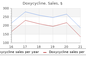
Doxycycline dosages: 200 mg, 100 mg
Doxycycline packs: 30 pills, 60 pills, 90 pills, 120 pills, 180 pills, 270 pills, 360 pills

Buy doxycycline 100 mg with visa
The deep lamina consists of a robust median band that ascends to the foramen magnum antibiotic resistance why is it a problem doxycycline 200 mg order, and two lateral bands that move to infection xpk doxycycline 200 mg without a prescription, and blend with, the capsules of the atlanto-occipital joints as they attain the foramen magnum. The membrane is separated from the cruciform ligament of the atlas by a thin layer of unfastened areolar tissue, and typically by a bursa. Alar ligaments the alar ligaments are thick cords, about eleven mm lengthy, which cross horizontally and laterally from the longitudinally ovoid flattenings on the posterolateral side of the apex of the dens to the roughened areas on the medial facet of the occipital condyles. The primary perform of the alar ligaments is now thought of to be limitation of atlanto-axial rotation, the left changing into taut on rotation to the best and vice versa. The slightly downward movement of the atlas during rotation helps permit a wider vary of motion by decreasing rigidity in the alar ligaments, and within the capsules and accent ligaments of the lateral atlanto-occipital joint. Apical ligament of the dens the apical ligament of the dens followers out from the apex of the dens into the anterior margin of the foramen magnum between the alar ligaments, and represents the cranial continuation of the notochord and its sheath. It is separated for many of its extent from the anterior atlanto-occipital membrane and cruciform ligament by pads of fatty tissue, although it blends with their attachments at the foramen magnum, and with the alar ligaments on the apex of the dens. The ligamentum nuchae and the anterior longitudinal ligament additionally join cervical vertebrae with the cranium. The synovial cavity of the posterior element of the median joint complex is larger, lying between the horizontally oriented ovoid aspect, on the posterior surface of the dens and the cartilaginous anterior floor of the transverse ligament; communication usually exists with one or each of the atlanto-occipital joint cavities. Other ligaments connecting the axis and the occipital bone, the fibrous capsules, the ligamentum nuchae and the posterior neck muscle tissue additionally contribute to stability; the suboccipital muscle tissue play an necessary proprioceptive and postural role. Muscles producing movements Movement is simultaneous at all three joints and consists nearly solely of rotation across the axis. The form of the articular surfaces is such that, when rotation happens, the axis ascends barely into the atlantal ring, limiting stretch on the lateral atlanto-axial joint capsules. Rotation is limited primarily by the alar ligaments, with a minor contribution from the accent atlanto-axial ligament. The muscle tissue that produce atlanto-axial rotation act on the skull, transverse processes of the atlas and spinous process of the axis. They are mainly obliquus capitis inferior, rectus capitis posterior main and splenius capitis of 1 facet, and the contralateral sternocleidomastoid. Iliolumbar ligament the iliolumbar ligament is connected to the tip and anteroinferior facet of the fifth lumbar transverse process, and typically has a weak attachment to the fourth transverse process. A decrease band passes from the inferior aspect of the fifth lumbar transverse process and the body of the fifth lumbar vertebra across the anterior sacroiliac ligament to attain the posterior margin of the iliac fossa. An higher band, a half of the attachment of quadratus lumborum, passes to the iliac crest anterior to the sacroiliac joint, and is continuous above with the anterior layer of the thoracolumbar fascia. The decrease ligament has a more vertical component that reaches the posterior iliopectineal line; this part is a lateral relation of the L5 ventral ramus. A posterior element of the iliolumbar ligament passes behind quadratus lumborum to attach to the ilium. Anteriorly, the atlanto-axial articulations, capsules and ligaments are separated from the buccopharyngeal fascia and superior constrictor by longus capitis and longus colli, the prevertebral fascia and the retropharyngeal (potential) area. Vascularsupply the vascular provide of the lumbosacral junction is derived primarily from the iliolumbar and superior lateral sacral arteries. Many are minor segmentation anomalies and may be incidental radiological or osteological findings. More severe anomalies could lead to craniocervical instability (Piper and Traynelis 1998). Innervation the lumbosacral junction is innervated by branches derived from the fourth and fifth lumbar spinal nerves. The sympathetic trunks cross it anterolaterally, whereas the obturator nerves and lumbosacral trunks move shut laterally. The relations of the lumbosacral side joints are just like these of the lumbar side joints (see above). Its surfaces carry 738 Muscles hyaline cartilage that varies from thin veils to small islands. Semispinalis capitis Sternocleidomastoid Splenius capitis Rhomboid minor Rhomboid major Trapezius Deltoid Levator scapulae Supraspinatus Infraspinatus Ligaments the anterior sacrococcygeal ligament consists of irregular fibres that descend on the pelvic surfaces of both sacrum and coccyx, and is connected in the same way because the anterior longitudinal ligament. The deep posterior sacrococcygeal ligament passes from the again of the fifth sacral vertebral body to the dorsum of the coccyx and corresponds to the posterior longitudinal ligament. On all sides, the lateral sacrococcygeal ligaments connect the coccygeal transverse processes to the inferolateral sacral angles, finishing foramina for the fifth sacral spinal nerves. Similarly, the intercornual ligaments join the sacral and coccygeal cornua, and a fasciculus connects the sacral cornua to the coccygeal transverse processes. Vascularsupply the arterial provide of the sacrococcygeal junction is derived from the inferior lateral sacral and median sacral arteries. InnervationThe innervation of the sacrococcygeal junction is derived from the lower two sacral and the coccygeal nerves. Segments are additionally linked by extensions of the anterior and posterior sacrococcygeal ligaments. In grownup males all segments unite comparatively early, but in females union is later. The apex of the terminal phase is related to overlying skin by white fibrous tissue. The true back muscle tissue are characterized by their place and by their innervation by branches of the posterior (dorsal) rami of the spinal nerves. The true back muscle tissue below the neck lie deep to the posterior layer of the thoracolumbar fascia. In the lumbar region, the place the layers of the thoracolumbar fascia are well outlined, they occupy the compartment between its posterior and middle layers. The most superficial of these run between the higher limb and the axial skeleton, and include trapezius, latissimus dorsi, levator scapulae and the rhomboid muscular tissues. Beneath this layer lie the serratus posterior group, superior and inferior, that are variably developed however normally skinny muscle tissue whose operate could also be respiratory or probably proprioceptive. Trapezius, levator scapulae, rhomboid main, rhomboid minor and latissimus dorsi are described on pages 816, 818 and 821 respectively, serratus posterior superior and inferior are described on pages 941, 942. The more superficial layers comprise the splenius muscles in the neck and upper thorax, and the erector spinae group within the trunk as an entire. The deeper layers embrace the spinotransverse group, which is itself layered into semispinalis, multifidus and the rotatores, and the suboccipital muscle tissue. The lumbar intertransversarii mediales, thoracic intertransversarii and medial parts of cervical posterior intertransversarii are innervated by dorsal rami, however the others are supplied by ventral rami (Commentary 5. On the left solely the skin, superficial and deep fasciae (other than gluteofemoral) have been removed; on the right, sternocleidomastoid, trapezius, latissimus dorsi, deltoid and external indirect have been dissected away. It is a singular arrangement of tendons and fascia between the posterior muscle tissue of the neck. It is hooked up to the exterior occipital protruberance superiorly, and the tip of the C7 spinous process inferiorly. In its superior half it consists of the aggregated tendons of probably the most medial fibres of the cervical portion of trapezius. Because of their longitudinal association, these tendons have been described as forming the funicular portion of the ligamentum nuchae (Mercer and Bogduk 2003).
Doxycycline 200 mg order line
Ultrasound investigations support the function of motion in the improvement of these curvatures antibiotic iv generic 100 mg doxycycline with mastercard. The early look of the secondary curves might be accentuated by postnatal muscular and nervous system growth at a time when the vertebral column is very versatile and is able to assuming virtually any curvature virus nyc 200 mg doxycycline purchase mastercard. Neonatal curvatures Structural defects of the posterior bony elements 716 Deformity and bony deficiency might happen at several websites within the posterior parts. The laminae could additionally be wholly or partially absent, or the spinous course of alone may be affected, with no abnormalities within the overlying delicate tissues (spina bifida occulta). A defect might happen in the bone that joins the superior and inferior articular processes (pars interarticularis): this condition is spondylolysis, and may be developmental or result from acute or fatigue fracture. Abnormality of the laminar bone, or degenerative modifications in the side joints, may also lead to similar displacement within the absence of pars defects. It is particularly flexible and, if dissected free from the body, it could possibly simply be bent (flexed or extended) into a perfect half-circle. A slight sacral curvature may be seen, which develops as the sacral vertebrae ossify and fuse. The thoracic part of the column is the primary to develop a comparatively fastened curvature, which is concave anteriorly. An infant can usually assist its head at three or four months, sit upright at around 9 months, and will commence walking between 12 and 15 months. These useful changes exert a serious influence on the event of the secondary curvatures within the vertebral column and changes within the proportional size of the vertebrae, in particular within the lumbar region. The secondary lumbar curvature turns into necessary in maintaining the centre of gravity of the trunk over the legs when strolling begins, and thus modifications in physique proportions exert a serious influence on the next form of curvatures within the vertebral column. Adult curvatures In adults, the cervical curve is a lordosis (convex forwards), and the least marked. It extends from the atlas to the second thoracic vertebra, with its apex between the fourth and fifth cervical vertebrae. It extends between the second and the eleventh and twelfth thoracic vertebrae, and its apex lies between the sixth and ninth thoracic vertebrae. This narrowing, generally identified as lumbar foraminal stenosis, results in leg ache in an L4 sensory distribution. This condition is accompanied by huge superior articular side hypertrophy and subsequent critical central and lateral recess stenosis (arrowheads). The pelvic curve is concave anteroinferiorly and entails the sacrum and coccygeal vertebrae. The presence of these curvatures implies that the cross-sectional profile of the trunk modifications with spinal stage. The anteroposterior diameter of the thorax is way larger than that of the lower abdomen. Compensatory lateral curvature can also develop to deal with pelvic obliquity, corresponding to that imposed by inequality of leg size. These curvatures have developed with rounding of the thorax and pelvis as an adaptation to bipedal gait. A Precentral branches Lumbar artery Dorsal branch Spinal artery Spinal artery Postcentral branch Radicular (neural) department Vertebralcolumnintheelderly In older individuals, age-related modifications in the structure of bone result in broadening and lack of top of the vertebral bodies. The bony adjustments within the vertebral column are accompanied by changes within the collagen content of the discs and by decline in the activity of the spinal muscular tissues. This leads to progressive decline in vertebral column mobility, significantly in the lumbar backbone. Overall, these adjustments within the vertebral column lead directly to loss of complete height within the particular person. This, coupled with narrowing of the spinal canal, can lead to elevated threat of neurological damage of the cervical spinal cord, in addition to ache. Twomey et al (1983) noticed a reduction in bone density of lumbar vertebral our bodies with age, principally on account of a discount in transverse trabeculae (more marked in females because of postmenopausal osteoporosis), which was related to elevated diameter and growing concavity in their juxtadiscal surfaces (end-plates). Osteophytes (bony spurs) could type from the compact cortical bone on the anterior and lateral surfaces of the bodies. Although individual variations happen, these changes seem in most individuals from 20 years onwards. They are commonest on the anterior facet of the physique and by no means involve the ring epiphysis. Osteophytic spurs are frequently asymptomatic however might lead to diminished movements inside the spine. In the cervical and sacral areas, longitudinal anastomoses between the intersegmental vessels persist as longitudinal vessels, which themselves give spinal branches to the vertebral column. The ascending cervical artery and the lateral sacral artery are persistent elements of the precostal anastomosis. On all sides, the main trunk of the artery (posterior intercostal or lumbar) passes around the vertebral physique, giving off primary periosteal and equatorial branches to the physique, after which a major dorsal department. B, Arterial anastomoses between postcentral branches of spinal arteries inside the vertebral canal. There is free anastomosis between these dorsal articular and gentle tissue branches, extending over several segments (Boelderl et al 2002, Crock and Yoshizawa 1976). At cervical and sacral ranges, the longitudinally working arteries described above have direct spinal branches. The spinal branches are the principle arteries of supply to all bony elements of the vertebrae and to the dura and epidural tissues, and in addition contribute to the provision of the spinal wire and nerve roots by way of radicular branches. As they enter the vertebral canal, the spinal arteries divide into postcentral, prelaminar and radicular branches. The majority of the vertebral arch, the posterior epidural tissues and dura, and the ligamentum flavum are equipped by the prelaminar branches and their anastomotic plexus on the posterior wall of the vertebral canal. Fine anterior central and anterior laminar and posterior laminar vessels can be seen. The arteria radicularis magna (Arm), which varieties a significant contribution to the anterior spinal artery of the twine, can be seen arising at L2. The vascularity of the lumbar vertebra may be considered the archetypal sample from which different regions evolved variations. In this part, the arteria radicularis magna is seen as a neural branch on the right facet. Key: 1, meningeal artery in hypoglossal canal; 2, occipital artery; 3, apical arcade of odontoid process; 4, ascending pharyngeal artery giving collateral branch beneath anterior arch of atlas; 5, posterior ascending artery; 6, anterior ascending artery; 7, precentral and postcentral arteries to typical cervical vertebral physique; 8, anterior spinal plexus; 9, medullary department of vertebral artery: radicular, prelaminar, and meningeal branches are additionally discovered at each degree; 10, collateral to ascending pharyngeal artery passing rostral to anterior arch of atlas; eleven, left vertebral artery. Basivertebral veins the basivertebral veins are paired valveless veins that drain the pars spongiosa of the vertebral our bodies into the inner and exterior vertebral venous plexuses. Posteriorly, they drain into the transverse branches of the anterior inner vertebral plexuses. Anteriorly, they drain immediately into the anterior exterior vertebral venous plexus.
Order 200 mg doxycycline mastercard
Streptococci produce proteolytic enzymes that digest the unfastened connective tissue and so open up the tissue areas antibiotics news doxycycline 200 mg with mastercard. Understanding the configuration of the cervical fasciae and areas is important for the placement of native anaesthetic cervical plexus blocks in the neck to facilitate operations similar to thyroidectomy antibiotic eye drops for stye buy discount doxycycline 200 mg on-line, parathyroid ectomy and carotid endarterectomy (Pandit et al 2000). The areas are finest conceptualized by way of their location, remem bering that some communicate with one another and/or with the axilla or thorax, and that some include solely free areolar tissue while others comprise dense connective tissue. Thus, areas may be related to the face (buccal, canine, masticator, parotid), suprahyoid (peritonsillar, submandibular, sublingual, parapharyngeal) or infrahyoid (anterior visceral), or lengthen the size of the neck (retropharyngeal, danger, prevertebral, carotid). It is thicker around the arteries than the vein, an arrangement that allows the vein to expand. Peripherally, the sheath is linked to adjoining fascial layers by free areolar tissue. There is disagreement about whether or not the carotid sheath is shaped by all three layers of the deep cervical fascia, or solely by the deep or the superficial layer, or even that it lacks a demonstrable fascial ensheathment (Guidera et al 2012). It is hooked up ante riorly to the inside border of the primary rib, and posteriorly to the anterior border of the transverse strategy of the seventh cervical vertebra and is covered and strengthened by a couple of spreading muscular fibres derived from the scaleni. Tissue areas around the pharynx and larynx are described on pages 578 and 594, respectively. Cellulitis in the neck Visceral house or compartment the visceral house or compartment accommodates the pharynx, cervical oesophagus, larynx, trachea, thyroid and parathyroid glands, recurrent laryngeal nerves, and the cranial sympathetic trunk. It is bounded anter iorly by the fascia that encloses the infrahyoid strap muscular tissues (muscular layer of middle deep cervical fascia), posteriorly by the alar fascia, and laterally by the carotid sheath on either side. It could also be subdivided into an anterior visceral (pretracheal) area and a posterior visceral area; these areas communicate freely between the degrees of the midthyroid cartilage and the inferior thyroid artery, however are separated inferiorly by the fascia related to the artery. It incorporates the trachea, in addition to the thyroid and parathyroid glands, larynx, cervical oesophagus, recurrent laryngeal nerves and the cranial sympathetic trunks. The area extends superiorly from the hyoid bone and the attachments of the strap muscle tissue and their fasciae to the hyoid bone and thyroid cartilage down into the anterior portion of the superior mediastinum. It communicates freely with the posterior visceral space around the sides of the larynx, the caudal portion of the pharynx and the higher cervical oesophagus, but becomes separated from the pos terior visceral area at lower ranges by dense connective tissue related to the inferior thyroid artery as the latter approaches the thyroid gland. Infection often spreads into the anterior visceral area by either perforation of the anterior wall of the oesophagus by endoscopic instru mentation, international bodies or trauma or from the posterior visceral space. Radiologically, the portion of the anterior visceral area between the strap fascia and the fascia of the thyroid gland is referred to as the anterior cervical space; its posterolateral border is either the carotid sheath or the fascia covering sternocleidomastoid. The anterior cervical house usually supplies a symmetric landmark on transverse imaging (Smoker and Harnsberger 1991). Posterior visceral space the posterior visceral space lies posterior to the pharynx and cervical oesophagus, extending from the cranium base right down to the superior medi astinum, its caudal limit being the level of fusion between the alar and visceral layers of fascia. The posterior visceral space is commonly referred to as the retropharyngeal space in the higher neck. The most common causes of cellulitis of the neck are infections arising from the region of the mandibular molar enamel and the palatine tonsils. Several fascial areas are accessible from this space, and several anatomi cal factors contribute to the spread of an infection. Thus, the apices of the second and, extra particularly, the third, mandibular molar enamel are often near the lingual surface of the mandible. The apices of the roots of the third mandibular molars are often, and the second molars are often, below the attachment of mylohyoid on the inside facet of the mandible and so drain directly into the submandibular tissue house. The posterior free border of mylohyoid is near the sockets of the third mandibular molars, and at this level, the ground of the mouth consists solely of mucous membrane overlaying part of the submandibular salivary gland. Any virulent periapical infection of the mandibular third molar tooth may subsequently penetrate the lingual plate of the mandible and is then at the entrance to the submandibular and sublingual spaces anteriorly, and the parapharyngeal and pterygoid areas posteriorly. Infection in this space can also spread from an acute pericoronitis, par ticularly when the deeper tissues are opened to infection by extraction of the tooth through the acute section. Cellulitis within the region of the maxilla is much more uncommon, but fascial area infections might develop in various sites as the results of contaminated native anaesthetic needles. All types of cellulitides of the neck or deep neck area infections are potentially very critical. Obstruction of the upper airway develops on account of inflammation and oedema, compounded by salivary pooling consequent on dysphagia, and this could be fairly catastrophic. Increased rigidity and decreased compliance of the tissues make manoeu vres such as manual anterior jaw thrust or laryngoscopy nearly impos sible. Sternocleidomastoid is a key landmark as a result of it divides the neck into anterior and lateral areas (anterior and posterior triangles, respectively); the anterior region could also be further subdivided into a number of smaller named triangles (see above). Muscles in the anterior region are organized into supra and infrahyoid groups, and, with one exception, are all connected to the hyoid bone. The suprahyoid muscle tissue, which connect the hyoid bone to the mandible and the bottom of the cranium, include mylohyoid, geniohyoid, stylohyoid and digastric. The infra hyoid (strap) muscular tissues, which join the hyoid, sternum, clavicle and scapula, are organized in two planes: a superficial aircraft consisting of sternohyoid and omohyoid, and a deep plane consisting of sterno thyroid and thyrohyoid. The muscles that form a part of the musculoskeletal column in the neck are described in Chapter 43. They could be considered in three teams � anterior, lateral and posterior; very broadly speaking, the muscles in these teams lie anterior, lateral or posterior to the cervical vertebrae. The anterior and lateral teams embody longi colli and capitis; recti capitis anterior and lateralis; and scaleni anterior, medius, poste rior and minimi (when present). The deeper layers include the transversospinal group (semispi nales cervicis and capitis, multifidus and rotatores cervicis), interspinales and intertransversarii, and the suboccipital group (recti capitis posterior major and minor, and obliquus capitis superior and inferior). The muscle tissue related to the pharynx and larynx are described in Chapters 34 and 35, respectively. The prevertebral tissue house is the potential area mendacity between the prevertebral fascia and the vertebral column. It extends from the cranium base to the coccyx, and encloses the prevertebral muscle tissue. Almost all of the pathology that affects the prevertebral space arises from both the adjacent vertebrae or their intervertebral discs, or the spinal cord and related nerve roots and spinal nerves. Danger house the danger space lies between the alar and prevertebral fascia, and extends from the skull base right down to the posterior mediastinum, where the alar, visceral and prevertebral layers of deep cervical fascia fuse. The potential house so created is closed superiorly, inferiorly and lat erally; infections can only enter by penetrating its partitions. The danger house is so called as a end result of its unfastened areolar tissue provides a potential route for the fast downward spread of an infection, primarily from the retro pharyngeal, parapharyngeal or prevertebral spaces, to the posterior mediastinum. Carotid space the carotid sheath is a layer of unfastened connective tissue demarcated by adjacent parts of the investing layer of deep cervical fascia, the pre tracheal fascia and the prevertebral fascia. The literature concerning the existence of a carotid house is complicated: some authors dispute that a possible cavity exists throughout the carotid sheath that could allow the unfold of infections from the upper neck down into the lower neck and mediastinum, while others consider that the suprahyoid sheath must be considered a half of the parapharyngeal area (see discus sion in Som and Curtin (2011)).
Discount 100 mg doxycycline amex
The lateral finish of each canal is dilated to form an ampulla antibiotic walmart 200 mg doxycycline buy with visa, within the ampulla of the osseous canal bacteria listeria cheap doxycycline 100 mg online. The short section of duct between the ampullae and utricle is the crus ampullaris. Guinea pigs are one of the frequently used animal fashions of human listening to and their inner ear ultrastructure may be very similar. The utricular macula has been tilted in the plane of the page to emphasize that it lies horizontally, whereas the saccular macula lies vertically when the head is in an upright place. Note the different shapes of the maculae, the place of the striola as indicated by a curved line in every case, and the different orientations of their stereociliary bundles. The stereocilia are arranged in rows of increasing height in course of the tallest factor, the kinocilium. Deflection within the direction of the kinocilium leads to depolarization of the hair cell. The inset reveals a tip hyperlink connecting a short stereocilium tip to the tall stereocilium aspect behind. The membranous wall of every ampulla contains a transverse elevation (septum transversum), on the central area of which is a saddleshaped sensory ridge, the ampullary crest, containing hair cells and supporting cells. It is broadly concave on its free edge alongside most of its size and has a concave gutter (planum semilunatum) at either end between the ridge and the duct wall. Sectioned throughout the ridge, the crests of the lateral and anterior semicircular canals have smoothly rounded corners; the posterior crest is extra angular. The three semicircular canals thus detect angular accelerations during tilting or turning movements of the head in all three different planes of three-dimensional space. Microstructure of the vestibular system the maculae and crests detect the orientation of the top with respect to gravity and changes in head motion by the use of the mechanosensitive hair cells. These hair cells are in synaptic contact with afferent and efferent endings of the vestibular nerve on their basolateral facet. The whole epithelium lies on a bed of thick, fibrous connective tissue containing myelinated vestibular nerve fibres and blood vessels. The axons lose their myelin sheaths as they perforate the basal lamina of the sensory epithelium. Type I vestibular sensory cells measure 25 �m in size, with a free surface of 6�7 �m in diameter. The apical floor is characterized by 30�50 stereocilia (large, often arranged, modified microvilli about 0. The kinocilium is significantly longer than the stereocilia, and should attain forty �m, whereas the stereocilia are of graded lengths. The kinocilium emerges basally from a typical basal physique, with a centriole immediately beneath it. Close to the inner surface of their basal two-thirds, each cell contains quite a few synaptic ribbons with related synaptic vesicles. The postsynaptic floor of an afferent nerve ending encloses the larger a half of the sensory cell body within the form of a cup (chalice or calyx). Efferent nerve fibres make synapses with the external surface of the calyx, quite than immediately with the sensory cell. The kinocilium confers structural polarity on the bundle, which relates to practical polarity. The stereocilia and kinocilium are all interconnected by fine extracellular filaments of varied varieties, called cross links. The tip link is widespread to all forms of hair cell and is thought to play a central role in transduction; mutations within the proteins that comprise the tip hyperlink are significant in Usher syndrome, which is characterised by auditory and visual abnormalities. Deflection of the bundle in the course of the kinocilium ends in depolarization of the hair cell and increases the rate of neurotransmitter release from its base. Deflection away from the kinocilium hyperpolarizes the hair cell and reduces the release of neurotransmitter. Some are up to forty five �m lengthy and virtually span the whole thickness of the sensory epithelium, whereas others are shorter than type I cells. They are principally cylindrical, however in any other case resemble type I cells of their contents and the presence of an apical kinocilium and stereocilia. However, their kinocilia and stereocilia are inclined to be shorter and less variable in length. Polarization permits the hair cells to have specific orientations that optimize their function inside every sensory organ. In the utricle, the kinocilia are positioned on the facet of the sensory cell nearest to the striola so that the excitatory path is in the direction of the midline. In the ampullary crests, the cells are orientated with their rows of stereocilia at proper angles to the long axis of the semicircular duct. The otolithic membrane is a layer of extracellular materials divided into two strata. The external layer consists of otoliths or otoconia, which are barrel-shaped crystals of calcium carbonate with angular ends, up to 30 �m long, and heterogeneous in distribution. The gelatinous materials consists largely of glycosaminoglycans associated with fibrous protein. The epithelial lining and subepithelial connective tissue become more advanced where the duct dilates to type the endolymphatic sac. In the intermediate phase, the epithelium consists of light and dark cylindrical cells. Light cells are regular in type and have numerous lengthy surface microvilli with endocytic invaginations between them and huge clear vesicles of their apical region. In distinction, darkish cells are wedge-shaped and have a slender base, few apical microvilli and dense, fibrillar cytoplasm. The endolymphatic sac has important roles within the upkeep of vestibular perform. Endolymph produced elsewhere within the labyrinth is absorbed in this area, most likely primarily by the light cells. Damage to the sac, or blockage of its connection to the the rest of the labyrinth, causes endolymph to accumulate; this produces hydrops, which affects each vestibular and cochlear perform. The epithelium can be permeable to leukocytes, together with macrophages, which may take away cellular particles from the endolymph, and to various cells of the immune system that contribute antibodies to this fluid. The osseous spiral lamina initiatives for a half of the space between the modiolus and the outer wall of the cochlea and is connected to the inside fringe of the basilar membrane. The endosteum of the outer wall is thickened to type a spiral cochlear ligament that tasks inwards as a triangular basilar crest hooked up to the outer rim of the basilar membrane. Immediately above this may be a concavity, the external spiral sulcus (sulcus spiralis externus), above which the thick, extremely vascular periosteum projects as a spiral prominence. Above the prominence is a specialized, thick epithelial layer, the stria vascularis. The facet facing the scala vestibuli bears flattened perilymphatic cells, with tight junctions between them, creating a diffusion barrier.
Generic 200 mg doxycycline amex
It bends over the joint antibiotic prophylaxis for colonoscopy cheap doxycycline 200 mg without prescription, persevering with forwards over the lateral cricoarytenoid muscle before terminating inside thyroarytenoid antibiotics z pack dosage buy doxycycline 100 mg amex. The anterior branch of the recurrent laryngeal nerve first innervates posterior cricoarytenoid by one or more branches, then innervates interarytenoid and lateral cricoarytenoid, and terminates in thyroarytenoid, which it also supplies (see additionally Maranillo et al 2005). The posterior branch of the recurrent laryngeal nerve ascends deep to posterior cricoarytenoid to be part of the descending branch of the internal laryngeal nerve. There can be a posh anastomosis within and over the posterior surface of the interarytenoid muscle tissue, and, much less regularly, anastomoses the consequences of vagal nerve lesions are complicated, reflecting the lengthy course of the nerve and the possible involvement of three of its branches, namely: the pharyngeal, superior laryngeal and recurrent laryngeal nerves. Collectively, these nerves innervate muscular tissues of the larynx, soft palate and pharynx; damage may therefore have deleterious results on phonation and/or taste bud movements and/or swallowing. A lesion of the vagus above the level at which the pharyngeal department is given off will affect both the superior and recurrent laryngeal nerves. This causes immobility of the vocal folds on the affected aspect and imparts a breathy voice with lack of pitch and restricted loudness. There may even be a level of hypernasality due to the consequences on actions of the taste bud caused by paralysis of levator veli palatini. Unilateral palsies influence very significantly on the quality of lifetime of the patient because of impaired vocalization and a tendency to aspirate. Bilateral palsies are extraordinarily serious and a tracheostomy is required to shield the airway. Complete part is most likely through the ligation of the vessels forming the vascular pedicle of the thyroid gland during thyroid lobectomy. Unilateral lesions could result in the vocal folds appearing relatively normal and the effect on voice is barely noticeable and is usually ignored. A more detailed examination could detect some shortening of the vocal folds on the affected aspect with uneven tilt of the epiglottis and the anterior larynx canted towards the unaffected facet. Bilateral superior laryngeal nerve lesions lead to shortening of both vocal folds, the overhanging of the epiglottis over the folds and lowered tilt between the thyroid and cricoid cartilage. Effects on the voice are correspondingly higher, with decreased loudness and pitch but variable breathiness. Damage to the inner laryngeal nerve causes lack of mechanoreceptive and proprioceptive sensation from the larynx. Unilateral loss produces a sense that something is stuck in the throat, whereas bilateral loss will lead to aspiration and can cause dysphagia, with a danger of choking. There is olated paralysis of all the laryngeal muscular tissues on the affected facet besides cricothyroid, which is innervated by the exterior laryngeal nerve. The patient may be asymptomatic or have a hoarse, breathy voice and there shall be diminished capacity to manipulate pitch. The hoarseness could also be everlasting or may turn out to be much less extreme with time as the contralateral twine develops the power to hyperadduct and appose the paralysed wire and thus shut the glottis during phonation and coughing, though that is seldom sufficient to restore full voice high quality. Bilateral lesions of the recurrent laryngeal nerve lead to both vocal cords being paralysed and taking over a paramedian position. Phonation could be nearly regular as a outcome of the vocal folds lie so close to the midline, however there might be audible stridor and a really compromised airway. Clinically, the place of the vocal wire within the acute part after section of the recurrent laryngeal nerve could be very variable. The cords are barely more widely separated in persistent lesions, which renders the voice weaker however with a much less precarious airway. Atrophy and fibrosis of paraglottic muscle tissue in all probability affect the place of paralysed vocal cords in continual lesions to a greater degree than variations in the power of the apposing adductor and abductor muscle teams. This effect was thought to reflect the interior segregation inside the recurrent laryngeal nerve of axons supplying the laryngeal abductor muscle tissue; the thought was later undermined by the demonstration that axons destined for explicit laryngeal muscular tissues are randomly distributed inside the nerve. It is likely that predicting the impact of partial lesions of the recurrent laryngeal nerve is complicated by the variable patterns of anastomosis that happen between the laryngeal nerves. Central management of voice production entails two parallel pathways: a limbic pathway liable for the control of innate non-verbal and emotional vocalizations, and a larynx-specific motor cortical pathway responsible for regulating the motor management essential for voluntary voice production. For further studying, see Clark et al (2007), Kaplan (1971), Laver (1980), Atkinson and McHanwell (2002), Titze (1994), Zemlin (1998). For all sounds in Western European languages, and most sounds in other languages, this energy takes the form of a pulmonary expiration. This steady airflow is converted into a vibration inside the larynx by a mechanism referred to as phonation, in which the vocal folds vibrate periodically, interrupting the column of air because it leaves the lungs and changing it into a series of discrete puffs of air. Speech sounds which are produced by vocal fold vibration in this way are stated to be voiced. Speech sounds which are produced without vocal fold vibration are termed unvoiced sounds. Amplification and modification of the sound happen in the supralaryngeal vocal tract, which can be thought-about as a 17 cm lengthy tube, slender on the larynx and broadening out proximally because it passes by way of the pharynx, and oral and nasal cavities. The range of sounds that the human vocal tract is able to producing is very wide, though anybody human language will make use of a subset of those sounds to convey which means. The thorax is capable of responding mechanically to broadly varying demands for oxygen. From a tidal volume at remainder of 500 ml and a respiratory price of 12 per minute, air flow can increase in match individuals throughout vigorous train to tidal volumes of four. Normal ventilatory patterns are considerably modified throughout speech, reflecting the particular demands that speech locations on air flow. The primary source of energy for the manufacturing of speech sounds is a pulmonary expiration, although different mechanisms are possible. In order for speech to be produced, sufficient pressure has to be generated beneath the vocal folds. This subglottal stress (the difference between the air pressures above and beneath the vocal folds) has to be sustained above a minimum degree throughout an utterance. It units the vocal folds into vibration if the sound is to be voiced or generates airflow for an voiceless sound. The minimum subglottal pressure needed for speech manufacturing is 7 cmH2O, and this increases when loud sounds are produced or when sounds are confused. The have to generate adequate sustained strain implies that speech ventilation is markedly non-rhythmical. Expiration is much longer than regular, perhaps lasting as a lot as 30 seconds, reflecting the truth that the vocal tract is extra constricted at the larynx to be sure that pauses for additional inspirations are made at suitable points in an utterance. Conversational speech usually takes place at the next vary of lung capacities than function in normal quiet ventilation. The non-rhythmical sample during speech requires greater inspiratory effort, and for most people it involves a larger use of the Autonomic innervation Parasympathetic secretomotor fibres run with each the superior and the recurrent laryngeal nerves to mucous glands throughout the larynx. Postganglionic sympathetic fibres run to the larynx with its blood provide; they originate within the superior and middle cervical ganglia. A variable number of paraganglia are situated on the inner laryngeal and recurrent laryngeal nerves. Histochemical proof suggests that one inhabitants of the neurones in these ganglia have a parasympathetic perform while a second inhabitants might secrete dopamine (Maranillo et al 2008, Ibanez et al 2010).
Syndromes
- What medications do you take?
- Vomiting
- Dizziness
- Loss of appetite
- Nasopharyngeal culture
- Fuse joints to prevent movement
- Decreased blood pressure and high heart rate
100 mg doxycycline discount with amex
Nerves to thyrohyoid and geniohyoid the nerves to thyrohyoid and geniohyoid come up near the posterior border of hyoglossus infection next to fingernail order doxycycline 100 mg. Lesions of the hypoglossal nerve Lesions affecting the accent nerve Lesions of the accent nerve may be sustained centrally antibiotics quizlet buy discount doxycycline 100 mg on-line, at its exit from the cranium or within the neck. The supranuclear fibres that affect motor neurones innervating sternocleidomastoid decussate twice; a lesion of the pyramidal system above the pons due to this fact produces weakness of the ipsilateral sternocleidomastoid and contralateral trapezius. Episodic contraction of sternocleidomastoid and trapezius, usually accompanied by contraction of other muscle groups. In jugular foramen syndrome, attributable to pathologies together with nasopharyngeal carcinoma or a jugular paraganglioma, lesions of the glossopharyngeal, vagus and accent nerves coexist. The accessory nerve could be injured extra distally in the neck by trauma or by surgical exploration in the posterior triangle. If the accessory nerve is sacrificed as part of a radical neck dissection and the innervation of trapezius is lost, the patient develops intractable neuralgia due to traction on the brachial plexus brought on by the unsup ported weight of the shoulder and arm. Com plete hypoglossal division causes unilateral lingual paralysis and occasion ual hemiatrophy; the protruded tongue deviates to the paralysed facet, and, on retraction, the wasted and paralysed facet rises greater than the unaffected side. The larynx could deviate in the direction of the active facet in swal lowing, due to unilateral paralysis of the hyoid depressors related to loss of the primary cervical spinal nerve, which runs with the hypoglos sal nerve. Taste and tactile sensibility are unaffected however articulation is sluggish and swallowing very troublesome. The hypoglossal nerve is commonly used to reanimate the face in sufferers, significantly the elderly, whose facial nerves have been transected. Direct endtoend, endtoside or splitnerve grafts are dependable and provide a robust, efficient and fast alternative to more complicated reanimation procedures. The cervical sympathetic ganglia ship grey rami communicantes to all the cervical spinal nerves but receive no white rami communicantes from them. Their spinal preganglionic fibres emerge in the white rami communi cantes of the upper 5 thoracic spinal nerves (mainly the upper three), and ascend within the sympathetic trunk to synapse within the cervical ganglia. In their course, the grey rami communicantes could pierce longus capitis or scalenus anterior. Hypoglossal nerve the hypoglossal nerve is motor to all of the muscles of the tongue, except palatoglossus. The hypoglossal rootlets run laterally behind the verte bral artery, collected into two bundles that perforate the dura mater individually reverse the hypoglossal canal within the occipital bone, and unite after traversing it. Here, it makes a halfspiral turn round the inferior vagal ganglion and is united with it by connective tissue. It then descends virtually vertically between the vessels and anterior to the vagus to a degree stage with the angle of the mandible, turning into superficial below the posterior stomach of digastric and emerging between the inner jugular vein and inside carotid artery. It lies on the transverse processes of the second and third cervi cal vertebrae and is probably formed from four fused ganglia, judging by its gray rami to C1�4. The inside carotid artery inside the carotid sheath is anterior, and longus capitis is posterior. The decrease end of the ganglion is united by a connecting trunk to the center cervical gan glion. Postganglionic branches are distributed within the inside carotid nerve, which ascends with the inner carotid artery into the carotid canal to enter the cranial cavity, and in lateral, medial and anterior branches. They supply vasoconstrictor and sudomotor nerves to the face and neck, dilator pupillae and easy muscle within the eyelids and orbitalis. Communicating branches the hypoglossal nerve communicates with the sympathetic trunk, the lingual, glossopharyngeal and vagus nerves and the pharyngeal plexus. The vagal connections are close to the cranium, and numerous filaments pass between the hypoglossal nerve and the inferior vagal ganglion in the connective tissue uniting them. As the hypoglossal nerve curves around the occipital artery, it receives the ramus lingualis vagi from the pharyngeal plexus. Lateral branches the lateral branches are grey rami communicantes to the upper four cervical spinal nerves and to some of the cranial nerves. Branches pass to the inferior vagal ganglion, the hypoglossal nerve, the superior jugular bulb and associated jugular glomus or glomera, and to the meninges in the posterior cranial fossa. Another branch, the jugular nerve, ascends to the cranial base and divides into two; one part joins the inferior glossopharyngeal ganglion and the opposite joins the superior vagal ganglion. Medial branches the medial branches of the superior cervical gan glion are the laryngopharyngeal and cardiac. The laryngopharyngeal branches supply the carotid body and move to the side of the pharynx, becoming a member of glossopharyngeal and vagal rami to form the pharyngeal plexus. A cardiac branch arises by two or extra filaments from the decrease part of the superior cervical ganglion and occasionally receives a twig from the trunk between the superior and middle cervical ganglia. It is assumed to comprise solely efferent fibres, the preganglionic outflow being from the upper thoracic segments of the spinal wire, and to be devoid of ache fibres from the guts. It descends behind the common carotid artery, in entrance of longus colli, and crosses anterior to the inferior thyroid artery and recurrent laryngeal nerve. The right cardiac branch usually passes behind, but typically in entrance of, the subclavian artery and runs posterolateral to the brachiocephalic trunk to be part of the deep (dorsal) a part of the cardiac plexus behind the aortic arch. Branches of distribution the branches of distribution of the hypoglossal nerve are meningeal, descending, thyrohyoid and muscular nerves. Meningeal branches Meningeal branches depart the nerve within the hypoglossal canal and return by way of it to supply the diplo� of the occipital bone, the dural partitions of the occipital and inferior petrosal sinuses, and far of the floor of the anterior wall of the posterior cranial fossa, probably through pathways aside from that of the hypoglossal nerve. Descending department the descending branch (descendens hypo glossi) contains fibres from the primary cervical spinal nerve. It leaves the hypoglossal nerve when it curves across the occipital artery, and runs down on the carotid sheath. The left cardiac branch, within the thorax, is anterior to the left widespread carotid artery and crosses in front of the left facet of the aortic arch to attain the superficial (ventral) a part of the cardiac plexus. Sometimes, it descends on the proper of the aorta to end within the deep (dorsal) a half of the cardiac plexus. It communicates with the cardiac branches of the center and inferior cervical sympathetic ganglia and sometimes with the inferior cervical cardiac branches of the left vagus, and branches from these mixed nerves kind a plexus on the ascending aorta. It is often thought to be a indifferent part of the middle cervical or inferior cervical ganglion. Like the middle cervical ganglion, it could provide gray rami communicantes to the fourth and fifth cervical spinal nerves. The inferior cervical ganglion sends grey rami communicantes to the seventh and eighth cervical and first thor acic spinal nerves, and gives off a cardiac department, branches to nearby vessels and generally a branch to the vagus nerve. The grey rami communicantes to the seventh cervical spinal nerve vary from one to five (two being the usual number). A third typically ascends medial to the vertebral artery in entrance of the seventh cervical transverse process. It connects with the seventh cervical nerve, and sends a filament upwards through the sixth cervical transverse foramen in company with the vertebral vessels to be part of the sixth cervical spinal nerve because it emerges from the intervertebral foramen. The cardiac department descends behind the subclavian artery and alongside the entrance of the trachea to the deep cardiac plexus. Behind the artery, it connects with the recurrent laryngeal nerve and the cardiac branch of the center cervical ganglion (the latter is usually changed by fantastic branches of the inferior cervical ganglion and ansa subclavia). The branches to blood vessels form plexuses on the subclavian artery and its branches.
Discount doxycycline 100 mg line
As it pierces the deep layer of the temporal fascia antibiotic resistance obama order 100 mg doxycycline otc, it sends a slender twig between the two layers of the fascia towards the lateral angle of the attention antibiotics for sinus infection augmentin buy doxycycline 100 mg line. Zygomaticofacial nerve the zygomaticofacial nerve traverses the inferolateral angle of the orbit and emerges on the face by way of a foramen within the zygomatic bone. It perforates orbicularis oculi to supply the skin on the prominence of the cheek and types a plexus with zygomatic branches of the facial nerve and palpebral branches of the maxillary nerve. Infraorbital nerve the infraorbital nerve emerges on to the face on the infraorbital foramen, the place it lies between levator labii superioris and levator anguli oris. The palpebral branches ascend deep to orbicularis oculi, pierce the muscle to supply the skin within the decrease eyelid, and join with the facial and zygomatico facial nerves close to the lateral canthus. Nasal branches provide the skin of the side of the nose and of the movable part of the nasal septum, and be part of the exterior nasal branch of the anterior ethmoidal nerve. Superior labial branches, that are massive and numerous, descend behind levator labii superioris to supply the pores and skin of the anterior part of the cheek and higher lip. They are joined by branches from the facial nerve to type the infraorbital plexus. The infraorbital nerve is commonly implicated in trigeminal neuralgia, and is amenable to cryotherapy where medical remedy fails. Mandibular nerve the mandibular nerve provides pores and skin over the mandible, the decrease lip, the fleshy a half of the cheek, a half of the auricle of the ear and part of the temple through the buccal, mental and auriculotemporal nerves. Buccal nerve the buccal nerve emerges on to the face from behind the ramus of the mandible and passes laterally in entrance of masseter to unite with the buccal branches of the facial nerve. Mental nerve the mental nerve is the terminal branch of the inferior alveolar nerve. Surg Gynecol Obstet 102:385�412, with permission from the American College of Surgeons. The cervical branch emerges from the lower a part of the parotid gland and runs anteroinferiorly under platysma to the entrance of the neck. Typi cally single, it provides platysma and communicates with the transverse cutaneous cervical nerve. Lesions of the facial nerve Facial paralysis could also be due to an higher motor neurone lesion (when frontalis is partially spared because of the bilateral innervation of the muscular tissues of the higher a half of the face) or a lower motor neurone lesion (when all branches could additionally be involved). A, the branches given off immediately after the nerve exits the stylomastoid foramen. Only these muscular tissues innervated by degenerating or demyelinating branches of the facial nerve turn into weak. The facial nerve is routinely isolated as a part of a superficial parotid ectomy operation, typically in the remedy of parotid tumours, when that part of the gland mendacity superficial to the plane of the facial nerve is removed along with the tumour. Although this can affect all the branches of the facial nerve, the weakness is commonly confined to the ter ritory innervated by the marginal mandibular department, which is most probably to be stretched throughout surgical intervention and is manifested by a weak spot of the lower lip on the affected aspect. Each has a mean weight of 25 g and is an irregular, lobulated, yellowish mass, lying largely under the external acoustic meatus between the mandible and sternocleidomastoid. Sometimes, a small, usually detached, part referred to as the accessory parotid gland (pars accessoria or socia parotidis) lies between the zygomatic arch above and the parotid duct below (Fromer 1977). Sometimes, however, the gland is more or less of even width all through, and the upper and lower poles are rounded. In its ordinary inverted pyramidal kind, the parotid gland presents a small superior surface, and superficial, anteromedial and posteromedial surfaces. The concave superior surface is said to the cartilaginous a part of the exterior acoustic meatus and posterior side of the temporomandibular joint. Here, the auriculotemporal nerve curves round the neck of the mandible, embed ded within the capsule of the gland. The apex overlaps the posterior belly of digastric and the carotid triangle to a variable extent. The superficial floor is roofed by skin and superficial fascia, which incorporates the facial branches of the nice auricular nerve, superficial parotid lymph nodes and the posterior border of platysma. It extends upwards to the zygomatic arch, backwards to overlap sternocleidomas toid, downwards to its apex posteroinferior to the mandibular angle, and forwards to lie on masseter beneath the parotid duct. The anteromedial floor is grooved by the posterior border of the mandibular ramus. It covers the posteroinferior part of masseter, the lateral side of the temporomandibular joint and the adjoining part of the mandibular ramus. It passes forwards, medial to the ramus of the mandible, to attain medial pterygoid. Branches of the facial nerve emerge on the face from the anterior margin of this floor. The posteromedial surface is moulded to the mastoid course of, sternocleidomastoid, posterior belly of the digastric, and the styloid process and its related muscles. The anteromedial and posteromedial surfaces meet at a medial margin that may project so deeply that it contacts the lateral wall of the pharynx. The posterior auricular artery can also branch from the exterior carotid artery within the gland, leaving by its posteromedial surface. It descends within the parotid gland and emerges behind the apex of the gland, where it usually divides into an anterior department, which passes forwards to be a part of the facial vein, and a posterior branch, which joins the posterior auricular vein to form the external jugular vein. Acute inflam mation of the parotid gland (acute sialadenitis) may trigger beautiful ache within the preauricular region as a end result of stretching of the capsule and stimulation of the nice auricular nerve. The ache is normally exacer bated at mealtimes when the gustatory stimulus to the gland ends in further turgor inside the capsule. Causes of acute sialadenitis include parotid duct obstruction (calculus, mucus plug and duct stricture) and mumps. It crosses masseter, turns medially at its anterior border at virtually a proper angle, and traverses the buccal fat pad and buccinator reverse the crown of the higher third molar tooth. The duct then runs obliquely forwards for a brief distance between buccinator and the oral mucosa earlier than it opens on a small papilla reverse the second higher molar crown. The submucosal passage of the duct serves as a valvular mechanism, preventing inflation of the gland with raised intraoral pressures. While crossing masseter, the duct lies between the higher and lower buccal branches of the facial nerve, and may obtain the accessory parotid duct. The accent a half of the gland (Fromer 1977) and the transverse facial artery lie above the parotid duct; the buccal branch of the person dibular nerve, rising from beneath temporalis and masseter, lies just below, at the anterior border of masseter. The parotid duct could also be crossed by anastomosing branches between the zygomatic and buccal branches of the facial nerve. The ramifications of the ductal methods, and their patterns and cali bres, may be demonstrated radiographically by injecting a radioopaque substance into the parotid duct through a cannula. In a lateral parotid sialo gram, the main duct could be seen to be shaped close to the centre of the posterior border of the mandibular ramus by the union of two or three ducts that ascend or descend, respectively, at proper angles to the principle duct. Deep lacerations of the cheek, where the integrity of the parotid duct is in doubt, must be explored and repaired using microsurgical strategies, to prevent saliva leaking into the soft tissues of the cheek and subsequent sialocele formation. Lymph nodes occur in the skin overlying the parotid gland (pre auricular nodes) and within the substance of the gland.
100 mg doxycycline effective
The glottis is typically divided into two regions: an anterior intermembranous half antibiotics diabetes 200 mg doxycycline visa, which makes up about three-fifths of its anteroposterior length and is fashioned by the underlying vocal ligament; and a posterior intercartilaginous part antibiotics for steroid acne discount doxycycline 100 mg visa, formed by the vocal processes of the arytenoid cartilages. It is the narrowest part of the larynx, having a median sagittal diameter in adult males of 23 mm, and in grownup females of 17 mm; its width and shape range with the movements of the vocal cords and arytenoid cartilages during respiration and phonation. Its partitions are lined by respiratory mucosa, and are supported by the cricothyroid ligament above and the cricoid cartilage below (Reidenbach 1998). The partitions of this a half of the laryngeal cavity are stated to be exponentially curved, a function that will serve to speed up the airflow in course of the glottis with the minimum lack of energy (Lenneberg 1967). Nodules enhance vocal fold mass and affect vocal fold closure; the persistent posterior glottal opening causes hoarseness, a breathy voice, decreased vocal intensity and an incapability to produce larger frequencies of vibration. These adjustments could cause a cycle during which rising vocal effort is required by means of compensation, and this exacerbates the issue (Aronson and Bless 2009). From the epiglottic margins, the aryepiglottic folds could be traced posteromedially and the cuneiform and corniculate elevations acknowledged. Smaller articles could enter the trachea or bronchi, or lodge within the laryngeal ventricle and trigger reflex closure of the glottis with subsequent suffocation. Laryngotomy under the vocal cords through the cricothyroid ligament, or tracheotomy below the cricoid cartilage, could additionally be essential to restore a free airway. The consequences of trauma to the larynx ensuing from either blunt or penetrating injuries could result in any or all the following: oedema, haematoma, fracture, dislocations or paralysis. Trauma at the degree of the rima glottidis could lead to thyroid cartilage fracture and displacement of the fragments into the vocal folds posteriorly with consequent oedema. Suicidal wounds are normally made through the thyrohyoid membrane, damaging the epiglottis, superior thyroid vessels, exterior and internal carotid arteries, and inner jugular veins. Less regularly, these wounds are inflicted above the hyoid, in order that the lingual muscle tissue and lingual and facial vessels are damaged. Caustic substances swallowed accidentally or throughout a suicide attempt, the ingestion of scorching liquids and the inspiration of hot gases could all end in serious laryngeal damage. Radiotherapy as a part of the remedy for neck most cancers may cause radiation burns within the larynx (Myer 2004). For further studying, see Adewale (2009), Hudgins et al (1997), Pracy (1983) and Sapienza et al (2004). The three main areas are the pre-epiglottic, the paraglottic and the subglottic spaces. Their exact definition, and the extent to which they communicate with one another, remain controversial. An awareness of the anatomy of these areas, and the potential pathways of tumour unfold from them, have significantly influenced the surgical approach to disease on this region (Welsh et al 1983). The higher part of this area additionally extends beyond the lateral margins of the epiglottis, an arrangement that offers the house the type of a horseshoe and has led to the suggestion that periepiglottic house can be a more acceptable time period for this area (Reidenbach 1996a). The higher boundary is formed by the weak hyoepiglottic membrane, strengthened medially because the median hyoepiglottic ligament; the anterior boundary is the thyrohyoid membrane, strengthened medially because the median thyrohyoid ligament; and the decrease boundary is the thyroepiglottic ligament, steady laterally with the quadrangular membrane behind. It can be in continuity with the mucosa of the laryngeal surface of the epiglottis via a quantity of perforations within the cartilage of the epiglottis (Reidenbach 1996a). At rest, the upper border of the toddler epiglottis is at the stage of the second or third cervical vertebra; when the larynx is elevated, it reaches the extent of the first cervical vertebra. This high position permits an infant to use its nasal airway to breathe whereas suckling. The epiglottis is X-shaped, with a furled petiole, and the laryngeal cartilages are softer and more pliable than in the grownup larynx (which may predispose to airway collapse in inspiration, resulting in the scientific picture of laryngomalacia). The thyroid cartilage is shorter and broader, and lies nearer to the hyoid bone in the neonate, which signifies that the thyrohyoid ligament is relatively short. Calcification of the cricoid cartilage has been famous as early as 7 years of age (Strauss 2000). The arytenoid cartilages are larger in proportion to the larynx as an entire and so are more outstanding; the aryepiglottic folds are disproportionally massive. The time at which the toddler larynx assumes adult characteristics varies within the literature from 5 to 12 years; some adults retain a funnelshaped larynx (Wheeler et al 2009). Further discussion is past the scope of this book; the interested reader ought to consult Wheeler et al (2009) and Holzki et al (2010). The mucosa is also lax in the infraglottic cavity and swelling here quickly causes severe respiratory obstruction due to the disproportionally narrower lumen (3. The neonatal infraglottic cavity extends posteriorly as well as inferiorly, which is an important consideration when passing an endotracheal tube (Litman et al 2003). The infraglottic airway rapidly increases in size in the course of the first two years of life (Eckel et al 1999). By in regards to the third 12 months, sexual variations become apparent: the larynx is bigger in boys, though the angle between the thyroid laminae is more pronounced in ladies. Congenital anomalies of the larynx embody aplasia and hypoplasia of the epiglottis, high-rising epiglottis, bifid epiglottis, saccular cysts, vocal cord palsy, laryngeal atresia, laryngocele, laryngo-tracheooesophageal cleft and laryngeal internet. It is bounded laterally by the thyroid cartilage and thyrohyoid membrane, superomedially by the quadrangular membrane, inferomedially by the conus elasticus, and posteriorly by the piriform fossa. The lower border of the thyroid cartilage is inferior, and the paraglottic house is steady inferiorly with the house between the cricoid and thyroid cartilages. Anteroinferiorly, there are deficiencies in the paramedian gap at the side of the median cricothyroid ligament, and posteroinferiorly, adipose tissue extends in the direction of the cricothyroid joint. Superiorly, the paraglottic space is often continuous with the pre-epiglottic house, although the 2 areas could additionally be separated by a fibrous septum. There is disagreement between authors as to the exact boundaries between these two spaces. Some authorities exclude thyroarytenoid from the paraglottic space and embrace it throughout the preepiglottic space, forming its inferior border posterolaterally (Reidenbach 1996b). It is steady below with the inner surface of the cricoid cartilage and its mucosa (Reidenbach 1998). The extrinsic muscles join the larynx to neighbouring buildings and are responsible for transferring it vertically during phonation and swallowing. They include the infrahyoid strap muscles, thyrohyoid, sternothyroid and sternohyoid, and the inferior constrictor muscle of the pharynx. The position of the extrinsic muscle tissue during respiration seems to be variable; the larynx has been seen to rise, descend or barely transfer throughout inspiration. The extrinsic muscle tissue can affect the pitch and the standard of the voice by raising or decreasing the larynx, and geniohyoid elevates and anteriorly displaces the larynx, particularly throughout deglutition. Elevation of the larynx decreases the length and calibre of the laryngopharynx and thus shortens the vocal tract total. The elevating of the larynx may also be accompanied by forward or backward motion of the tongue due to the attachment of the tongue to the hyoid bone.
Buy 200 mg doxycycline with visa
Bony anomalies involving this embryologically complex area could produce altered n-922 antimicrobial 100 mg doxycycline cheap with visa, unstable anatomical relationships that result within the compression of underlying neural and/or vascular structures and may also compromise cerebrospinal fluid dynamics (Menezes 2008 treatment for dogs eating cane toads doxycycline 200 mg discount on-line, Pang and Thompson 2011, Pang and Thompson 2014). The improvement of the cervical backbone, notably of the upper cervical vertebrae, is carefully associated to the event of the basiocciput and exocciput; anomalous growth will affect each regions. For instance, abnormal prolapse of the dens into the foramen magnum, a situation termed basilar invagination, has been attributed variously to hypoplasia of the basiocciput (clivus), occipital condyles or atlas; deficiencies within the bony arches of the atlas with spreading of the lateral plenty; and atlanto-occipital assimilation (Smith et al 2010). Malfusions of the caudal portion of occipital sclerotome four and the cranial portion of cervical sclerotome 1 may produce defects of the occipital condyles. The proatlas, a transient bony construction derived from the fourth occipital sclerotome, often fuses with the three higher occipital sclerotomes to kind the occipital bone and the dorsal a half of the foramen magnum. Very rarely, remnants of the proatlas persist into adult life; several malformations and anomalies of probably the most caudal of the occipital sclerotomes have been attributed to proatlas segmentation failures (Menezes and Fenoy 2009, Muhleman et al 2012). A, Stage 12: occipital somites innervated by hypoglossal fibres (small yellow circles). Neural crest associated with the occipital somites is hypoglossal and maybe accent (also shown in green). The less dense cranial and dense caudal parts of the sclerotomes and occipital sclerotomes 1�4 are indicated. Hypoglossal fibres and cervical ventral rami migrate via the less dense components of the sclerotomes. A perinotochordal sheath could be seen extending rostrally to the termination of the notochord. Intersegmental arteries are seen within the much less dense areas of the sclerotomes, as are the spinal nerve fibres. B, A dorsal view of the creating vertebrae, with the centra within the center and the bilateral parts of the neural processes laterally. X, Y and Z are three centra that may produce the tip of the dens of the axis (X), the bottom of the dens of the axis (Y) and the centrum of the axis (Z). The costal process, similar to the pinnacle, neck and tubercle of a rib, limits the foramen ventrolaterally. The seventh cervical vertebra is transitional in form between cervical and thoracic vertebrae. The laminae are longer than other cervical vertebrae within the neonate and lie almost perpendicular to the basal plane; the inferior articular facets are more upright, and resemble those of thoracic vertebrae; and, within the lateral view, the superior articular sides prolong transversely to the highest of the transverse processes. Anomalies of the lower cervical vertebrae are generally brought on by inappropriate cervical vertebral fusion; collectively, that is termed Klippel�Feil syndrome. This time period consists of all congenital fusions of the cervical backbone, from two segments to the complete cervical backbone. Affected people have a low posterior hairline, short neck and limitations of head and neck motion. Hypoplasia and aplasia of the X and Y centra, and aplasia of the Z centrum, will all lead to reduced size of the dens. There are widely differing views about whether or not this can result in atlanto-axial instability. For further particulars of the event of the human craniovertebral joints and associated ligaments, see Hita-Contreras et al (2014). Development of sclerotomes Thoracic vertebrae At stage 23, the neural processes of thoracic vertebrae are brief, barely bifurcated and joined by collagenous fibres. The three aspects for articulation with the ribs on the costovertebral and costotransverse joints are current. However, in course of the tip of the second month, ossification begins within the cartilaginous vertebrae in a craniocaudal progression. Very hardly ever, important malformation of the sacral or lumbosacral vertebrae might develop, typically in association with a maternal history of diabetes. Spina bifida cystica happens where the meninges have developed adjacent to or over the faulty neural tissue. Local accumulation of cerebrospinal fluid might push a faulty neural plaque or spinal cord superficial to the extent of the vertebrae, so forming a meningomyelocele. Alternatively, if one or two spinous processes are absent, a meningeal sac might protrude in the midline above a fashioned spinal twine, producing a meningocele. In spina bifida aperta, the uncovered spinal cord has no cystic masking, although it might be coated by skinny membranes. Meningomyelocele occurs in thoracolumbar, lumbar or lumbosacral regions; sacral lesions are much less widespread. The vertebral lesion normally extends cranially further than the neural lesion, exhibiting deformities of the vertebral bodies and laminae. It often occurs in the lumbosacral or sacral segments; affected individuals have bifid spinous processes usually at the lumbosacral junction and some have a pigmented naevus, angioma, hirsute patch, dimple or dermal sinus on the overlying pores and skin. Prior to antenatal prognosis of spina bifida by ultrasonography, most reside births with spina bifida cystica had a meningomyelocele. Prenatal prognosis of meningomyelocele and termination of affected fetuses have led to a major lower within the incidence of live births with this condition. The costal processes attain their maximum size as the ribs within the thoracic area. The head of the rib develops from somitocoele cells from one somite, which migrate with the caudal half of the sclerotome. The distal portion of the rib types from caudal and cranial sclerotomal halves; these cells mix as the rib extends into the ventral body wall and segmentation diminishes. The ribs arise anterolaterally from the ventral a half of the pedicles, and form bilateral costal processes that broaden to meet the ideas of the transverse processes. As they elongate laterally and ventrally, they come to lie between the myotomic muscle plates. The transverse processes develop laterally behind the vertebral ends of the costal processes, at first linked by mesenchyme that later becomes differentiated into the ligaments and different tissues of the costotransverse joints. The capitular costovertebral joints are equally fashioned from mesenchyme between the proximal end of the costal processes and the perichordal disc, and the adjacent components of two (sometimes one) vertebral bodies, which are derived from the neural arch. Ribs 1�7 (vertebrosternal) curve round the body wall to attain the developing sternal plates. Ribs 8�10 (vertebrochondral) are progressively extra indirect and shorter, solely reaching the costal cartilage of the rib above, and contributing to the costal margin. Ribs 11�12 are free (floating), and have cone-shaped terminal cartilages to which muscles become connected. Occasionally, movable ribs may develop in association with the first lumbar vertebra. By stage 23, the anulus fibrosus may be seen in the peripheral part, and internally, the notochordal cells are increasing to kind the nucleus pulposus.
Doxycycline 200 mg generic on line
The mandibular part of the maxillary artery has five branches that all enter bone antibiotics for acne and scars cheap 200 mg doxycycline fast delivery, particularly: deep auricular bacteria 4 in urinalysis doxycycline 200 mg purchase on-line, anterior tympanic, center meningeal, accent meningeal and inferior alveolar arteries. The branches of the pterygopalatine part of the artery accompany equally named branches of the maxillary nerve (including those associated with the pterygopalatine ganglion) and are described on page 552. Mirroring masseter (which lies laterally), its fibres descend posteroinferiorly at an angle of about 10� to the vertical. The main component is the deep head that arises from the medial surface of the lateral pterygoid plate of the sphenoid bone and is due to this fact deep to the lower head of lateral pterygoid. The small, superficial head arises from the maxillary tuberosity and the pyramidal means of the palatine bone, and therefore lies on the lower head of lateral pterygoid. The fibres of medial pterygoid descend posterolaterally and are connected by a strong tendinous lamina to the posteroinferior part of the medial floor of the ramus and angle of the mandible, as excessive as the mandibular foramen and nearly as far forwards as the mylohyoid groove. Inferior alveolar nerve block injection can sometimes cause haemorrhage into the muscle, which can give rise to painful trismus. Medial pterygoid and masseter act collectively to assist the angle of the mandible as the pterygomasseteric sling. Their sturdy muscle attachments resist lengthening of the mandibular ramus surgically. Deep auricular artery the deep auricular artery pierces the osseous or cartilaginous wall of the exterior acoustic meatus and provides the skin of the exterior acoustic meatus and a half of the tympanic membrane. Note the pterygoid venous B, the maxillary artery passes medial to lateral pterygoid, and to the lingual and inferior alveolar nerves. E, the middle meningeal artery branches off distal to the inferior alveolar artery. It could arise either instantly from the primary a half of the maxillary artery or from a common trunk with the inferior alveolar artery. When the maxillary artery lies superficial to lateral pterygoid, the middle meningeal artery is normally the first branch of the maxillary artery. The middle meningeal artery ascends between the sphenomandibular ligament and lateral pterygoid, passes between the 2 roots of the auriculotemporal nerve, and leaves the infratemporal fossa by way of the foramen spinosum to enter the cranial cavity medial to the midpoint of the zygomatic bone. Inferior alveolar artery the inferior alveolar artery descends in the infratemporal fossa posterior to the inferior alveolar nerve, between the ramus laterally and the sphenopalatine ligament medially. Two anterior branches are given off the artery prior to its entry into the mandibular foramen. Deep temporal arteries Accessory meningeal artery the accent meningeal artery runs by way of the foramen ovale into the middle cranial fossa and should arise directly from the maxillary artery or as a branch of the middle meningeal artery itself. In its course in the infratemporal fossa, the accessory meningeal artery is closely associated to tensor and levator veli palatini and often runs deep to the mandibular nerve. Although it runs intracranially, its main distribution is extracranial, principally to medial pterygoid, lateral pterygoid (upper head), tensor veli palatini, the greater wing and pterygoid processes of the sphenoid, branches of the mandibular nerve and the otic ganglion. The arterial supply to the temporalis within the coronal airplane is concentrated primarily on its medial and lateral features. The anterior and posterior branches of the deep temporal arteries cross between temporalis and the pericranium, producing shallow grooves in the bone. They anastomose with the middle temporal branch of the superficial temporal artery situated laterally (Cheung 1996). The anterior deep temporal artery connects with the lacrimal artery by small branches which perforate the zygomatic bone and larger wing of the sphenoid. The masseteric artery anastomoses with the masseteric branches of the facial artery and with the transverse facial branch of the superficial temporal artery. A, the center meningeal artery branches off proximal to the inferior alveolar artery. A small lingual department could additionally be given off to accompany the lingual nerve and supply buildings within the ground of the mouth. It is shaped from the confluence of veins from the pterygoid plexus and passes again between the sphenomandibular ligament and the neck of the mandible, to enter the parotid gland. It unites inside the substance of the gland with the superficial temporal vein to form the retromandibular vein. Pterygoid venous plexus the pterygoid plexus of veins consists of copious large-calibre veins surrounding the pterygoid section of the maxillary artery. It is discovered partly between temporalis and lateral pterygoid, and partly between the two pterygoid muscle tissue. Sphenopalatine, deep temporal, pterygoid, masseteric, buccal, alveolar (dental), higher palatine and center meningeal veins and a department or branches from the inferior ophthalmic vein are all tributaries. The plexus connects with the facial vein by way of the deep facial vein, with the cavernous sinus through veins that move via the sphenoidal emissary foramen (of Vesalius), foramen ovale and foramen lacerum and with the orbit through the inferior ophthalmic vein(s). Its deep temporal tributaries often join with tributaries of the anterior diploic veins and thus with the center meningeal veins. The massive sensory root emerges from the lateral a part of the trigeminal ganglion and exits the cranial cavity through the foramen ovale. The small motor root passes under the ganglion and through the foramen ovale to unite with the sensory root simply outside the cranium. As it descends from the foramen ovale, the nerve is usually round 4 cm from the floor and a little anterior to the neck of the mandible. The mandibular nerve immediately passes between tensor veli palatini, which is medial, and lateral pterygoid, which is lateral, and offers off a meningeal department and the nerve to medial pterygoid from its medial side. The posterior division provides off three main sensory branches � the auriculotemporal, lingual and inferior alveolar nerves � and motor fibres that offer mylohyoid and the anterior stomach of digastric. As is the case with the extraocular muscles, the masticatory muscle tissue function as a bunch; solely when pathology intervenes do their individual actions, or lack of motion, turn into obvious. Wasting of the masticatory muscle tissue, most evident within the temporal fossa, occurs when tumours have an result on the motor root of the mandibular nerve. The mandible deviates to the aspect of the pathology on account of the unopposed motion of the contralateral lateral and medial pterygoids. It divides into anterior and posterior branches, which accompany the primary divisions of the center meningeal artery and provide the dura mater within the center cranial fossa and, to a lesser extent, within the anterior fossa and calvarium. Nerve to medial pterygoid Lymphatic drainage Lymphatic drainage of the infratemporal fossa (and the temporal fossa) is to the superior deep cervical nodes. The nerve to medial pterygoid is a slender ramus that enters the deep side of the muscle. It provides one or two filaments that cross via the otic ganglion with out interruption to provide tensor tympani and tensor veli palatini. Anterior trunk of mandibular nerve the anterior trunk of the mandibular nerve gives rise to the buccal nerve, which is sensory, and the masseteric, deep temporal and lateral pterygoid nerves, which are all motor. The main sensory branches of the mandibular nerve lengthen beyond the infratemporal fossa and their distribution to the face is described on page 502. It descends deep to the tendon of temporalis, passes laterally in entrance of masseter, and anastomoses with the buccal branches of the facial nerve. It carries the motor fibres to lateral pterygoid, and these are given off as the buccal nerve passes through the muscle.