

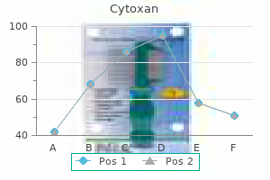
Cytoxan dosages: 50 mg
Cytoxan packs: 30 pills, 60 pills, 90 pills, 120 pills, 180 pills, 270 pills, 360 pills

50 mg cytoxan amex
A systematic review of danger components related to surgical web site infections among surgical patients asthma medications 7 letters cytoxan 50 mg order overnight delivery. Molecular epidemiology of microbial contamination in the working room setting: Is there a threat for infection Offset layered closure reduces deep wound infection in early-onset scoliosis surgical procedure medicine head cytoxan 50 mg order mastercard. Low recurrence price of a two-layered closure restore for main and recurrent midline incisional hernia with out mesh. Capsular contracture: results of 3002 sufferers with aesthetic breast augmentation. Surgical intervention and capsular contracture after breast augmentation: a prospective study of danger factors. The antibiotic prophylaxis guideline for prosthetic joints: making an attempt to do the proper factor. Evaluation of measures to lower intra-operative bacterial contamination in orthopaedic implant surgical procedure. Summary Dual-plane breast augmentation is a versatile technique that allows one to optimize long-term soft-tissue protection within the widest variety of breast sorts whereas minimizing trade-offs to the patient and maximizing advantages. Keywords: twin plane, breast augmentation, surgical technique, pocket plane Key Points � Dual plane is a versatile partial subpectoral implant pocket that has many advantages over the subglandular and subfascial pockets whereas minimizing the trade-offs of traditional subpectoral pockets. If needed, additional dissection between the pectoralis main and the parenchyma is completed to place more of the implant instantly beneath the breast to have a better growth of the parenchyma. Dual-plane breast augmentation is a partial subpectoral pocket plane for breast augmentation that was initially described in 2001. No division of the principle physique of the medial pectoralis main along the sternal border. Varying levels of parenchyma�pectoralis main muscle interface dissection to optimize implant breast tissue interface/dynamics. The term dual-plane implies a subglandular implant inferiorly and a subpectoral implant medially and superiorly. The aim of the dual-plane method is to optimize soft-tissue protection, which we know from the literature is all the time the primary priority in breast implant� primarily based surgical procedure. Risks and Benefits of Dual-Plane Approach � Trade-offs: Possible increased danger of palpable or seen implant edges inferiorly. This is essential as a outcome of inadvertent or intentional division of the medial sternal border of the pectoralis main muscle ends in uncorrectable deformities along the medial breast, together with implant palpability, rippling, traction rippling, nodularity, symmastia, and soft-tissue stretch. Contraindications to the dual-plane method include sufferers with extremely thin decrease pole tissue, which may be recognized with an inferior pole pinch of lower than 5 mm (see Chapter 3: Tissue-Based Planning). In these sufferers, a traditional subpectoral airplane is usually a better different, 70 7. The dual-plane seventy one Dual-Plane Breast Augmentation technique can be contraindicated in body builders for whom a pocket airplane above the pectoralis muscle is preferable. Of note, determination for the degree of parenchymal�pectoralis muscle interface division is the only intraoperative decision the surgeon makes during a routine main breast augmentation. Once the pectoralis muscle edge strikes superior to the higher areolar border, it turns into very difficult to acquire any muscle coverage at all over an implant. The placement of implant utilizing the new inframammary incision is detailed in Chapter 8: the Inframammary Approach and the Dual-Plane Pocket. This is the popular incision for the dual-plane technique; however, dual-plane breast augmentation may be performed by way of the periareolar technique and even the transaxillary method with correct instrumentation. Nevertheless, the data generated on the dual-plane technique have been primarily by way of the inframammary approach. The tenets of the initial pocket dissection are detailed in Chapter eight: the Inframammary Approach and the Dual-Plane Pocket. Briefly, the pocket is dissected under direct vision with precise a four step traumatic method utilizing potential hemostasis to remove bleeding within the pocket. The preliminary a half of the dissection is the division of the inframammary pectoral origin, and that is taken to a degree no further than the medial parasternal border. The main body of the pectoralis along the medial sternal border is left intact fully to maximize coverage over the implant. Using the nondominant hand, the overlying breast parenchyma is palpated bimanually, and the degree of expansion of this parenchyma and vertical laxity is assessed. Of note, the boundaries of dual-plane augmentation are when the overlying pores and skin and parenchymal laxity is bigger than may be expanded with a reasonable size implant. With the use of the nondominant hand, the overlying breast parenchyma is palpated bimanually, and the degree of expansion of this parenchyma and vertical laxity is assessed. Once the surgeon has performed this maneuver, if it is deemed that an increased direct interface within the lower pole of the breast is desired then dissection is commenced between the breast parenchyma within the anterior side of the pectoralis major muscle. This is an incremental division of the attachments to avoid over-release, and the surgeon can continually, utilizing seventy four 7. The goal is to get the specified implant�parenchymal muscle interface with probably the most minimal amount of dissection. Once the dual-plane fine-tuning has been accomplished, the pocket is prepared with betadine triple or non-betadine triple antibiotic irrigation, and the implant is then placed into the pocket, oriented, and checked. This technique may be employed simply in virtually all breast augmentations, and the decision for the exact kind of dual-plane dissection is the one intraoperative determination made throughout a standard main breast augmentation. Using this method, the scientific outcome data have been unsurpassed compared to any other pocket aircraft. Of note, there was additionally some radial scoring accomplished of the breast parenchyma in the lower pole of the breast to additional allow growth of the lower pole. Dual aircraft breast augmentation: optimizing implant-soft-tissue relationships in a variety of breast varieties. Summary this article particulars the state-of-the-art templated method for inframammary breast augmentation. The steps are exact, systematic, carried out atraumatically under direct vision with prospective hemostasis. Keywords: breast augmentation, inframammary method, inframammary fold, twin airplane, surgical approach features, potential hemostasis Key Points � the brand new inframammary fold incision can be easily planned preoperatively lies exactly within the postoperative fold. Virtually every examine that has published outcomes has used this strategy and one of the best outcome knowledge produced have used the inframammary incision. This article particulars the technical pearls for optimizing the surgical features of the inframammary incision. The width of the pocket is marked with a caliper with a medial and lateral dot that corresponds to the width of the selected implant. The height of the pocket is marked once more utilizing a caliper with a dot on the upper pole based mostly on the height of the chosen implant utilizing the new inframammary position for the implant decrease border. The key relationship for incision planning is the breast base width (see Chapter three: Tissue-Based Planning) and nipple-to-fold measurement on maximal stretch.
Buy cytoxan 50 mg cheap
Imaging Studies Imaging of the brain is necessary to consider nearly all of the potential etiologies in this class treatment 0f osteoporosis cytoxan 50 mg buy low price. Differential Diagnosis the differential analysis includes such diverse etiologies as stroke (Chapter 14) treatment arthritis order 50 mg cytoxan fast delivery, demyelinating disease (Chapter 20), traumatic harm (Chapter 17), brain tumor (Chapter 19), and an infection (Chapter 21). Parasagittal lesions lead primarily to leg weak point, extra lateral lesions lead primarily to face and arm weak point, and deep lesions may lead to weakness of all three elements. Cerebral hemisphere lesions could have accompanying cognitive signs, similar to aphasia or neglect. Abnormalities of sensation could be characterized by a rise, decrease, impairment, or loss of feeling. The prognosis of sensory problems requires an understanding of the anatomy and an evaluation of the presentation, location, characteristics, and distribution of signs. Pain and temperature sensation is carried by thinly myelinated (A-) and unmyelinated slowly conducting (C) fibers that synapse as they enter the dorsal horn of the spinal wire. Proprioception, vibration, and light-weight contact run ipsilaterally in closely myelinated (A- and A-) fibers in the dorsal column system, reaching the second-order neuron on the level of the medulla in the nuclei gracilis and cuneatus. At the level of the medial lemniscus, the upper body fibers turn out to be medial and people of the decrease physique lateral. The analysis of different primary sensory modalities, together with temperature, pain (or pinprick), light contact, vibration, and proprioception, is critical to characterize sensory loss and its extent. Thermal modalities are examined utilizing objects with a temperature vary between 10�C and 50�C as a end result of beyond those limits the stimulus becomes painful. Moving the nice toe (or a finger) up and down, by just some millimeters, and asking the patient to indicate the course of motion (with the eyes closed) tests proprioception or joint position sense. Vibration sense requires a tuning fork (128 Hz) to be utilized to the toes and different bony prominences. The subsequent step is to document the sensory signs and findings utilizing accepted definitions. Not only the presence or absence of sensation but also slight variations and gradations ought to be recorded. This leads to loss of pain and temperature sensation within the dermatomes concerned but preservation of posterior column function and, therefore, a traditional response to light touch and normal proprioception. Such dissociated sensory loss occurs frequently with central cord syndromes (see Chapter 22). Patients complaining of sensory dysfunction can report unfavorable signs, optimistic signs, or both. Negative signs embody numbness, loss of chilly or heat sensation, blindness, and deafness. Positive signs embody ache, paresthesias (tingling, pins, and needles), visible sparkles, and tinnitus. For instance, negative signs normally indicate disruption of nerve excitation (such as in a stroke), whereas optimistic signs refer normally to excitation or disinhibition (as seen with seizures or migraine). Once the history is obtained, the subsequent task is to establish the presence or absence of a neurologic lesion or deficit. If a lesion is identified, the extent or location of the lesion and its impact on completely different sensory modalities ought to be mapped out by a careful and detailed sensory examination. This is necessary as a end result of totally different pathologic processes can have an result on completely different sensory signs and result in specific patterns of sensory loss. In basic, compression of a peripheral nerve causes sensory loss in the territory of that particular nerve. Spinal cord illness leads to a characteristic loss of sensation beneath a sure spinal degree (sensory level). With brainstem lesions, the sensory abnormalities could occur on the ipsilateral side of the face and contralateral side of the physique. Central sensory loss involving the thalamus or sensory cortex will usually have an effect on the contralateral face, arm, and leg. Because there are numerous primary neurologic diseases in addition to systemic illnesses that may current with sensory symptoms, placing the sensory examination into the context of the rest of the physical examination could make potential etiologies extra obvious. Sometimes, the sensory issues are accompanied by different signs such as weakness, neglect, visual field cuts, cognitive or behavioral problems, or seizures that may help to determine the location of the lesion. Examples of different patterns of sensory loss and the location of the respective neurologic problem are proven in Table 6-1. This desk serves as a information to the process of localization and diagnosis based on clinical symptoms and the neurologic examination, without the necessity for additional technologic assets. The final step in evaluating these sensory abnormalities is to determine the cause. There are many major neurologic ailments as nicely as systemic diseases that can present with sensory symptoms; many are explored in additional element in Chapter 23 on peripheral neuropathies. The dorsal columns carry well-localized contact, pressure, vibration, and aware proprioception. Damage to a peripheral nerve produces a sensory deficit within the territory innervated by that nerve. Damage to the brachial or lumbar plexus produces sensory loss in a number of nerve territories. Examples of sensory dissociation include syringomyelia (loss of ache and temperature sensation, with preserved proprioception); Brown-Sequard syndrome (loss of proprioception on the aspect ipsilateral to the lesion, and loss of ache and temperature sensation on the contralateral side); and subacute combined degeneration (loss of proprioception, however preserved pain and temperature sensation). As a result, the differential diagnosis for selfreported "dizziness" is very broad. As with all of drugs, obtaining a transparent history of what "dizziness" actually means to the affected person, adopted by a careful neurologic examination, is significant to establish the diagnosis and keep away from unwarranted testing. The following sections detail several types of dizziness, including the important thing options and underlying etiologies. This is mostly a sensation of the room spinning in a clockwise or counterclockwise course. The particular explanation for the vertigo have to be established so as to establish a prognosis. This can occur at any level along the vestibular pathway from the vestibular nerve or labyrinth to the central vestibular constructions. Vestibular problems can subsequently be divided into peripheral and central classes, based on the trigger. Patients could describe a staggering gait or that they have to maintain on to close by objects to keep away from falling. Depending on the etiology, patients may also have signs of diplopia (double vision). The length of signs and associated options assist in narrowing the diagnostic possibilities. It is attributable to an otolith (also referred to as a canalith) of calcium carbonate particles in the semicircular canal. The semicircular canals are within every internal ear and are lined with cilia and endolymph.
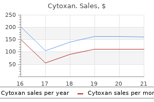
Buy cheap cytoxan 50 mg online
Intracranial hypertension caused by a depressed skull fracture resulting in superior sagittal sinus thrombosis in a pediatric patient: treatment with ventriculoperitoneal shunt insertion medications side effects cytoxan 50 mg purchase free shipping. Variations of the superior sagittal sinus and bridging veins in human dissections and computed tomography venography symptoms 11dpo buy cytoxan 50 mg with mastercard. The chordae Willisii in the superior sagittal sinus: morphology and classification. The bovine cerebral venous system: comparative anatomy, visualization, and implications for translational research. Transarterial intravenous coil embolization of dural arteriovenous fistula involving the superior sagittal sinus. The anatomical relationship between the superior sagittal sinus and the sagittal suture with surgical considerations. Radical resection of superior sagittal sinus meningioma with venous interposition graft and reimplantation of the rolandic veins. Then, a capillary plexus varieties from the aortic arch and creates a vascular community that spreads upward to invest the fore and midbrains. In the early section of formation of the superficial dural venous plexus, channels at the ventral midline coalesce to form larger venous lakes. Tributaries that drain into the primary head vein be a part of it primarily along the dorsal margin. Rostrally, this community dips between the growing cerebral hemispheres, and the superior and inferior sagittal sinuses originate in this mesh. In addition, it could form a gradual hook, with large veins draining the medial hemisphere extra anteriorly than traditional. It may help localize the anterior midline of the mind through the venous part of a cerebral angiogram, in addition to the place of the anterior portion of the callosal cistern and inferior margin of the falx cerebri. The presence of part shift�induced move artifacts propagated for the vein with a shiny luminal signal confirms the presence of no less than some circulate. Hemodynamic and parenchymal effects of dural sinus thrombosis depend on many factors, similar to website extent, rapidity of occlusion, and anatomy of collateral pathways. Lumbar puncture was important for elevated intracranial pressure and xanthochromia. There was a plicalike fold over the superior half of the opening on the junction with the anterior-inferior side of the cavity in the falx cerebri. Isolated inferior sagittal sinus thrombosis attributable to a uncommon combination of elevated lipoprotein (a) and iron deficiency anemia. The transverse sinus runs throughout the cavum epiptericum between the 2 layers of the tentorium cerebelli, and as a result of the cavum epiptericum reduces in size, it comes to lie within the hooked up margin of the tentorium cerebelli. This is thought to be as a result of the natural continuation of the sagittal sinus to the proper aspect. They noticed that, in circumstances by which the distal 1�3 cm of the superior sagittal sinus was doubled, the smaller channel was the origin of the smaller transverse sinus. In transverse sinus aplasias general, both the superior sagittal and straight sinuses often drain into the contralateral transverse sinus, and in such instances, the superior petrosal sinus can continue instantly into the sigmoid sinus. Rarely, the transverse sinus is severely hypoplastic and the contralateral transverse sinus is absent. In such cases, the superior sagittal and straight sinuses can each drain by way of an enlarged occipital sinus into the superior jugular bulbs. In the latter case, the superior sagittal sinus continued as a large occipital sinus that joined the marginal sinus and then the distal transverse sinuses at the jugular foramina. Hoople31 found that the jugular foramen usually was contracted and the sigmoid sinus was lowered or absent when this occurred. Knott22 described two situations of complete absence of the best transverse sinus with only a small venous channel terminating via the mastoid foramen. A case was reported during which the sigmoid sinus ended in a blind pouch and drained via the large mastoid foramen. Furstenberg34 described a variation within the "lateral sinus" by which it was positioned in the delicate tissues completely outdoors the cranium. Overlapping lambdoidal sutures in infants could result in septation of the transverse sinus, normally at the proximal end. Occasionally, the affected facet is associated with the absence of the transverse sinus, sigmoid sinus, or both. To forestall intracranial hemorrhage, a mix of arterial and venous surgery, forty four Anatomy, Imaging and Surgery of the Intracranial Dural Venous Sinuses be accessed, the transverse or sigmoid sinus typically is ligated. However, some have reported instant or delayed complications (24�48 h later) after sinus ligation and resection. Venograms have been reviewed to determine relative tumor places and the drainage patterns of the confluence of sinuses. Stimulation of the superior surface of the transverse sinus resulted in orbital and supraorbital ache. Considering that the width of the proximal transverse sinus is 6 mm on average, the first and most superior burr hole within the midline infratentorial supracerebellar method may be positioned safely roughly 1 cm below the inferior nuchal line. Longitudinal changes in the ductus venosus, cerebral transverse sinus and cardiotocogram in fetal progress restriction. Color Doppler study of the venous circulation in the fetal brain and hemodynamic study of the cerebral transverse sinus. Cerebral dural arteriovenous fistulas: medical and angiographic correlation with a revised classification of venous drainage. Early rebleeding from intracranial dural arteriovenous fistulas: report of 20 cases and review of the literature. Intraarterial and intravenous remedy of transverse/sigmoid sinus dural arteriovenous fistulas. Comparison of the infratentorial and transtentorial approaches to the pineal region. Quantitative anatomy of the occiput and the biomechanics of occipital screw fixation. Inconstant veins that drain into the sigmoid sinus include veins from the pons and medulla oblongata. The sigmoid sinus has anastomoses with the mastoid emissary and condylar emissary veins routinely. This most likely is attributable to the difference in cranial size base between the sexes. When evaluating either side, regardless of cranial width, the sigmoid sinus on the proper facet protrudes deeper, masking a greater cross-sectional space, and is situated more laterally. The asterion is the convergence level of the lambdoid, occipitomastoid, and parietomastoid sutures and is used because it overlays the region of the transverse-sigmoid junction. Of notice, when studying the older literature, some writers use the time period lateral sinus to describe the combined sigmoid and transverse sinuses. At the very starting of the fetal stage, when the embryo continues to be 40 mm, changes happen within the configuration of the sinuses that establish lots of the morphologic features which are present within the grownup brain.
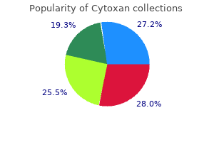
Buy cytoxan 50 mg overnight delivery
This molecule is converted into a 4carbon molecule known as succinylCoA 94 medications that can cause glaucoma discount 50 mg cytoxan overnight delivery, which gets oxidized in the citric acid cycle medications with aspirin cytoxan 50 mg cheap with mastercard. The enzyme that creates succinylCoA needs vitamin B12 as a coenzyme to help with catalysis. Based on its role in energy manufacturing, vitamin B12 is commonly added to power drinks targeted at young folks. When muscle cells are working heavily, they run low on oxygen, and when this occurs, they rely on fermentation, which requires rather more glucose and glycolysis than when oxygen is abundant. Describe how its manufacturing will affect oxygen release based on the Bohr impact, which is that protons and carbon dioxide have an result on hemoglobin on binding by favoring the release of oxygen. People with deficiencies within the enzymes essential to convert galactose to glucose can readily form cataracts as a end result of a galactoserelated crystal that may kind in the eyes. Describe a dietary modification that might be recommended to someone with this deficiency. Even more than glycolysis, the citric acid cycle is a central metabolic pathway the place everything comes collectively. And this one pathway is utilized by each cell, from the cells in bacteria to amoebas to you. This is the place the citric acid cycle is obtainable in, offering a second stage for the environment friendly manufacturing of vitality from food. Like glycolysis, the citric acid cycle extracts energy and supplies intermediates for different pathways. And molecules from different pathways can enter it at multiple points and leave as needed. Most commonly, the glycolysis pathway that oxidized sugars to yield pyruvate has been traveled, or acetylCoA has been chewed off in several successive oxidations of fatty acids. But pyruvate made in glycolysis needs an additional step to get transformed to acetylCoA, and then it, too, can enter. To start the process of coming into the pathway, pyruvate must transfer from the cytosol (the cytoplasm), where glycolysis produced it, to an innermost part of mitochondria referred to as the (mitochondrial) matrix. To get converted to acetylCoA, pyruvate is first transported into mitochondria, the place an enzyme complex known as pyruvate dehydrogenase acts on it. Remember, the conversion of pyruvate to acetylCoA is important for the entry of pyruvate into the cycle. Lecture thirteen Metabolism Meets on the Citric Acid Cycle 133 the citric acid cycle has traditionally also been called the Krebs cycle, named for its discoverer, Sir Hans Krebs, who won the Nobel Prize in Physiology or Medicine in 1953 for his work. The enzyme complex makes use of 5 different cofactor molecules that act as helpers for the reactions. The primary events that the enzymes and their cofactors bring about are as follows: w A molecule of carbon dioxide is launched from pyruvate. If you switch cash in the financial institution from one account to one other, one account gets debited, and the other will get credited. Almost all cellular oxidations occur this way: Molecules lose electrons in oxidations, and electron carriers receive the electrons in reductions. As a results of all of this, the 3carbon molecule of pyruvate is made into the 2carbon acetyl group in acetylCoA, which can then enter the citric acid cycle. In addition to sources from sugar oxidation and fatty acid oxidation, acetylCoA can be produced by breaking down amino acids and ketone bodies, which might join the citric acid cycle, just like sugars and fatty acids. And that is why these molecules can function energy sources when glucose supplies are low. Though the pathway is a circle, the reactions are numbered ranging from the entry level for acetylCoA. This reaction to create citrate is very favorable as a result of breaking the bond between the acetyl group and coenzyme A releases plenty of power. This seems to Without the citric acid be important when the circle is accomplished to make oxaloacetate. The lack of a carbon as carbon dioxide converts the 6carbon isocitrate right into a 5carbon molecule, alphaketoglutarate. The product of the reaction, a 4carbon compound, hyperlinks to a CoA, forming succinylCoA. The enzyme 136 to harvest enough Biochemistry and Molecular Biology right here, alphaketoglutarate dehydrogenase, is intently associated to pyruvate dehydrogenase; it uses the same 5 coenzymes and has an analogous reaction mechanism. The removing of the CoA from succinylCoA is also a very energetically favorable reaction, just like when the CoA was launched from acetylCoA in the first reaction. The reaction, catalyzed by malate dehydrogenase, is notable for going backward under standard circumstances. The product gets removed, and the reaction will get pulled forward by the citrate synthase response, which follows in the next round of step 1. This reaction is energetically favored and removes the product, oxaloacetate, as it reacts Not solely is citrate a supply with acetylCoA to type citrate. With of the raw materials needed oxaloacetate being rapidly eliminated to make citrate, the manufacturing of for making fatty acids, however oxaloacetate can proceed. Connections to Other Metabolic Pathways the citric acid cycle hyperlinks to many pathways, feeding intermediates into some and being fed by others. In its position as a hub for so much of pathways, portions of the cycle can operate independently, and a few molecules can enter and exit the cycle without going all the way round. When extra citrate is made than is needed for quick power manufacturing, the citric acid cycle gets overloaded. When this happens, citrate accumulates and gets transported into the cytoplasm, where the citrate gets split back into oxaloacetate and acetylCoA. In the cytoplasm, the acetylCoA is used to construct fatty acids, whereas oxaloacetate gets recycled back into mitochondria. When fatty acids are synthesized, they get bundled together as triacylglycerols, or fats, to be stored. Lecture thirteen Metabolism Meets at the Citric Acid Cycle 139 w the cycle intermediate alphaketoglutarate has several roles in addition to being an intermediate within the citric acid cycle. It can seize excess nitrogen and easily be made into the 5carbon amino acid glutamic acid. This is solely one of a minimum of 10 situations the place an intermediate of the citric acid cycle is linked to amino acid synthesis. Glutamic acid and glutamine can be transformed to alphaketoglutarate; thus, glutamic acid and glutamine enter the citric acid cycle through alphaketoglutarate. Fumarate and oxaloacetate even have quite a few connections to different pathway molecules. It also can easily mop up an excess amino group to become the amino acid aspartate, or it could possibly mop up an extra 2 amino groups to turn into asparagine. In the reverse reactions, the breakdown of those amino acids can give rise to oxaloacetate, offering entry points for two more amino acids. However, none are linked to so many pathways as the citric acid cycle is, with its direct connections to amino acid metabolism, the urea cycle, sugar metabolism, fatty acid metabolism, nucleotide metabolism, and the synthesis of heme. So, reactions run in reverse, beginning with alpha-ketoglutarate and resulting within the launch of acetylCoA into the cytoplasm, the place it can make fatty acids and ldl cholesterol.
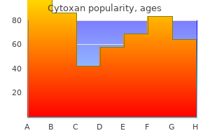
Generic cytoxan 50 mg visa
Cellular communication is determined by particular molecular interactions medicine x 2016 purchase 50 mg cytoxan mastercard, where the message and the receiver are biomolecules symptoms meningitis cytoxan 50 mg discount otc. Binding of a message by the mobile receptor molecule triggers information transmission inward to the cell and changes its actions. It has long been identified that even bacteria sense and transfer toward meals sources and away from toxic substances. Even extra fascinating, bacteria talk to one another, type of like youngsters at a mall, to find out how many "associates" are close by. Plants sense and talk in response to mild, gravity, temperature, and water. Such plants share assets with family members by limiting the quantity of soil vitamins they take up. If they sense a stranger, though, they compete by placing out extra roots to take up extra nutrients. Not surprisingly, the most complex communication techniques are present in multicellular animals. Many of the signaling molecules come from the endocrine system and are generally known as hormones. Hormones may be divided into 2 broad groups: 1 molecules that act within minutes by binding to receptors on the surface of cell membranes, and a pair of steroid hormones that act extra slowly and bind to receptors inside cells. In animals, hormones originate in a couple of specialized organs that make up the endocrine system. Major endocrine glands embrace the hypothalamus, pineal gland, pituitary gland, thyroid gland, parathyroid, thymus, adrenals, pancreas, and ovaries/testes. Lecture 21 Hormones, Stress, and Cell Division 217 How alerts get to their goal cells is diversified. A sign molecule that acts only on the outer floor of the identical cell the place a signal was created is called autocrine. An intracellular sign molecule that stays and signals inside the membrane of the identical cell known as intracrine. There are additionally exocrine glands that launch outside the body, corresponding to sweat glands that send alerts to talk with different organisms. But for steroid hormones, and also thyroid hormones, the receptor is located inside the cell, so such indicators should cross the membrane and enter the cell to meet up with their receptors. For intracellular receptors concerned in steroid signaling, a typical outcome is that cells adjust the quantity and/or timing of the synthesis of specific proteins. For signaling occurring via membrane receptors, the receptor protein conveys the message through a series of other mobile molecules into the inside of the cell. The Stress-Response System In some types of membrane receptor signaling, enzyme actions are altered. Affected enzymes embody protein kinases, which phosphorylate goal proteins, and phosphatases, which take away phosphates. Adding and eradicating phosphates is an easy way to control whether or not mobile proteins are energetic or not. A good instance of such signaling is the fightorflight response that advanced to prepare an organism in a dangerous scenario for combating with or fleeing from predators or opponents. That similar stressresponse system kicks in when we get minimize off in visitors by an aggressive driver, interview for a job, or even make a presentation earlier than the boss. It begins in a piece of the brain called the amygdala, which is involved in reminiscence, choice making, and feelings similar to concern, aggression, and anxiousness. When the brain perceives danger, it sends messages to the adrenal glands above the kidneys, stimulating them to secrete hormones: epinephrine (also often known as adrenaline), norepinephrine, and cortisol. Lecture 21 Hormones, Stress, and Cell Division 219 Adrenaline is familiar to most individuals and is usually credited with giving folks extraordinary power or endurance beneath stress. At the molecular degree, there are quite a few steps within the pathway from adrenaline binding at the cell surface to the ultimate mobile modifications, however there are three essential themes: 1 the sign arriving at a cell have to be certain by a receptor for any action to observe. As a results of adrenaline signaling, a collection of reactions is about in motion, culminating in offering loads of glucose to the physique, each by breakdown of glycogen and new synthesis of glucose within the liver. The enzymes involved are activated by the addition of a phosphate, so the off switch includes the removing of the phosphate that was added. In addition to the shortterm stress response for energy manufacturing by adrenaline, a longerterm response to stress comes with the production of cortisol, a steroid hormone that may be a member of the class known as glucocorticoids. Normally, as soon as adrenaline has carried out its factor, the system returns to the "no hazard" state. But if the menace is perceived to be ongoing, cortisol keeps the stress pedal depressed. When a steroid binds, the steroid hormone receptor acts to have an effect on the synthesis of particular proteins. This route of communication is comparatively sluggish and should require hours to totally activate. Among the proteins whose manufacturing is elevated by cortisol are the enzymes used in gluconeogenesis. To get hold of even more glucose, cortisol stimulates fat and muscle breakdown to present fatty acids and amino acids that might be funneled into glucose synthesis. In addition, cortisol can suppress activity of the immune system in an attempt to divert all resources to coping with quick threats. These actions of cortisol are helpful in continuing to provide power to an organism that should get its mind and body geared for action in the face of continued danger. The draw back is that low ranges of continual stress, similar to worrying about being able to pay your payments, additionally set off the cortisol response. The longterm use of glucocorticoids, although, does have potential adverse results, corresponding to loss of bone density, weight gain, cataracts, and glaucoma. Research into drugs that might ship the benefits without the unwanted aspect effects is ongoing. Norepinephrine can be produced at low levels usually and acts equally to epinephrine. Cell Division There is one other class of signaling by which external alerts can induce changes in cells that tell them to divide. This pathway also relies on receptor binding, relaying of the message via varied gobetweens, and at last activating kinases that bring about changes within the cell. All of the receptors are transmembrane proteins which might be anchored in the cell membrane. One end of the receptor peptide sticks out of the cell, while the other end extends into the cytoplasm. Predict the physiological results it might have based on the data in this lecture.
Myroxylan toluiferum (Tolu Balsam). Cytoxan.
- How does Tolu Balsam work?
- What is Tolu Balsam?
- Dosing considerations for Tolu Balsam.
- Are there safety concerns?
- Bedsores, bronchitis, cancer, cough, cracked nipples, lips, reducing lung swelling (inflammation), and minor skin cuts.
Source: http://www.rxlist.com/script/main/art.asp?articlekey=96373
50 mg cytoxan cheap amex
And repeated communication between neurons ends in physical in addition to biochemical changes within the brain symptoms night sweats cheap cytoxan 50 mg without prescription. Lecture 22 Neurotransmitters symptoms 6 week pregnancy buy 50 mg cytoxan fast delivery, the Brain, and Addiction 233 the 2013 update to the Diagnostic and Statistical Manual of Mental Disorders added gambling dysfunction to the identical class as substance abuse. At the same time, even though the drug might not provide the pleasure it as soon as did, drug craving then begins in the rewired mind, leaving the user unsuccessfully trying to obtain the original excessive. Meanwhile, the conventional, smaller elevations of dopamine from everyday feelgood activities not satisfy. This makes addicts lose curiosity in issues that after mattered to them, whereas drug cravings drive them to give attention to obtaining the drug. A variation on this theme results in drug dependence on opioid drugs and prescription painkillers. The most necessary group of biochemical signals that we notice is the molecular reactions that give rise to our 5 senses-the signals that give us virtually all of our conscious experiences. The savory taste is usually called umami, derived from a Japanese word that means "delicious. Much of the sensory data we get about food comes not simply from our taste buds, but additionally from our noses. The capacity to distinguish smells is rather more finely tuned than our capability to style. In contact with the sensory cells are the dendrites of the sensory nerves that carry taste info to the brain. For detecting salt, sensory cells with a sodium channel depolarize when sodium enters them. The positively charged protons trigger membrane depolarization once they enter the cell and set off an motion potential. Smell the sense of smell, known as olfaction, is enormously necessary to animals- not only for having fun with meals, but additionally for warning of danger and even selecting mates. It occurs because of the binding of odorant molecules to olfactory receptor cells in the nasal cavity. A second one is situated the place the roof of the mouth connects the throat to the nostril. Chewing meals releases odorants that can travel the second path to the olfactory receptors. Mucus-a viscous fluid full of glycoproteins called mucins-covers the lining of the nostril, the nasal epithelium, and serves as both a solvent for odorant molecules and an essential safety towards infection, because nasal neurons connect on to the mind. But every receptor can bind several different odorants-some weakly and others extra tightly. A explicit odorant might bind a couple of kind of receptor, once more with totally different affinities. These combos give humans the ability to distinguish many greater than 350 smells. In fact, a 2014 study put the number of smells people can tell aside at one trillion. Lecture 23 the Biochemistry of Our Senses 237 Mammals have about a thousand to 1500 olfactory receptor genes. In humans, solely about 350 of them make energetic receptors, but rats have almost 1500 genes that make energetic olfactory receptors. The sense of odor might play a role in the alternative of sexual companions, even in people. Children of such couples might have a more sturdy immune system, which might give them an evolutionary benefit. Vision the detection of light and the power to distinguish element by our eyes includes a tremendous convergence of optics, lightsensitive proteins, nerve signaling, and brain processing. About a hundred thirty million photoreceptors within the retina absorb light and transmit visible indicators to the brain. The retina is the placement of photoreceptor cells, which include proteins referred to as opsins that detect mild. This is just like the style receptors for sweet, bitter, and umami; the distinctive function of opsins is that they maintain a molecule of vitamin A that gets altered when exposed to gentle. Biochemistry and Molecular Biology Thanks to opsins, photoreceptor cells detect mild and move signals on to other neurons, called ganglion cells. There are 2 forms of retinal photoreceptor cells involved in light detection: rods and cones. But what rod cells lose in color detection, they acquire in sensitivity; a rod cell can detect a single photon of light. Humans have 3 forms of cone cells, each specialised to absorb wavelengths similar to purple, green, or blue. Though injury to any of the units of cones is possible, the most typical kind of colour blindness is redgreen colour blindness, and it outcomes from the shortage of both a purple or a green opsin. Lecture 23 the Biochemistry of Our Senses 239 Genes encoding the opsins are on the X chromosome. Human males have just one X chromosome, whereas females have 2, so the loss of coding for an opsin on its single X is a bigger deal for men than for women. People with regular listening to can detect sounds of pitches, or frequencies, between 20 and 20,000 hertz. The length of time it takes our nervous system to reply to a sound sign is on the order of tens of microseconds. Sound detection happens when tiny hair cells referred to as stereocilia transfer on the membrane of an ear structure called the cochlea. Each of the stereocilia is attached to an adjoining one by a filament that controls the opening of an ion channel. And when this happens, the upanddown motion of the basilar membrane sets up sidetoside motion of the fluid between the basilar membrane and the tectorial membrane. The sensitivity of the mechanism is astounding: Movements as small as 1/2 the diameter of an atom may be detected. Hearing loss is complex, however one factor that commonly occurs is harm to the hair cells of the inner ear-whether because of age, an infection, or exposure to loud noises. Cells responding to excessive frequencies are located in the lower cochlea and are among the most simply broken, thus explaining why highfrequency hearing loss is A 2016 report shows that men usually the first to happen. Touch stimuli may be mechanical signals, the place lowthreshold mechanoreceptors sense contact with the pores and skin. Lowthreshold mechanoreceptors detect strain, vibration, stretching of the skin, and movement of hair follicles in the skin. These receptors reply by triggering action potentials and communicate with sensory neurons to send data to the mind. For example, the "taste" of a meal is actually made by combining data from scent receptors, taste buds, and even texture or touch. Pain receptors that detect dangerous stimuli and signal the spinal cord and mind are referred to as nociceptors, related to the word noxious. Nociceptors for temperature are activated by dangerous heat or chilly circumstances with separate sensing cells for every.
Syndromes
- The time it was swallowed
- Double vision or difficulty with side (peripheral) vision
- Widespread bone pain, especially in the hips
- Inhalers to relieve nasal congestion
- Bilirubin in the blood
- Stool softeners help reduce straining and constipation
- Free T4 test
Cheap cytoxan 50 mg online
Immediate imaging is warranted if infection or malignancy is a concern medications bipolar generic cytoxan 50 mg on line, if ache is accompanied by saddle anesthesia and urinary retention treatment glaucoma cytoxan 50 mg cheap without prescription, or if symptoms are acute in onset and related to progressive deficits. Structural lesions, together with disk herniation and degenerative modifications, are the most common causes of radiculopathy. Radiculopathy is often associated with pain or sensory symptoms in the distribution of a given nerve root. Corresponding energy, sensory, and reflex abnormalities on exam can help medical hypotheses. All of the most important nerves in the legs arise from the lumbosacral plexus, a mix of the L1-S3 nerve roots. The lumbosacral plexus lies behind the psoas muscle tissue in the retroperitoneum before persevering with under the pelvic outlet. Specifically, motorbike and other motorized vehicle accidents can lead to injury from abrupt traction when the head is pulled away from the shoulder. Inflammatory brachial plexopathies, also referred to as brachial neuritis or ParsonageTurner syndrome, are also usually acute in onset. Triggers can embrace antecedent sickness, vaccination, surgical procedures, or train; some triggers are tough to determine. An apical lung (Pancoast) tumor can lead to a more insidious growth of a brachial plexopathy from local infiltration or direct compression. Similarly, radiation remedy could also be associated with a slowly creating brachial plexopathy, even a yr following the conclusion of remedy to the shoulder or chest region. In neonates, brachial plexopathies could result from traction accidents at delivery, particularly within the setting of shoulder dystocia. Type 2 diabetes, within the type of diabetic amyotrophy, is the commonest cause of lumbosacral plexopathy. As with brachial plexopathies, neoplasias can be associated with lumbosacral plexopathies. It could be useful to take into consideration brachial plexus lesions in two major etiologic classes: (1) structural. In a basic Parsonage-Turner syndrome, sufferers awaken from sleep in the early hours of the morning with ache in the periscapular and shoulder girdle regions. Typically, ache is severe and can last as lengthy as four weeks earlier than abating spontaneously. The pain is ultimately associated with weakness (sometimes leading to scapular winging) and muscle atrophy. Typically, patients with lumbosacral plexopathies current with leg pain, sensory change, atrophy, and uneven weak spot. Weakness of knee extension, adduction hip flexion, or combinations of those, points to a lumbar plexus drawback. In sure situations, imaging is essential to consider for structural compression of the plexus. An evaluation of serology for infectious, autoinflammatory, and metabolic disturbances and, less commonly, cerebrospinal fluid evaluation, could assist decide a trigger and guide administration. Ultimately, plexopathies as a result of irritation often get well spontaneously over months, whereas compressive lesions might require intervention. Trauma is the most typical explanation for brachial plexus lesions, whereas sort 2 diabetes is often associated with a type of lumbosacral plexopathy. Plexus lesions current with ache and patchy sensorimotor deficits; the pattern of abnormalities can help localize the components of the plexus affected. In basic polyneuropathies, the most distal elements of the nerves are initially and preferentially affected. A mononeuropathy is a dysfunction of an individual nerve, as can occur in the setting of focal compression, such as in the carpal tunnel syndrome. In the rare case that separate individual nerves are affected concurrently or sequentially, the condition is characterised as a mononeuropathy multiplex. Peripheral nerve deficits could be categorised based on whether or not they affect motor, sensory, autonomic, or combos of fibers. Further, neuropathies might preferentially affect large-diameter fibers answerable for proprioception, or small-diameter fibers that relay pain and temperature data. The pattern of nerve involvement can give hints to underlying pathophysiology (Box 23-1). Infectious, inflammatory, poisonous, and metabolic circumstances might play roles in acquired conditions; diabetes is a very common reason for neuropathies (Table 23-2). Accordingly, key information gleaned from the history, exam, and investigative studies assist characterize the sort of peripheral nerve lesion. For instance, tingling and "pins and needles" sensations, together with reduced joint place sense and reflexes recommend large fiber dysfunction. Burning, capturing, and jabbing pain, combined with deficits in ache and temperature sensation, often point out a small fiber neuropathy. Diabetic Neuropathies Clinical Features Mixed sensory�autonomic�motor polyneuropathy; variants embrace small fiber (painful, normally spontaneous burning pain), massive fiber (ataxic), and autonomic Diabetic proximal motor neuropathy (diabetic Severe thigh ache, adopted within weeks by mild-toamyotrophy) extreme hip and thigh muscle weak point with muscle atrophy; often impacts older type 2 diabetic patients Acute axonal diabetic polyneuropathy (intensely painful Diabetic neuropathic cachexia usually associated with acute or subacute progressive symmetric sensory axonal weight reduction; "insulin neuritis" (with improved management of peripheral neuropathy) hyperglycemia) Diabetic mononeuropathy, radiculopathy, and Can current with cranial neuropathy (third, fourth, or polyradiculopathy sixth nerves), multisegmental truncal radiculopathy, or limb mononeuropathy Focal compression neuropathies related to diabetes Diabetic sufferers are more susceptible to compression neuropathies, similar to in the median nerve at the wrist, the ulnar nerve at the elbow, and the peroneal nerve at the knee Type Chronic progressive distal symmetric diabetic polyneuropathy On exam, distal symmetric sensory or sensorimotor deficits are the most common presentation of a polyneuropathy. If the presenting symptoms and signs follow an atypical sample, that may narrow the differential, as shown in Table 23-3. More common mononeuropathies, similar to carpal tunnel syndrome, ulnar neuropathy at the elbow, radial neuropathy on the spiral groove ("Saturday evening palsy"), lateral femoral cutaneous neuropathy ("meralgia paresthetica"), peroneal neuropathy on the fibular neck, and facial neuropathy. Common Mononeuropathies Nerve Upper extremity Median nerve: on the wrist is known as carpal tunnel syndrome Clinical Features Numbness or tingling involving a number of of the first 4 digits. Weakness consists of triceps, brachioradialis, supinator, and wrist and finger extensors. The triceps is affected by axillary compression however spared by spiral groove compression. Compression of the radial nerve on the stage of the axilla ("Saturday evening palsy"); within the spiral groove, or within the forearm (posterior interosseous neuropathy). Lower extremity Meralgia Burning sensation and variable paresthetica lack of sensation over the anterolateral thigh. Femoral neuropathy Peroneal neuropathy Area of sensory change over the lateral side of the thigh. Usually trauma from surgery, stretch harm (prolonged lithotomy place in childbirth), diabetes mellitus, and different inflammatory processes. Entrapment of the peroneal nerve between the neck of the fibula and the insertion of the peroneus longus muscle. Depending on the clinical context, extra research designed to assess for toxic�metabolic abnormalities. Therefore, if a affected person is assumed to have a small fiber neuropathy, a skin biopsy may be essential. In the setting of small fiber neuropathy and important autonomic dysfunction, a fat pad biopsy or genetic testing could also be warranted to search for amyloidosis.

Purchase 50 mg cytoxan fast delivery
Sometimes treatment juvenile rheumatoid arthritis cytoxan 50 mg buy cheap, the occipital sinus actually features as the main drainage of the venous sinuses treatment water on the knee cytoxan 50 mg generic with mastercard. The occipital sinus also features as the principle drainage when the lateral sinus is rudimentary. Three of these cases drained into the sigmoid sinus, whereas seven others drained into the marginal sinus. Another indirect occipital sinus also was reported that terminated within the straight sinus, the place it functioned as the primary drainage in the case of a rudimentary transverse sinus. Interestingly, one reported variant of the superior sagittal sinus is direct drainage into the jugular bulb. In kind 2, more than two occipital sinuses open to the confluence, while the transverse and sagittal sinuses exhibit the same sample as in type 1. However, within the majority of individuals, the occipital sinus exhibits a midline path,four which permits the world that should be averted to protect the occipital sinus to be decided extra simply. Surgeries that pose a high risk of sacrificing the occipital sinus primarily are posterior cranial fossa surgical procedures. Yaargil35 described multiple surgical procedures carried out with the median suboccipital method that pose a excessive danger of injuring the occipital sinus. A more careful evaluation ought to be carried out in cases with an indirect occipital sinus. The growth of alterations in the vascular system of the mind of the human embryo. Cranial venous system in man in reference to improvement of grownup configuration and relation to arteries. The cross section areas of the vessels that kind the torcular and the way in which flow is distributed to the best and to the left lateral sinus. Blood drains from the capillary network by way of this major head vein, which serves as the place to begin of venous drainage from the mind tube. The anastomosing venous loops stemming from the capillaries drain within the form of three plexuses, the anterior, middle, and the posterior dural. As the embryo matures, the anterior plexus loses much of its plexiform architecture. This marks the delivery of the torcular Herophili, the last hint of the anterior and center dural plexuses. These meshes have been thought to be the relics of the venous plexuses from which the torcular Herophili originated. It is lined by endothelial cells and lies between the tough, fibrous meningeal and periosteal layers of the dura. After Testut; (B) Variations of the torcular Herophili distilled from various sources by Lang J. In 41% (66 of 160) of the instances, the proper dominant kind was observed most incessantly. The bifurcated type (seen in 14% or 22 cases) and the confluence kind (seen in 35% or fifty six specimens) made up the rest of the observations. This variant corresponds with the historic definition of the torcular Herophili. On the other hand, the StS drains into the left transverse sinus thrice as typically as the right. A uncommon variation on the torcular Herophili was noticed in considered one of his 12 specimens, by which an occipital emissary vein was noticed to join it. A skinny wall (laminar chorda) was found between these two channels, the flexibility of which suggests that distension of the bigger channel might occlude the smaller channel effectively. Further, distal obstruction of the outflow on the facet ipsilateral to the larger channel could result in signs and signs reflective of a bilateral occlusion, as a end result of the smaller outlet may become compressed ultimately, which may have some bearing on procedures or accidents in which one of the jugular veins is sacrificed. This doubtless is the embryological remnant of the venous plexus from which the sinuses developed. Obstruction of circulate at the stage of the jugular vein on the facet communicating freely is predicted to result in severe stasis in the superior cerebral veins. In some instances, the twins could share leptomeningeal and central nervous system tissue. This section evaluations the imaging methods used to get hold of info on the macroscopic construction of the torcular Herophili, and by extension, the pathology in this area. Also not the so-called accent torcular on the junction between the falcine sinus and superior sagittal sinus. Conventional Angiography Conventional angiography has lengthy served as the gold commonplace to visualize vascular constructions. Because venous sinuses drain from totally different sources, filling defects may be seen as distinction drains into some areas of the sinuses earlier than others. Aplasia and thrombosis of venous constructions also are just about unimaginable to distinguish from one another. The choice of machine settings and interpretation of results, nevertheless, depend highly on trained personnel. It appeared anechoic and was situated inferior to the occipital lobes, posterior to the cerebellum, and simply contained in the cranial vault. One additionally must be conscious of false-positive filling defects secondary to arachnoid granulations or cavernous nodules, luminal septa, or fenestrations. Both of these methods can doubtlessly yield 3-D photographs, which may be rotated for scrutiny in various views. This entails inserting a catheter into the entry of the systemic arterial system, advancing it into the region of the torcular Herophili, and releasing the embolization material into the arterial lumen. Once this is achieved, the sinus is packed with bigger fibered occlusion coils. This reality stems from the function the torcular Herophili performs as a typical point within the drainage of dural venous sinuses. This has the potential to progress quickly to clinically evident intracranial hypertension. Dural arteriovenous malformations positioned at the torcular Herophili actually have a poorer prognosis. Endoscopic and microscopic anatomy of the superior sagittal sinus and torcular Herophili. Other Less Common Pathologies Hemangiopericytomas are tumors which are composed of rounded or spindle-shaped cells proliferating round endothelial-lined capillaries which might be derived from the pericytes of Zimmerman. Thrombosis of an ectatic torcular Herophili: anatomic localization using fetal neurosonography. Prenatal thrombosis of torcular Herophili with spontaneous resolution and regular consequence. Combining endovascular and neurosurgical therapies of high-risk dural arteriovenous fistulas in the lateral sinus and the confluence of the sinuses. A unique type of dural arteriovenous fistula at confluence of sinuses handled with endovascular embolization: a case report. A case of dural arteriovenous fistula draining to the diploic vein presenting with intracerebral hemorrhage. Variations of the cerebral dural sinuses at the torcular Herophili: importance in radical neck dissection.
Cytoxan 50 mg online
Individual ailments in each category are discussed within the later chapters covering particular neurologic issues symptoms 0f parkinson disease 50 mg cytoxan safe. Associated Signs and Symptoms Associated signs and signs might occasionally include muscle ache if the muscle disorder is inflammatory symptoms zoloft withdrawal order 50 mg cytoxan mastercard, corresponding to polymyositis. Reflexes are characteristically preserved until the process is so extreme that the muscle tissue are almost paralyzed. Differential Diagnosis Primary muscle issues, mentioned in Chapter 24, include each acquired problems (myopathies), which might end result from inflammatory or toxic etiologies amongst other causes, and congenital issues (muscular dystrophies). Depending on the particular disease, power could additionally be worse after utilizing the muscle tissue or towards the tip of the day; it could improve after resting or in the morning (fatigability). Alternatively, strength may improve paradoxically after train in different circumstances. Some of the illnesses in this class have specific serum markers, similar to anti-acetylcholine receptor antibodies and muscle particular kinase in myasthenia gravis. A lesion involving a selected peripheral nerve will result in weakness in the muscle tissue innervated by that nerve whereas sparing different, typically neighboring muscle tissue. Certain systemic circumstances can result in dysfunction of a quantity of peripheral nerves in succession, a disorder generally identified as mononeuropathy multiplex. Finally, when peripheral nerves are all affected diffusely, in a polyneuropathy, dysfunction typically happens within the longest nerves first. Thus, weakness from a polyneuropathy often seems first in the distal muscle tissue, symmetrically. Associated Signs and Symptoms Mononeuropathies might cause sensory symptoms-such as numbness, tingling, or pain- within the distribution of the relevant peripheral nerve. Polyneuropathies, relying on etiology, normally have related sensory loss and depressed or absent reflexes, significantly in the distal extremities. Differential Diagnosis Mononeuropathies mostly happen as a outcome of entrapment (as in carpal tunnel syndrome). Mononeuropathy multiplex is related to systemic vasculitis and other metabolic or rheumatologic ailments. Demyelinating polyneuropathies could be hereditary (such as Charcot�Marie�Tooth disease) or acquired (as in Guillain�Barr� syndrome), whereas axonal polyneuropathies have many potential underlying causes from systemic circumstances or ingestions. Polyneuropathies first affect the muscles of the distal extremities symmetrically. In any case, a lesion of a single nerve root will trigger weak spot in the muscles innervated predominantly by fibers from that root, while leaving other, usually neighboring muscles unaffected. Some processes lead to dysfunction of a quantity of nerve roots directly (polyradiculopathy), leaving a pattern of weak spot that may be more diffuse and tough to type out as a outcome of multiple muscular tissues associated to a quantity of nerve roots can be weak bilaterally. Associated Signs and Symptoms Radiculopathies typically have related tingling or ache, incessantly radiating out from the neck or back. If the nerve root is one that subserves a specific muscle stretch reflex (Table 5-2), that reflex may be depressed or absent. Cervical radiculopathies (causing symptoms within the arms) and lumbar radiculopathies (causing signs in the legs) are the commonest radiculopathies. Commonly Tested Muscle Stretch Reflexes Reflex Biceps Brachioradialis Triceps Finger flexor Patellar (knee jerk) Hip adductor Ankle jerk Root C5 C6 C7 C8/T1 L4 L3 S1 Differential Diagnosis Single radiculopathies can be brought on by herniated disks or by reactivation of varicella zoster virus (shingles), for instance. If the nerve root subserves a selected muscle stretch reflex, that reflex may be depressed or absent. A polyradiculopathy could result in weakness of multiple muscles related to a quantity of nerve roots bilaterally. In the leg, for example, weakness in each hip flexors and hip adductors must contain the L1, L2, and L3 roots or both the nerve to the iliopsoas and the obturator nerve (Table 5-1); a much extra likely rationalization is a lesion in the higher a half of the lumbosacral plexus. Differential Diagnosis Plexopathies can be attributable to idiopathic irritation, radiation, infiltration by metastases, hemorrhage, or trauma, and are mentioned in Chapter 23. Diabetic sufferers are susceptible to develop a characteristic lumbosacral plexopathy generally recognized as diabetic amyotrophy. First, the anterior horn cells positioned on the degree of the lesion are affected, leading to weakness of the muscle tissue innervated by the nerve root at that stage. Associated Signs and Symptoms Depending on the extent of the lesion, there may be sensory findings due to the interruption of the ascending tracts. There could also be a sensory level (loss of sensation beneath a selected dermatomal level) on the torso. The findings in patients with a spinal cord lesion may differ relying on the mechanism and acuity of the lesion. Patients with acute spinal wire accidents can also have spinal shock which is manifested by loss of reflexes, flaccid paralysis, and lack of sensory features beneath the extent of the damage. They may have neurogenic shock because of the impaired autonomic operate resulting in hypotension, bradycardia, and hypothermia. In the later phases of a spinal cord damage, the neurologic findings change considerably. Muscle stretch ("deep tendon") reflexes under the level of a spinal twine lesion are elevated, and there could additionally be Babinski signs. Spasticity ensues, and patients with lesions above T6 may develop autonomic dysreflexia after the first month from the onset of the harm. This condition is characterized by paroxysmal profound hypertension, bradycardia, flushing, and headache. It may be triggered by almost any physical or metabolic stimuli and ends in important morbidity and cardiovascular mortality for sufferers with spinal twine injuries. Differential Diagnosis Spinal cord problems are discussed in Chapter 22; they may stem from inflammation (transverse myelitis), infarction, compression, or other causes. Amyotrophic lateral sclerosis causes degeneration of each the corticospinal tracts and anterior horn cells. The sample of weakness and associated findings corresponding to tone and reflexes might differ depending on the acuity and mechanism of the spinal twine lesion. There could also be sensory loss below the extent of the lesion because of the interruption of ascending tracts. Reflexes beneath the extent of the lesion are typically increased, and Babinski signs may be present. Autonomic dysreflexia may develop in sufferers with lesions above T6 a few month after the initial spinal wire trauma. Deep hemispheric lesions, as within the inner capsule, may result in weak spot of all three components of the contralateral physique (face, arm, and leg), because motor fibers from all areas of the motor strip be a part of together as they travel toward the brainstem. Associated Signs and Symptoms Lesions of the cerebral hemispheres frequently have related cognitive signs, such as these described in Chapter eleven. Left hemisphere lesions may trigger aphasia or apraxia, whereas right hemisphere lesions might cause neglect or visuospatial dysfunction. Lesions of the brainstem could trigger cranial nerve problems, such as extraocular motion disorders. Each time the head strikes, the endolymph moves, which in flip causes the cilia to transfer and ship indicators to the central vestibular buildings. When an otolith is in the semicircular canal, it disrupts the movement of the endolymph and creates a sensation of spinning.