

Suprax dosages: 200 mg, 100 mg
Suprax packs: 30 pills, 60 pills, 90 pills, 120 pills, 180 pills, 270 pills, 360 pills

Suprax 100 mg discount amex
The pattern of respiration during successful and unsuccessful trials of weaning from mechanical air flow first line antibiotics for sinus infection generic suprax 100 mg without prescription. Respiratory fee predicts cardiopulmonary arrest for inside medicine inpatients infection thesaurus suprax 200 mg generic with amex. The want for chest roentgenograms in adults with acute respiratory illness: scientific predictors. Clinical standards for the detection of pneumonia in adults: guidelines for ordering chest roentgenograms within the emergency department. Community-acquired pneumonia in adults in British hospitals in 1982�1983: a survey of aetiology, mortality, prognostic components and end result. Comparative validation of prognostic guidelines for community-acquired pneumonia in an aged population. Association between hemodynamic impairment and Cheyne-Stokes respiration and periodic breathing in persistent stable congestive coronary heart failure secondary to ischemic or idiopathic dilated cardiomyopathy. Hemodynamics, cerebral circulation, and oxygen saturation in Cheyne-Stokes respiration. Cheyne-Stokes respiration: a evaluate of scientific manifestations and critique of physiological mechanisms. Cheyne-Stokes respiration as the cause of paroxysmal dyspnea at the onset of sleep. Deep breathing in youngsters with severe malaria: indicator of metabolic acidosis and poor outcome. Prospective research of controlled oxygen therapy: poor prognosis of patients with asynchronous respiration. Asynchronous respiratory actions in patients with continual obstructive pulmonary illness. Bilateral diaphragmatic paralysis: scientific spectrum, prognosis, and diagnostic approach. Orthopnea: Its relation to ventilation, important capability, oxygen saturation and acid-base condition of arterial and jugular blood. The effect of physique place change on lung compliance in regular subjects and in patients with congestive heart failure. Clinical, radiographic, and hemodynamic correlations in continual congestive coronary heart failure: conflicting results may lead to inappropriate care. The tolerance of sure cardiac patients for varied recumbent positions (trepopnea). The effect of lateral positions on gas exchange in pulmonary disease: a potential evaluation. Trepopnea could clarify right-sided pleural effusion in sufferers with decompensated heart failure. Trepopnea resulting from massive aneurysm of sinus of Valsalva and descending aorta. Positional dyspnea and oxygen desaturation related to carcinoma of the lung: up with the good lung. Trepopnea related to paroxysmal severe tricuspid regurgitation triggered at left lateral decubitus position. An evaluation of platypnea-orthodeoxia syndrome, including a "new" therapeutic method. Platypnea-orthodeoxia: clinical profile, diagnostic workup, administration, and report of seven circumstances. Eosinophilic endomyocardial disease presenting as cyanosis, platypnea, and orthodeoxia. Reversible orthodeoxia and platypnea as a end result of right-to-left intracardiac shunting associated to pericardial effusion. Platypnea-orthodeoxia syndrome as a presentation of hereditary hemorrhagic telangiectasia. It is regarded the fifth important signal,1,2 though some clinicians argue that pulse oximetry is a diagnostic test, not a physical signal, as a outcome of it requires particular gear. Consequently, pulse oximetry has become indispensable in the monitoring of sufferers in emergency departments, recovery and operating rooms, pulmonary clinics, and intensive care models, where measurements typically reveal unsuspected oxygen desaturation, leading to changes in diagnosis and therapy. The use of pulse oximetry to diagnose aspiration in patients with stroke (during swallowing) is discussed in Chapter 60. Because pulse oximetry readings indicate only the diploma of oxygen saturation of hemoglobin, they fail to detect problems of poor oxygen supply. In patients with methemoglobinemia, the coronary heart beat oximetry readings lower initially but finally plateau at around 85%, regardless of true oxyhemoglobin levels that proceed to lower to a lot decrease ranges. Darker colors of nail polish cut back oxygen saturation readings, though the error is small with trendy oximeters. Meta-analysis of arterial oxygen saturation monitoring by pulse oximetry in adults. Long time period domiciliary oxygen therapy in persistent hypoxic cor pulmonale complicating persistent bronchitis and emphysema. Continuous or nocturnal oxygen remedy in hypoxemic continual obstructive lung disease: a scientific trial. A physiologically-based early warning rating for ward sufferers: the association between score and outcome. Risk components for dying in aged emergency department patients with suspected infection. Derivation of a triage algorithm for chest radiography of comunity-acquired pneumonia sufferers within the emergency department. Vital-sign abnormalities as predictors of pneumonia in adults with acute cough sickness. Only the swinging flashlight test uncovers the afferent abnormality in these sufferers. Possible causes embrace parasympathetic or sympathetic denervation, pharmacologic mydriasis, or issues of the iris. Possible diagnoses are third nerve palsy, tonic pupil, pharmacologic mydriasis, or irregular iris. This restless undulation, known as hippus or pupillary unrest, is more distinguished in younger patients and during publicity to brilliant gentle. Clinicians of the nineteenth century associated hippus with various disorders, starting from myasthenia gravis to brain tumors, however hippus is now known to be a standard phenomenon. Because both pupillary constrictor muscles usually receive equivalent alerts from the midbrain, they constrict the identical quantity, which can be small or giant depending on the summation of sunshine intensity coming into each eyes. For instance, both pupils dilate the identical amount in darkness, constrict an equivalent small amount when a dim mild is held in entrance of one eye, and constrict an identical larger quantity when a bright gentle is held in entrance of 1 eye. With a lightweight held in front of 1 eye, ipsilateral pupillary constriction known as direct response to mild and contralateral constriction known as consensual reaction. The anatomy of the conventional gentle reflex has two essential medical implications: 1.
Suprax 200 mg buy low price
When there are three radial pulse beats between each pause xyrem antibiotics suprax 200 mg low cost, the suitable term is trigeminal pulse or trigeminal rhythm bacterial 2 hybrid order 100 mg suprax mastercard. The finding of several beats between each pause is normally known as group beating, and even longer intervals of regular rhythm interrupted by the rare pause are generally referred to as pulse intermissions. Because the cadence of those rhythms becomes predictable after quick intervals of observation, the time period often irregular is sometimes used. This time period, nonetheless, inaccurately conveys to others what is actually happening and is best discarded. The two most important questions that distinguish these mechanisms are the next: (1) Is there a untimely radial pulse instantly previous the pause The radial pulse tracing and heart tones are offered, illustrating the three mechanisms for the pause: (1) premature contraction that opens the aortic valve, (2) untimely contraction that fails to open the aortic valve, and (3) heart block. This early beat is always evident within the type of a palpable apical impulse or additional heart tones, although it is most likely not felt within the radial artery. If so, the clinician will really feel a quick beat within the radial pulse simply previous the pause, although the short beat is usually not as robust as a standard sinus beat. When listening to the center tones, the clinician will hear each the first and second coronary heart sounds of the early beat, which produces the next attribute cadence: lub dup lub dup lub dup lub dup lub dup (In this and the following two examples, lub is the first coronary heart sound and dup is the second sound; each rhythm begins with three normal beats, i. Listening to the heart, she or he will only hear the primary sound of the premature beat (S2 is absent as a outcome of the aortic valve by no means opens): lub dup lub dup lub dup lub lub dup B. The cadence of coronary heart tones contrasts with those of the untimely beat: lub dup lub dup lub dup lub dup 3. This happens as a end result of the best atrium, still beating beneath the path of the uninterrupted sinus impulses, contracts after the ventricular untimely contraction has closed the tricuspid valve. Rarely, extraordinarily premature ectopic atrial beats can also produce cannon A waves, however these waves precede the first coronary heart sound of the premature contraction, whereas cannon A waves from ventricular untimely contractions at all times comply with the primary heart sound of the premature beat. There are three causes of normal bradycardia that are recognizable at the bedside: sinus bradycardia, full heart block, and halved pulse. Atrioventricular dissociation causes two essential bedside findings: altering intensity of the first heart sound and intermittent cannon A waves within the venous pulse. Intermittently, however, the atrium contracts simply before the ventricle contraction, which leads to a first heart sound of booming intensity (named bruit de canon because of its explosive high quality; see Chapter forty for the pathophysiology of S1 intensity). If cannon A waves appear intermittently, nonetheless, in a affected person whose ventricular pulse is regular, the one potential prognosis is atrioventricular dissociation. All of these findings represent common atrial contractions that continue during the long ventricular diastoles. A rare signal of complete heart block is an intermittently audible summation gallop (or third heart sound; see Chapter 41). This is usually as a outcome of untimely contractions that seem every other beat however are too weak to open the aortic valve and attain the radial pulse. The common tachycardias that sometimes are recognizable on the bedside embody sinus tachycardia, atrial flutter, paroxysmal supraventricular tachycardia, and ventricular tachycardia. The bedside observations that distinguish these arrhythmias are response to vagal maneuvers, signs of atrioventricular dissociation, and abnormalities of the neck veins. Even so, bedside examination is diagnostic in solely the minority of patients with speedy charges, and the cautious clinician all the time relies on electrocardiography for diagnosis. To perform the Valsalva maneuver, the clinician asks the affected person to bear down and pressure towards a closed glottis as if "having a bowel motion. In sufferers with supraventricular tachycardia, 15 seconds of straining is as effective as 30 seconds. Abrupt termination of the tachycardia indicates paroxysmal supraventricular tachycardia (which occurs with each nodal reentry tachycardias and reciprocating tachycardias from accent pathways). No response is unhelpful, being attribute of ventricular tachycardia13 but in addition occurring with each other regular tachycardia. These findings embrace the intermittent look of cannon A waves within the neck veins, changing depth of the first coronary heart sound, and changing systolic blood stress (usually detected with the blood strain cuff). Other causes of normal tachycardias are much less prone to create neck palpitations as a result of the atrial and ventricular contractions occur at slightly completely different times. Definition of findings: For various arterial pulse, various amplitude of radial or carotid pulse by palpation. Frequent multifocal untimely contractions might sometimes appear chaotic at the bedside, however two findings distinguish this rhythm from atrial fibrillation: (1) Venous pulse. In atrial fibrillation, the venous pulse is straightforward and consists of only one wave per cardiac cycle. Definition of findings: For chaotic pulse, "frequent or continuous irregularity" during 20-second examination of the radial pulse. The irregularly irregular or chaotic rhythm might represent atrial fibrillation (top) or sinus rhythm with a quantity of extrasystoles (bottom). This distinction in rhythm, which again focuses on the ventricular rhythm at the apex, not the radial pulse, is sort of conspicuous once the clinician is aware of it. The Study of the Pulse: Arterial, Venous, and Hepatic and of the Movements of the Heart (facsimile by the Classics of Cardiology Library). Arhythmia of the Heart: A Physiological and Clinical Study (facsimile by the Classics of Cardiology Library). The first heart sound in complete heart block: phono-echocardiographic correlations. Relative efficacy of varied bodily manoeuvres in the termination of junctional tachycardia. Comparison of remedy of supraventricular tachycardia by Valsalva maneuver and carotid sinus massage. Carotid sinus massage: is it a secure way to terminate supraventricular tachycardia The hemodynamic mechanism of pounding within the neck in atrioventricular nodal reentrant tachycardia. Identification of sufferers with atrial fibrillation generally follow: a examine of screening strategies. Atrial fibrillation: a comparison of methods to establish circumstances normally apply. Only the auscultatory technique detects pulsus paradoxus, pulsus alternans, and pulsus bisferiens. The oscillometric technique, in contrast, reduces observer biases and avoids the error of the auscultatory hole. In patients with the murmur of aortic regurgitation, a large pulse strain (80 mm Hg or more) will increase the chance of moderate-to-severe regurgitation. Pulse strain could additionally be regular, abnormally small (narrow), or abnormally massive (wide; see the section on Abnormal Pulse Pressure). The imply arterial strain could be estimated by (S + 2D)/3, the place S is systolic blood stress and D is diastolic blood pressure. In 1901, after Harvey Cushing first brought the blood pressure cuff to America and inspired its use in neurosurgical sufferers, most clinicians resisted utilizing it because they believed palpation of pulse revealed far more info, including its "fullness," "tension," "price," "rhythm," "size," "force," and "length". Janeway confirmed, for example, that the first signal of intestinal perforation or hemorrhage in patients with typhoid fever was progressive hypotension. The oscillometric technique, in distinction, has some nice advantages of comfort, decreased observer bias, and elimination of the auscultatory hole.
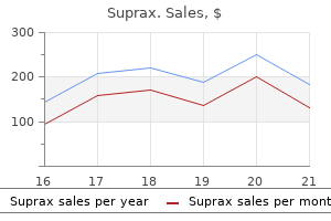
Buy 200 mg suprax overnight delivery
Auramine stain (tuberculous) and Indian ink stain (cryptococcal infection) in immunocompromised or different atrisk particular person antibiotics not working for uti 200 mg suprax purchase amex. Unlike meningitis antibiotic resistance risk factors suprax 100 mg buy generic line, cerebral function is usually abnormal, with altered mental status, motor and sensory deficits. It is brought on by all kinds of viruses and can also occur in bacterial and other infections. Acute viral encephalitis A viral aetiology is usually presumed, although not confirmed serologically or by culture. Japanese encephalitis (arbovirus) in South-East Asia, West Nile encephalitis in Egypt and Sudan. An inactivated vaccine is out there for travellers spending time in endemic areas. Clinical options Many of those infections trigger a mild self-limiting sickness with fever, headache and drowsiness. If the affected person is in a coma, the prognosis is poor, whether or not remedy is given. Brain and spinal abscesses An intracranial abscess might develop within the epidural, subdural or intracerebral websites. Epidural abscesses are unusual; subdural abscess presents equally to intracerebral abscess. Cerebral abscess Infection follows the direct spread of organisms from a parameningeal infective focus. Infection with tubercle bacilli might end in continual caseating granulomata (tuberculomas) presenting as intracranial mass lesions. Clinical features these embrace headache, focal neurological signs, seizures and sometimes evidence of raised intracranial strain (p. Management Treatment is with a combination of intravenous antibiotics and sometimes surgical decompression. Spinal epidural abscess Back ache and fever are adopted by paraparesis and/or root lesions. Encephalitis and mind abscess Toxoplasma, cytomegalovirus, herpes simplex and other organisms trigger severe encephalitis. Primary intracranial tumours are derived from the cranium itself, or from any of the constructions lying within it, or from their tissue precursors. They could also be malignant on histological investigation however rarely metastasize outside the brain. Clinical features the medical options of a cerebral tumour are the result of the following: Progressive focal neurological deficit Raised intracranial strain Focal or generalized epilepsy. Neurological deficit is the end result of a mass impact of the tumour and surrounding cerebral oedema. Subsequent involvement of the frontal speech area and motor cortex produces expressive aphasia and hemiparesis. Parietal lobe tumours trigger a homonymous field defect, cortical sensory loss, hemiparesis and partial seizures on the side contralateral to the tumour. Rapidly rising tumours destroy cerebral tissue and loss of function is an early feature. The headache usually adjustments with posture and is made worse by coughing, sneezing, bending and straining. Distortion of regular structures at a distance from the rising tumour leads to focal neurological indicators (false localizing signs). Hydrocephalus 777 Differential diagnosis the primary differential is from different intracranial mass lesions (cerebral abscess, tuberculoma, subdural haematoma and intracranial haematoma) and a stroke, which may have an equivalent medical presentation. Benign (idiopathic) intracranial hypertension presents with headache and papilloedema in younger obese females. Surgical exploration, and both biopsy or elimination of the mass, is usually carried out to confirm its nature. Selected centres supply stereotactic (gamma knife) radiotherapy to deliver high doses of radiation to small targets with precision. Cerebral oedema surrounding a tumour is quickly reduced by corticosteroids � intravenous or oral dexamethasone. Chemotherapy has little real worth within the majority of primary or secondary mind tumours. The prognosis is very poor in sufferers with malignant tumours, with only 50% survival at 2 years for high-grade gliomas. Aetiology In youngsters, hydrocephalus could also be caused by a congenital malformation of the brain. Clinical options There is headache, vomiting and papilloedema attributable to raised intracranial pressure. Management Treatment is by the surgical insertion of a shunt between the ventricles and the best atrium or peritoneum (ventriculoatrial or ventriculoperitoneal). Tension headache Most continual every day and recurrent complications are rigidity headaches. They are thought to be generated by neurovascular irritation and referred to scalp muscles and delicate tissues. Treatment consists of explanation and reassurance, analgesic withdrawal (to keep away from analgesic overuse headache) and tricyclic antidepressants in some instances. Migraine Migraine is recurrent headache associated with visible and gastrointestinal disturbance. It is a typical condition, occurs extra frequently in ladies and onset is normally earlier than forty years of age. Changes in brainstem blood circulate result in an unstable trigeminal nerve nucleus and nuclei within the basal thalamus. Cortical spreading depression is a selfpropagating wave of neuronal and glial depolarization that spreads throughout the cerebral cortex. It is proposed to cause the aura of migraine and result in launch of inflammatory mediators which influence on the trigeminal nerve nucleus. Precipitating elements include stress, an extreme quantity of or too little sleep, noise and aggravating lights, hormonal factors. Clinical features Migraine is classed into three sorts: Migraine with aura (classic migraine) Migraine without aura (common migraine) Migraine variants (unilateral motor or sensory signs resembling a stroke). Premonitory signs of fatigue, nausea, modifications in mood and appetite could occur within the hours or days before the headache. Auras are related to depression of visible cortical function or retinal operate and persist for minutes to hours before the headache. There may be scotomata, unilateral blindness, hemianopic subject loss, flashes and fortification spectra. Other aura embrace aphasia, tingling, numbness and weak spot of one facet of the body. Differential diagnosis A sudden migraine headache could resemble meningitis or subarachnoid haemorrhage. They inhibit the discharge of vasoactive peptides, promote vasoconstriction and block pain pathways within the brainstem. They range of their onset of motion, recurrence fee and route of administration.

100 mg suprax otc
However xeno antibiotics suprax 200 mg order line, we may also use the term anterior to describe a construction whose anatomical course takes it toward the entrance of the physique antibiotic resistance japan 100 mg suprax order otc. Posterior can discuss with the again facet of the physique or of a physique part, or to a construction that travels towards the back facet of the physique (such as the posterior rami). The directional term superior is used to describe constructions that are toward or closer to the pinnacle. The reverse time period of superior is inferior, which means away from the head or toward the tail. For example: the 2 largest veins in the body are the superior vena cava and the inferior vena cava. The superior vena cava is the vessel situated nearer the pinnacle, and the inferior vena cava is the one positioned farther away from the top (or nearer to the tail). We can describe the pinnacle as being superior to the neck, and we are in a position to say that the abdomen is inferior to the chest. A essential rule to bear in mind with superior and inferior is that these terms are used to describe buildings solely on the pinnacle, neck, and trunk, that are positioned in the part of the physique often identified as the axial region. The upper and lower limbs are the a half of the physique generally identified as the appendicular region. We use the terms proximal and distal on the appendicular area instead of using superior and inferior. The reverse time period, distal, refers to the farness-or distance-of a structure from its level of origin. Superior and inferior are straightforward sufficient on the head, neck, and trunk, however many college students overlook the rules and want to use them on the upper and decrease limbs. Here are a couple of straightforward ones: the extra proximal construction is the one in the closest proximity to the hip (or shoulder). The terms medial and lateral reference an imaginary line working down the center of the physique known as the midline. A construction is described as medial when its position is closer to this midline, and its place turns into more lateral because it moves farther away from the midline. We can describe the ears as being lateral to the eyes as a end result of the ears are farther away from the midline than are the eyes. Procedure 2 Directional Terms Fill within the right directional time period for every of the next objects. For instance, you likely know the places of the "oral," "nasal," and "stomach" regions. Between the Dissection kits/dissection trays parietal and visceral layers is a skinny potential space that incorporates serous Laminated define of the human body fluid. It has two major divisions: the thoracic and abdominopelvic cavities, every of which is split into smaller subcavities. We find the following smaller cavities throughout the thoracic cavity: (1) Pleural cavities. The pleural cavities are situated between two layers of a serous membrane referred to as the pleural membrane. The parietal pleurae are hooked up to the body wall and the surface of the diaphragm, and the visceral pleurae are attached to the floor of the lungs. The parietal pericardium is connected to surrounding buildings, and the internal visceral pericardium is attached to the heart muscle. Between the two layers we discover the pericardial cavity, which is filled with a thin layer of serous fluid. There are three subcavities within the abdominopelvic cavity: (1) Abdominal cavity. The space superior to the bony pelvis, referred to as the abdominal cavity, houses many organs together with the liver, gallbladder, small intestine, stomach, pancreas, kidneys, adrenal glands, spleen, and far of the colon (large intestine). The cavity housed throughout the bony pelvis, the pelvic cavity, accommodates certain intercourse organs, the urinary bladder, the rectum, and a part of the colon. The outer parietal peritoneum is connected to the body wall and surrounding structures, and the inner visceral peritoneum is connected to the floor of lots of the abdominal and pelvic organs. Between these two layers of peritoneal membrane we find the peritoneal cavity, which is full of serous fluid. Using a blunt dissection probe, peel back the pores and skin of the stomach fastidiously to expose the abdominopelvic cavity. Use scissors or a scalpel to rigorously cut by way of the sternum to expose the thoracic cavity. Mark each quadrant of the abdominopelvic cavity with a pin or marking tape (if working with a torso model). As you dissect the preserved mammal, search for the serous membranes listed in Table 1. Take care not to tear the fragile membranes, which include just some layers of cells. As you establish every membrane, name the structure to which the membrane is connected (the lungs, heart, stomach wall, and so on. Label each body cavity, in addition to the serous membranes surrounding the cavity, the place relevant. Exploring Anatomy & Physiology within the Laboratory: Core Concepts 18 n Procedure three Applications of Terms, Cavities, and Membranes Remember this from the unit opener The bullet entered the proper posterior scapular area, 3 centimeters lateral to the vertebral region, four centimeters inferior to the cervical area, and penetrated deep to the muscle and bone, but superficial to the parietal pleura. In this situation, your victim will be your fetal pig or a human torso model that your teacher has "shot. As coroner, remember to hold your affected person in anatomical place and that you should be as particular as attainable. The transverse plane, also referred to as a cross part or the horizontal plane, divides the specimen into a superior (or proximal) part and an inferior (or distal) part. In your study of anatomy and physiology, you will notice many alternative examples of those planes of part. Procedure 1 Identifying Organs Identify the following organs on your preserved mammal specimen or human torso models. Check off every organ as you determine it, and document the organ system to which it belongs in Table 1. Then, use your textbook or different sources to analysis the principal features of each system. Toward the entrance of the physique Toward the surface/skin three Which of the following is a correct use of a directional time period It is 8 centimeters lateral to the vertebral area and 3 centimeters superior to the scapular region. The wound is situated on the left anterior, medial femoral area, 10 centimeters distal to the inguinal region. Your job is to read her operative report and decide the place the incisions had been made. The cut prolonged vertically in an inferior path, ending 2 centimeters superior to the umbilical region.

Cheap 200 mg suprax otc
Correlation of prevalence and severity of aortic regurgitation detected by pulsed Doppler echocardiography with the murmur of aortic regurgitation in aged sufferers in a long-term health care facility virus 5 hari suprax 100 mg order mastercard. Diagnosis and quantification of aortic regurgitation by pulsed Doppler echocardiography in patients with mitral valve illness antibiotics for acne forum suprax 200 mg cheap with visa. An help to identification of the murmur of aortic stenosis with atypical localization. Haemodynamic explanation of why the murmur of mitral regurgitation is unbiased of cycle size. Diagnosis of left-sided regurgitant murmurs by transient arterial occlusion: a brand new maneuver utilizing blood strain cuffs. The murmur of papillary muscle dysfunction in acute myocardial infarction: medical features and prognostic implications. Effects of prompt squatting on the systolic murmur in idiopathic hypertrophic obstructive cardiomyopathy. The absence of this murmur is a compelling argument against the diagnosis of aortic stenosis. Its attribute findings are a systolic murmur, abnormal carotid pulse, and sustained apical impulse. The pathology of aortic stenosis was acknowledged in the 1600s, nevertheless it was James Hope who in 1832 first clearly described the characteristic murmur. Radiation of sound in the neck first seems on the proper side (clavicle and neck), however as the stenosis worsens the sound appears on each side of the neck and over both clavicles. In calcific aortic stenosis, the most typical fashionable etiology, the murmur at the higher sternal borders incorporates each high- and low-frequency vibrations, giving it a harsh or rough sound, like that of an individual clearing the throat. In distinction, at the apex the murmur of calcific aortic stenosis typically loses low-frequency components and instead consists of a slim band of high-frequency sound, thus making it sound like mitral regurgitation. This harmonic distortion of sound-the lack of low-frequency components of sound when the stethoscope is moved "upstream" toward the apex-is called the Gallavardin phenomenon. Chapter forty three discusses additional the differential prognosis of systolic murmurs and how the clinician-by observing the situation of sound, the second heart sound, the quality of the murmur, and how the murmur responds to irregular heart beats and different maneuvers- may be more assured a systolic murmur indeed represents aortic stenosis and never another valvular lesion. After clinicians are confident a murmur represents an aortic flow murmur, they have to decide whether or not the patient has significant aortic stenosis. Significant aortic stenosis refers to these lesions with such extreme obstruction that, if the patient has symptoms of angina, syncope, or dyspnea, valvular replacement is indicated. In comparison to congenital and rheumatic illness, calcific aortic stenosis impacts older sufferers, who generally have aortic move murmurs without stenosis. Importantly, all had aortic flow murmurs, and the bedside query was whether or not the murmur represented extreme aortic stenosis. Although some had gentle aortic regurgitation, different significant valvular disease was excluded from most of those research. Brachioradial delay and apical-carotid delay had been every investigated in solely single studies and thus require affirmation by others. The reason for false-positive results in the research of aortic stenosis is principally moderate aortic stenosis (defined as aortic valve area of 0. This diagnostic scheme distinguishes moderate-to-severe aortic stenosis from other causes of aortic circulate murmurs. Severe valvular aortic stenosis in sufferers over sixty five years of age: a clinicopathologic examine. The role of routine pre-operative bedside echocardiography in detecting aortic stenosis in sufferers with hip fracture. Prevalence and severity of valvular aortic stenosis decided by Doppler echocardiography and its association with echocardiographic and electrocardiographic left ventricular hypertrophy and bodily signs of aortic stenosis in elderly sufferers. A novel and simple technique using pocket-sized echocardiography to screen for aortic stenosis. Clinical clue of severe aortic stenosis: simultaneous palpation of the carotid and apical impulses. The apical A wave versus the fourth coronary heart sound in assessing the severity of aortic stenosis. Clinical and haemodynamic options in relation to severity of aortic stenosis in adults. A medical examine of the brachial arterial pulse form: with particular reference to the diagnosis of aortic valvular disease. A bedside clinical prediction rule for detecting average or extreme aortic stenosis. It is heard finest with the diaphragm of the stethoscope positioned close to the lower left sternal edge and with the patient sitting up, leaning forward, and holding his or her breath in exhalation. A diastolic blood strain higher than 70 mm Hg and pulse strain lower than 60 mg lower chance of moderateto-severe regurgitation. In patients with vital chronic regurgitation, the normal bodily findings are a diastolic murmur, dilated apical impulse, and abnormally forceful and collapsing arterial pulses (pulsus celer). In the 1700s clinicians related the postmortem discovering of damaged aortic valves with hearts "bigger than that of an ordinary ox" (the origin of the phrase cor bovinum) and the finding throughout lifetime of "violently throbbing" carotid arteries. In 1832 Sir Dominic John Corrigan, a Dublin surgeon, taught clinicians how to diagnose the illness during life, by emphasizing the significance of those dramatic arterial pulsations and the associated diastolic murmur. In some patients the murmur is audible only when the patient sits up, leans forward, and holds his or her breath in exhalation. The combination of this murmur and the early diastolic one causes a characteristic "to-fro" sound close to the sternum (see Chapter 43). Approximately half of Austin Flint murmurs have two diastolic components (mid-diastolic and presystolic), whereas the opposite half have only a presystolic element. Although all hypotheses assume the murmur is determined by a powerful regurgitant stream of blood being directed again toward the left ventricle throughout diastole, these hypotheses differ in how this regurgitant stream causes an apical rumbling sound. Proposed mechanisms include fluttering of the anterior leaflet of the mitral valve, untimely closure of the mitral valve from elevated left ventricular end-diastolic strain, collision of the regurgitant stream with the anterior mitral leaflet, ventricular vibrations attributable to the regurgitant stream itself, and harmonic distortion of the aortic regurgitant murmur. Excellent videos of patients with bounding carotids,21 Quincke pulse,22 and M�ller sign23 can be found. They are similar in high quality to the Korotkoff sounds heard when measuring blood pressure. For the Duroziez signal to be optimistic, each a systolic and diastolic murmur have to be current (many normal individuals develop systolic murmurs with pressure on the stethoscope). The diastolic component usually becomes louder with stress utilized distal to the stethoscope. In some sufferers the murmur could additionally be loudest to the right of the sternum, which suggests an eccentric regurgitant stream from dilation of the aortic root. Distinguishing aortic from pulmonary regurgitation was significantly related in sufferers with rheumatic mitral stenosis, who often had associated aortic valve illness however who also might develop pulmonary hypertension and the early diastolic murmur of pulmonary insufficiency. In patients with mitral stenosis who also have an early diastolic murmur of regurgitation heard subsequent to the sternum, the extra lesion is aortic regurgitation no less than 80% of the time. Many of the sufferers enrolled within the studies additionally had additional murmurs of aortic stenosis or mitral regurgitation.
Plantago Major (Great Plantain). Suprax.
- Common cold, ongoing (chronic) bronchitis, bladder infections, hemorrhoids, skin conditions, eye irritation, and other conditions.
- Dosing considerations for Great Plantain.
- What is Great Plantain?
- How does Great Plantain work?
- Are there safety concerns?
- Are there any interactions with medications?
Source: http://www.rxlist.com/script/main/art.asp?articlekey=96666
Cheap suprax 200 mg mastercard
Motor to the muscular tissues of swallowing; style to the posterior one-third of the tongue F antibiotic resistance why does it happen suprax 200 mg cheap line. Motor to the muscles of facial features; taste to the anterior two-thirds of the tongue J virus film suprax 200 mg mastercard. Motor to four of six extrinsic eye muscular tissues; dilates the pupil; opens the attention; changes the shape of the lens K. Motor to the muscle tissue of swallowing and talking; motor to the thoracic and belly viscera L. Motor certainly one of six extrinsic eye muscular tissues (lateral rectus muscle) 8 thirteen the receptor that detects the stretch in a stretch reflex is called a(an) a. Sensation could also be aware or unconscious, relying on the vacation spot of the sensory data. However, data finally taken to the cerebral cortex for integration and interpretation. The following workout routines ask you to study the anatomy and physiology of the particular senses: vision, listening to and equilibrium, style, and scent. You also will look at the final senses in this unit, which include touch, pain, and temperature. Many of the exterior constructions of the attention shield the fragile eyeball, and others move the eyeball. The palpebrae meet medially and laterally at the medial and lateral canti (or commissures), respectively. There are a quantity of structures in and around the palpebrae that include sebaceous or mucous glands to lubricate the palpebrae and anterior floor of the eyeball. One of probably the most prominent external structures of the eye is the lacrimal equipment, which produces and drains tears. The lacrimal apparatus consists of the lacrimal gland, situated in the superolateral orbit, and the ducts that drain the tears it produces. From here, tears journey via the nasolacrimal duct and, lastly, empty into the nasal cavity simply inferior to the inferior nasal concha (which is why your nostril runs when you cry). In addition, two muscle tissue that open and shut the eye insert into and around the palpebrae: the levator palpebrae superioris muscle and the orbicularis oculi muscle. The boundary for the cavities is a crystalline structure generally recognized as the lens, which is probably certainly one of the constructions within the eye that refracts (bends) mild coming into the eye to focus it. The anterior cavity is anterior to the lens and is crammed with a watery fluid referred to as aqueous humor. Aqueous humor is produced comparatively continuously and drained by a structure called the scleral venous sinus. Unlike aqueous humor, vitreous humor is present at start and stays relatively unchanged throughout life. The outermost layer of the eyeball is the fibrous tunic, which consists principally of dense irregular collagenous connective tissue. It is white due to quite a few collagen fibers that contribute to its thickness and toughness (in the identical way a joint capsule or a ligament is hard and white). It is made mainly of the ciliary muscle, easy muscle fibers that control the shape of the lens. The photoreceptors generally identified as rods are scattered all through the retina and are responsible for imaginative and prescient in dim mild and for peripheral vision. Cones, the second type of photoreceptor, are concentrated on the posterior portion of the retina. It is also called the blind spot as a end result of its lack of photoreceptors means that this area can produce no images. When viewing nearer objects, nevertheless, additional "fine-tuning" refraction is required by the lens. This is completed with the help of the ciliary muscle-when its clean muscle fibers contract, the ciliary physique strikes closer to the lens and removes rigidity on the suspensory ligaments. This causes the lens to turn out to be rounder, an adjustment referred to as accommodation which allows the lens to present the additional refraction essential to focus light on the retina. When the attention switches to a distant object again, the ciliary muscle relaxes, which strikes the ciliary body farther away from the lens and places tension on the suspensory ligaments. This flattens the lens and permits the cornea to again turn out to be the first refractive medium. Sclera Iris 2 3 Use scissors to take away the adipose tissue surrounding the eyeball. Hold the eyeball at its anterior and posterior poles, and use a pointy scalpel or scissors to make an incision in the frontal plane. Take care to preserve the fragile retina-the thin, delicate yellow-tinted inside layer. In this process, you will see (no pun intended) firsthand the differences in vision produced by the rods and the vision produced by the cones. Have your associate stand 20 ft in front of a Snellen eye chart and read down the chart, stopping with the smallest line read precisely. For instance, an individual with 20/40 imaginative and prescient can see at 20 ft what somebody with perfect imaginative and prescient could see at 40 ft. Visual acuity: 9 5 With the lights still dimmed and your partner standing in the same place, maintain a chunk of darkish green or darkish blue paper over the Snellen chart. Visual acuity: Paper colour: 7 In which scenario have been visible acuity and shade vision higher Have your lab partner measure with a ruler the distance from the web page to your eye with a ruler. These irregularities decrease visual acuity as a end result of these buildings are unable to focus gentle precisely on the retina. The ossicles transmit vibrations from the tympanic membrane to the inner ear via a construction called the oval window, to which the stapes is hooked up. Situated at proper angles to each other, the semicircular canals work along with the organs of the vestibule to keep equilibrium. Their orientation allows them to be answerable for a kind of equilibrium called rotational equilibrium. The cochlea has a hole in its lateral wall referred to as the round window, which performs a role in allowing the perilymph in the cochlea to vibrate. The constructions of the cochlea transmit sound stimuli by way of the cochlear portion of the vestibulocochlear nerve. Procedure 1 Model Inventory for the Ear Identify the next constructions of the ear on fashions and diagrams, utilizing this unit and your textbook for reference. As you look at the anatomical fashions and diagrams, report the name of the mannequin and the constructions you have been in a place to determine on the model stock in Table 9. Conductive hearing loss results from interference of sound conduction by way of the outer and/or middle ear. Sensorineural hearing loss outcomes from harm to the inner ear or the vestibulocochlear nerve. Two medical tests may help a healthcare professional decide whether or not listening to loss is conductive or sensorineural: the Weber take a look at and the Rinne (rinn-ay) test. The tuning forks are positioned either instantly on the bones of the skull to evaluate bone conduction-the ability to hear the vibrations transmitted by way of the bone-or in entrance of the ear, to consider air conduction-the capability to hear the vibrations transmitted via the air.
100 mg suprax discount
When calculating the entire magnification bacterial vaginosis home remedies buy 100 mg suprax overnight delivery, you must multiply the magnification of the ocular lens (10) by the power of the objective lens virus 404 error suprax 200 mg purchase line. Remember that the magnification of the objective lens is usually printed on the facet of the lens itself. Move the slide using the stage adjustment knob until the "e" is over the condenser. Note that you just may need to adjust the iris diaphragm to allow extra gentle to move by way of the specimen, as the newsprint is comparatively thick. You ought to only need to modify the main focus with the nice adjustment knob; no adjustment of the coarse focus ought to be needed. These "dirt discoveries" may be prevented by appreciating what is named depth of area. Thicker specimens will require you to focus up and down so as to see all ranges of the specimen. Switch the nosepiece to the low-power objective, place the slide on the stage, and secure it with the stage clips. Move the slide utilizing the stage adjustment knob until the threads are in your subject of view. Move the objective up and down slowly with the coarse adjustment knob, specializing in each individual thread. Well, these certainly are harder, however if you hold the following hints in thoughts, the task becomes a lot less complicated: Always begin on low power. You are supposed to start on low energy, anyway, to keep away from damaging the objective lenses. Bear in thoughts that most slides could have more than one histological or cellular construction on every slide. Starting on low power permits you to scroll through a large area of the slide, after which focus in on the specified part of the section. The simple solution to this downside is to examine the slide with the naked eye first, and then place it on the stage so that the specimen is true over the condenser (the light). Other good sources for micrographs include atlases, your textbook, and pictures on the Internet. Although you might are inclined to resist drawing, even essentially the most basic image is useful for two reasons. Also, drawing is helpful in that you simply really have to take a look at the specimen lengthy enough to draw it. By the end of the semester, you may come to respect the microscope and the fascinating world it reveals. To prepare a cell smear, acquire a pattern of cells with a cotton applicator swab and "smear" these cells on a clean slide. In our procedure, we shall be utilizing a cell pattern obtained from the mouth and making use of the stain methylene blue. If the blue areas remain, get hold of a brand new pattern of cheek cells and repeat the procedure. Place a coverslip over the stained space, and place the slide on the stage of a microscope. Focus the image grossly on low power, then switch to the high-power objective lens to discover particular person cells (use oil immersion if available). Identify and draw a few of the cells from your smear in the space supplied with coloured pencils. It is a dynamic, fluid structure that acts as a selectively permeable barrier, meaning that it only allows sure solutes to pass into or out of the cell. There are multiple elements combined in with the phospholipids, together with proteins, ldl cholesterol, and carbohydrates. The cytoskeleton is a group of protein filaments including actin filaments, intermediate filaments, and microtubules. Actin filaments are small protein strands, lots of which are positioned alongside the plasma membrane and in the core of microvilli, that help keep the form of the cell and performance in cell motion. The larger intermediate filaments are ropelike buildings that assist preserve the shape of the organelles and the nucleus and provides the cell mechanical power. Microtubules, the largest filaments, are hollow tubes that maintain the shape of the cell, hold organelles in place, move substances within the cell, and performance in cell division. In addition, microtubules type the core of motile extensions from the cell called cilia and flagella. Organelles are specialized mobile compartments that carry out a big selection of capabilities. Some ribosomes float freely within the cytosol, whereas others are bound to the membrane of another organelle or the nucleus. They metabolize fatty acids, synthesize certain phospholipids, and comprise enzymes that catalyze reactions to detoxify chemical compounds produced by cellular reactions. Enzymes inside the sacs catalyze reactions that process, modify, and kind these products, most of which then exit the Golgi equipment through new vesicles. These enzymes catalyze many types of reactions, together with people who break down substances brought into the cell, elements within the cell corresponding to old and worn-out organelles, and even the cell itself. The nucleus is surrounded by a double membrane referred to as the nuclear envelope, which incorporates holes referred to as nuclear pores. For instance, the cells of the liver include a large amount of clean endoplasmic reticulum, and immune cells referred to as phagocytes home many lysosomes. As you study the anatomical models and diagrams, report the name of the model and the constructions that you simply were in a position to determine on the mannequin stock in Table three. Cells differ not only in size and shape but also within the types and prevalence of organelles within the cell. Use the strategies you discovered in Exercise 3-1: Begin your statement on low power, and advance to excessive energy for each slide. Note that sperm cells could be tough to find, so an oil-immersion lens is helpful for locating the tiny cells. Draw, colour, and label the mobile buildings and organelles you see on each slide. Following are some suggestions for producing effective drawings that may assist you to study the fabric and study for sensible exams. First have a glance at a diagram of the cell or tissue in question in this book or your textbook to get oriented and get an idea of what you should look for within the subject of view. As always, start on low power, after which advance to higher-powered aims as wanted. You ought to goal for a magnification about the same because the magnification in the diagram. Reproduce as closely as potential what you see in the subject of view utilizing a pencil. You could draw in the circles in this guide, or on a piece of white paper for larger diagrams. Some slides may have uniquely coloured stains or staining patterns, and studying the colored photographs can show quite useful.

Suprax 200 mg discount without a prescription
Landmarks that define the four quadrants of the stomach; names of the quadrants; and a few organs that lie within every quadrant 6 antibiotics japan over counter suprax 200 mg. Landmarks that divide the stomach into nine regions; names of those regions; and a few organs that lie within each 7 virus yahoo email generic suprax 200 mg with mastercard. Differences between the serous and mucous membranes of the physique and examples of locations 10. The principal physique cavities; names of the membranes that line them; and the most important viscera found in every eleven. The location, look, and performance of the mesenteries, and how the mesenteries are associated to the peritoneum and the serosa of the viscera thirteen. The distinction between intraperitoneal and retroperitoneal organs, and a few examples of each 14. The 11 organ techniques of the human physique, features of each system, and principal organs of each 1. The meaning of physiology, and the significance of studying the physiology of other species for understanding people 2. The properties that outline something as alive and the difficulty of defining life or the moment of death 3. The which means of homeostasis, the position of unfavorable suggestions loops in maintaining homeostasis, and the distinction between the set level and a dynamic equilibrium 4. How positive suggestions differs from unfavorable suggestions, and examples of useful and harmful effects of optimistic suggestions 7. Examples of why precision is important in anatomical terminology, and why spelling errors can be extra significant than they could appear 2. Relationships between the singular and plural forms of the identical biomedical nouns four. The distinctions between dorsal and ventral; anterior and posterior; superior and inferior; medial and lateral; proximal and distal; and superficial and deep 5. When a well being care provider presses on the abdomen to really feel the scale and texture of the liver, he or she is using a technique of bodily examination referred to as. A method of medical imaging that makes use of X-rays and a pc to generate an image of a thin slice of the physique is called. Most physiological mechanisms serve the aim of maintaining a stable internal setting in the physique. A/an is the best physique construction to be composed of two or extra forms of tissue. The carpal area is extra commonly often known as the and the tarsal region is extra commonly known as the sixteen. The layer of peritoneum dealing with the physique wall is called the layer, and the layer on the surface of an inner organ is called the layer. Homeostasis is maintained by a cycle of occasions known as a, by which the physique senses a change and prompts mechanisms to minimize or reverse it. The directional terms of human anatomy assume that a person is in, which suggests standing upright with the feet collectively and the palms, face, and eyes forward. Abnormal pores and skin colour or dryness is one piece of information that could presumably be obtained by auscultation. Identify which anatomical plane-sagittal, frontal, or transverse-is the one one that would not present (a) both the cerebrum and tongue, (b) both eyes, (c) both the hypogastric and gluteal regions, (d) both the sternum and vertebral column, and (e) both the heart and uterus. Name one construction or anatomical function that might be found in every of the next areas relative to the ribs: medial, lateral, superior, inferior, deep, superficial, posterior, and anterior. Urea, a product of protein metabolism, is the principal natural waste product of urine. Why does an iron deficiency cause anemia and a calcium deficiency weaken the bones None of these questions can be answered, nor would the remainder of this book be intelligible, without understanding the chemistry of life. A little knowledge of chemistry might help you choose a healthy diet, use drugs extra properly, consider health fads and avoid frauds, and perceive medical treatments and procedures. ChemicalElements A chemical component is the best type of matter to have unique chemical properties. Water, for example, has unique properties, but it can be damaged down into two components, hydrogen and oxygen, every with unique properties of their own. If we break them down any further, however, we discover that hydrogen and oxygen are made from protons, neutrons, and electrons-none of which are distinctive. Therefore, the weather hydrogen and oxygen are the best distinctive parts of water. There are 91 naturally occurring parts on earth, 24 of which play regular roles in people. Other components with out natural physiological roles can contaminate the body and severely disrupt its features, as in heavy metallic poisoning with lead or mercury. The components are represented by one- or two-letter symbols, usually based mostly on their English names: C for carbon, Mg for magnesium, Cl for chlorine, and so forth. A few symbols are based mostly on Latin names, corresponding to K for potassium (kalium), Na for sodium (natrium), and Fe for iron (ferrum). The periodic desk of the elements (appendix E) summarizes info on all the pure chemical parts and their relative importance in human physiology. Several components are categorised as minerals-substances extracted from the soil by vegetation and handed up the meals chain to people and different organisms. Nearly three-quarters of that is Ca and P; the remainder is principally Cl, Mg, K, Na, and S. Third power level First power level Sodium (Na) 11p+, 11e-, 12n0 Atomic number = eleven Atomic mass = 23 Key Carbon (C) Atomic quantity = 6 Atomic mass = 12 6p+, 6e-, 6n0 = Proton (p+) = Neutron (n0) = Electron (e�) AtomicStructure Each chemical component consists of a unique type of atom. At the middle of an atom is the nucleus, composed of positively charged protons (p+) and uncharged neutrons (n0). The nucleus is orbited by electrons (e-), tiny particles with a single negative cost and really low mass (fig. They determine the chemical properties of an atom, governing chemical reactions and formation of molecules. Electrons swarm about the nucleus in concentric areas known as power levels (electron shells). Those of the outermost shell, referred to as valence electrons, decide the chemical bonding properties of an atom. IsotopesandRadioactivity Not each atom of a component is identical; all parts have two or extra varieties called isotopes,1 which differ from one another only in variety of neutrons. Most hydrogen atoms, for example, have a nucleus composed of only one proton; this isotope is symbolized 1H. Hydrogen has two other isotopes: deuterium (2H) with one proton and one neutron, and tritium (3H) with one proton and two neutrons. Over 99% of carbon atoms have a nucleus of six protons and 6 neutrons, and are known as carbon-12 (12C), but a small proportion of carbon atoms are 13C, with seven neutrons, and 14C, with eight. Many isotopes are unstable and decay (break down) to extra steady isotopes by giving off radiation.