

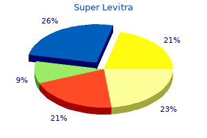
Super Levitra dosages: 80 mg
Super Levitra packs: 10 pills, 20 pills, 30 pills, 60 pills, 90 pills, 120 pills, 180 pills

Super levitra 80 mg low cost
Injuries or damages are usually recognized within the medical record and infrequently confirmed via independent medical examinations impotence exercises for men super levitra 80 mg cheap with amex. Expert witnesses had been appointed by the court docket and rendered goal opinions based on medical information to aid the actual fact finder (juror or judge) in understanding the sophisticated details of the case at hand impotence young purchase super levitra 80 mg line. As such, the testament may, in concept, be utilized by both or each parties to assist its case. To encourage open and trustworthy testament, the skilled witness was granted immunity for something mentioned on the witness stand. Without this protection, experts might have been reluctant to communicate openly for fear of legal retaliation. This menace of attainable sanction was felt to be enough of a deterrent to prevent dishonest, negligent testament. The American judicial system has evolved from the usage of impartial, court-appointed specialists to the reliance on (presumably) neutral witnesses recruited by each side to clarify the technical aspects of the case. This departure from the English system transfers the inherently adversarial relationship between opposing counsel to the consultants retained by each side. There is an unstated obligation of advocacy on the part of the professional witness to the authorized staff for which she or he was retained. With reimbursements declining, many physicians are actively seeking such alternate sources of income. Less scrupulous clinicians may indeed provide false, misleading, or misleading testament for the express function of influencing the result of a case. These embody a want to discredit knowledgeable adversary or competitor, a quest for personal acclaim and elevated professional stature, and a private campaign to protect fellow doctors from a tort environment seemingly stacked in opposition to healthcare suppliers. Key Features � � Expert testimony is critical to outline the standard of care and to determine if that normal has been met. This creates an inherent conflict of curiosity for the physician witness who is expected by the judicial system to stay impartial and objective. The skilled witness is important to make certain that victims of medical negligence obtain truthful compensation, but also that competent, certified physicians are protected against frivolous claims of medical malpractice. Professional societies have begun to outline acceptable codes of ethical conduct for expert witnesses, and to impose sanctions for physicians who run afoul of those standards. Certain groups, notably the trial bar and affected person advocacy groups, but additionally some physicians, see such initiatives as an effort by organized medication to intimidate medical doctors and forestall them from testifying for the patient in medical liability instances. The American Academy of Ophthalmology enacted a rule governing the Ethics of Expert Witness testament in 2004. Given the obligation to society to present expert witness testament, how can well-intentioned physicians fulfill their obligation with out operating afoul of established ethical standards To fulfill this duty, medical societies have begun to outline acceptable codes of ethical conduct for forensic medical consultants and impose sanctions for those members who run afoul of these standards. In a landmark case, the American Association of Neurological Surgeons handed down a 6-month suspension to a member neurosurgeon for providing conflicting and false testament in a number of related malpractice suits against fellow neurosurgeons. Posner of the United States Seventh Circuit Court of Appeals, opined that membership in medical societies conferred certain privileges and obligations. This sort of professional self-regulation furthers quite than impedes the trigger of justice. Reprimands and censures are sometimes printed in trade journals providing a really public discussion board for skilled humiliation. Doctors who incessantly present expert testimony can see their credibility � in addition to the demand for his or her companies � evaporate. Who, in any case, would wish to retain a witness who had been found guilty of providing false and deceptive testimony in other instances Sopulos M: Addressing false professional witness testimony in medical malpractice litigation. Milunsky A: Lies, damned lies and medical experts: the abrogation of responsibility by specialty organizations and a name for motion. One examine means that over 50% of the United States population take dietary dietary supplements or use some different therapy. The resultant whole bills incurred with using alternativemedical practices and therapies was about 27 billion dollars. In ophthalmology, there are tons of conditions that lack long-term palliation or treatment, similar to age-related macular degeneration and primary open-angle glaucoma. The use of alternative therapies, as a substitute of or in addition to traditional medical approaches, is often undertaken by patients within the hope of achieving an improved consequence. Key Features Examples of complementary and various medication: � Vitamins � Herbs � Dietary dietary supplements � Homeopathic cures � Folk drugs � Faith healing � Spiritual therapeutic � Acupuncture of medical literature and medical trials so as to in the end improve the overall quality of care provided to patients. Interestingly, a similar dedication has not been embraced by most practitioners of complementary and various medication. As a result, only a few well-done, randomized, placebo-controlled scientific trials have examined using alternative therapies. One randomized managed double-blind research examines using acupuncture for dry eye. A statistically important difference was discovered between the needle acupuncture group and the management group (p <0. However, this examine lacked the appropriate sham control group wanted to validate the results. Do they forgo evidence-based medicine in favor of anecdotal proof and private experience This nationwide, randomized scientific trial showed a big danger reduction for growth of superior age-related macular degeneration with high-dosage antioxidant nutritional vitamins. It is hoped that the future of Key Features Aspects of complementary and alternative medicine that create moral points: � Unregulated standing � Unproven profit � Unknown security profile � Potential for monetary conflicts of interest Limited studies of complementary and alternative drugs present an ethical dilemma to ophthalmologists training evidence-based drugs. Parallel investigations of complementary and alternative processes would also serve societal interests. The American Academy of Ophthalmology commissioned a Task Force on Complementary Therapies to asses the scientific merit of nontraditional practices such as marijuana use for glaucoma and microcurrent stimulation for age-related macular degeneration, and there are other examples. Thus, there are quite a few avenues for gaining or sustaining competence in the realm of other therapies, but pursuing them requires both interest and a commitment to persevering with training. Lack of information about complementary and alternative medicines and practices raises the plain issues with any incompetent doctor: the failure to acknowledge potential interactions with prescribed medications, to establish potential dangerous ocular unwanted effects, or to avoid increased surgical dangers. A examine printed in the Journal of the American Medical Association reviewed 443 websites advertising dietary supplements and noted that 55% of them made unlawful claims about remedy, prevention, prognosis, and remedy of specific diseases. Key information are lacking on product safety, efficacy, proper dosage, manufacturing, common unwanted facet effects, drug interactions, risks to pregnant ladies, results on systemic diseases, pharmacokinetics, etc. In addition, ophthalmologists have a fiduciary accountability to the patient to monitor any adverse occasions associated with their various practices. This is, more particularly, a challenge to the open communication integral to the patient�physician relationship.
Buy 80 mg super levitra otc
Research into neuroprotective brokers has not been clinically applicable presently erectile dysfunction pump images super levitra 80 mg buy discount line. Mechanisms for chiasmal harm embrace mechanical stretch and tear green tea causes erectile dysfunction 80 mg super levitra order with visa, contusion hemorrhage, contusion necrosis, and compression necrosis. Diabetes insipidus is reported to occur in 30% of sufferers with a traumatic chiasmal syndrome. Retro-chiasmal lesions result in contralateral homonymous visible subject defects, aside from these involving the monocular temporal crescent. Fibers from the superior hemiretina course immediately posteriorly into the parietal lobe on their way to the occipital cortex. Therefore, damage to the temporal lobe produces homonymous visual-field defects which are denser above, and parietal lobe lesions produce homonymous defects which might be denser beneath. In common, lesions superior to the calcarine fissure end in inferior quadrantanopic defects, and those below the calcarine fissure produce superior quadrantic defects. More extreme lesions of the optic tract, temporal lobe, parietal lobe, and occipital lobe may produce an entire homonymous hemianopia, a nonlocalizing finding. Optokinetic nystagmus asymmetry could additionally be used to localize the lesion to the parietal lobe. For this purpose, sufferers with parietal lobe lesions (and not those with purely occipital lobe lesions) demonstrate optokinetic nystagmus asymmetry, or depressed pursuit movement because the optokinetic nystagmus drum (or tape) is rotated toward the facet of the lesion. In addition, spasticity of conjugate gaze (a tonic deviation of the eyes to the side opposite the lesion) can also be observed with parietal lobe injury. Interruption of the geniculocalcarine radiations within the parietal, temporal, or occipital lobe might occur on account of head trauma with cerebral contusion and infarction. Penetrating intracranial injuries, corresponding to gunshot wounds, may cause harm to the postchiasmal pathways. Patients are often symptomatic for 1�24 h, and spontaneous resolution follows in most cases. Patients sustaining extreme head trauma may expertise visual perceptive problems. Other neurologic deficits might embrace aphasia, agnosia, alexia, and hemifield neglect. Functional neuroimaging could also be helpful within the assessment of sufferers with retro-chiasmal injury. The abducens and trochlear nerves could be the most regularly injured in head trauma although some series report the incidence of third nerve paresis to be higher than that of fourth-nerve paresis. Head trauma severe sufficient to induce brainstem contusion, edema, and infarction causes damage to the oculomotor nucleus. Oculomotor nerve paresis as a end result of minor head trauma is rare and will alert the clinician to the chance of an occult intracranial course of such as neoplasm or aneurysm. For occasion, if the fascicle is broken close to its course via the purple nucleus, the affected person could current with contralateral tremor. Injury near the cerebral peduncle might result in contralateral hemiparesis, and injury near the superior cerebellar peduncle may trigger ataxia. The affected person might present with indirect diplopia and decreased near imaginative and prescient as a consequence of diminished lodging. Ptosis together with motility deficits together with adduction, depression, and elevation of the attention will be found on examination. Aberrant regeneration regularly occurs weeks to months after harm to the oculomotor nerve. The ciliary ganglion can also be selectively broken, resulting in an efferent pupillary defect with pupillary mydriasis and loss of accommodation. It may also be damaged at the point at which the nerve pierces the dura to enter the cavernous sinus. Uncal herniation brought on by cerebral edema or intracerebral hemorrhage might end in compression of the third nerve, as it traverses the subarachnoid area, against the tentorium and posterior clinoid course of. Strabismus as a end result of oculomotor paresis is difficult to appropriate because of incomitance throughout the fields of gaze and the number of extraocular muscle tissue involved. Photophobia and glare may be managed with dark glasses or contact lenses or surgically with pupilloplasty. As with different cranial neuropathies, observation for spontaneous enchancment within a 6�12 month interval is really helpful. In most circumstances, unless levator operate recovers, ptosis restore requires frontalis suspension procedures. Orbital trauma, most commonly penetrating in nature, could result in superior indirect dysfunction. Fractures involving the nasoorbital ethmoid complex and the superior orbital rim may be associated with direct injury. Posttraumatic scarring or tenosynovitis of the trochlea might limit movement of the superior indirect tendon and may cause Brown syndrome. Compared to the optic and ocular motor nerves, the trigeminal nerve is much less generally affected after blunt head trauma. Frontal branch injury ends in hypoesthesia of the ipsilateral brow and is commonly associated with fractures, lacerations, and contusions in the region of the superior orbital rim. The nasociliary and lacrimal branches are rarely broken by penetrating orbital damage. Injury to the ophthalmic division produces corneal hypoesthesia, which if unrecognized, could result in neurotrophic keratitis and corneal ulceration. This is very important in patients with concomitant facial nerve palsy with its resultant lagophthalmos from orbicularis oculi muscle paresis. Orbital ground fractures commonly injury the infraorbital nerve, the department of the maxillary division that courses through the orbital floor. The patient will complain of numbness of the skin overlying the maxillary region, higher lip, and gingiva. Lesions that contain solely the ophthalmic division are probably to be within the anterior cavernous sinus or superior orbital fissure region, whereas, if the maxillary division is also concerned, the lesion is prone to prolong to the posterior cavernous sinus. Trauma is the most common cause96 of isolated fourth nerve paresis, and in a single sequence of head trauma trochlear nerve palsy was the most common neuroophthalmic finding. With blunt head trauma, fast deceleration causes displacement of the brain and brainstem inside the skull. The subarachnoid section of the nerve is susceptible to damage from the free fringe of the tentorium cerebelli close to the purpose where the nerve pierces the dura to enter the cavernous sinus. The presence or absence of a head tilt in old pictures may be helpful in figuring out whether or not the lesion preexisted the trauma, including congenital fourth nerve palsy, or has been newly acquired. The patient may complain of vertical binocular diplopia with torsion, typically with signs worsening on downgaze. There is usually a head tilt to the alternative side of the paresis and impaired despair of the involved eye in adduction. The Bielschowsky head tilt test used in the prognosis of vertical strabismus is beneficial in localizing dysfunction to the superior oblique muscle.
80 mg super levitra purchase with visa
Moreover erectile dysfunction statistics nih super levitra 80 mg purchase amex, in vivo lens dehydration can compromise their rotational stability especially for higherwater-content lenses and in dry eyes erectile dysfunction exercises 80 mg super levitra order mastercard. Increased ocular mucus secretion happens in more superior phases of this dysfunction and may result in rapid lens floor coating, sticky lens surfaces and excessive blink-induced lens motion accompanied by lowered imaginative and prescient and elevated lens awareness. However, evidence suggests that it might also contain sort 1 and delayed type 1V sensitivity. Lens wear ought to be temporarily discontinued in superior instances with increased mucus secretion. Up to 50% of soppy lens overnight storage circumstances are contaminated with gram-negative organisms, regardless of the method of disinfection used. Yet, the expectation that frequent substitute contact lens carrying would reduce significantly the incidence of contact lens-related microbial keratitis has not been realized. Deep stromal neovascularization can occur and will rarely trigger stromal lipid leakage and hemorrhage. Exacerbation of Non-contact Lens-related Ocular Conditions Soft lens carrying can provoke nonspecific bulbar conjunctival injection in sufferers with pingueculae and in those with reactive bulbar conjunctival blood vessels that become dilated because of minimal irritation corresponding to occurs after the instillation of a topical anesthetic. Indeed, ocular redness brought on by any preexisting corneal or conjunctival illness may be exacerbated by contact lens put on. These limitations have essentially limited their use to pediatric aphakic eyes by which steady put on is required. This is best established by observing the fitting traits of trial lenses. In the previous, a horizontal line of staining was generally observed in clear cornea adjoining to the superior limbus and was typically symptomatic. T oxic- or immune-mediated reactions to lens care preservatives may cause nonspecific superficial punctate keratitis as can the desquamation of epithelia overlying epithelial microcysts. Central fluorescein staining associated with corneal epithelium desiccation has been associated with thin, high-water-content delicate lenses. Increasing the oxygen transmissibility of silicone-acrylate polymers to a physiologically applicable degree was enhanced by incorporating fluorine in the polymer matrix that reduces its affinity for hydrophobic tear particles. Edge clearance is controlled by the curvatures and widths of the peripheral curves. Digital lathes and acceptable software applications now allow the again surfaces of lenses to be minimize with a junctionless transition from base curve to edge. They are particularly useful for plus energy lenses since their smaller anterior optical zones reduce the center thickness of the lenses. The excessive becoming sample is most comfortable,seventy six particularly if the upper lid covers the superior fringe of the lens all through the reflex blink cycle (lid attachment). Low-riding lenses are usually the least comfortable as a result of they promote incomplete reflex blink patterns. Central positioning lenses are sometimes extra snug than low-riding lenses, especially if the upper lid covers their superior edge throughout the blink cycle. Low-riding minus lenses are the least snug due to their bulkier peripheries. Failing that, minimizing their peripheral and edge profiles turn into more and more important factors in the carrying comfort equation. Lens motion after each blink ought to be modest (but present), easy and uniform. Traditional keratometry estimates the curvature of a small central space (~3 mm) of the principal meridians across the visual axis,78 whereas Placedo disk videokeratoscopes capture computer-generated pictures of a sequence of concentric rings that are displayed as a color-coded map or numeric array that mirror native spherical approximations of the corneal surface curvature. Unfortunately, facial features obstruct the projection of the ring targets on the corneal periphery and limbal areas that largely management contact lens becoming patterns. These topographic traits are identified by observing the peripheral fluorescein patterns of lenses at the limits of their vertical excursions. These patterns encompass three ill-defined zones: central, paracentral, and peripheral. The optimal pattern exhibits minimal dye over the corneal apex when the lens is centered (minimal apical clearance). The depth of the dye will increase in the paracentral zone because the lens vaults over the flattening cornea and terminates in an annular zone of lens�cornea contact. Astigmatic fluorescein patterns show much less dye over the flatter corneal meridian (astigmatic band). Central to low-riding lenses could be tolerated nicely within the presence of full and sustained reflex blinking and a beneficiant tear meniscus. Because their interactive results on contact lens clinical performance are complex, probably the most correct methodology of predicting scientific efficiency is by observing the fitting characteristics of trial contact lenses. The choice of the initial trial lens base curve is based on the keratometry measurements and that of diameter (usually ~9. The lens is inserted following the administration of a topical anesthetic and its fluorescein pattern, position, and motion are evaluated after tearing has subsided. Central pooling is much less desirable due to the tendency of steep-fitting lenses to trap central air bubbles and resist upward motion. The optimum lens diameter is chosen by observing the excursion patterns of trial lenses following regular reflex blinks and is influenced by its becoming pattern and corneal diameter. Lens energy is decided by refracting over a trial lens having the appropriate base curve and taking into account the vertex distance if indicated. High-Riding Lenses this is the most comfortable fitting sample, particularly if the upper lid covers the superior edge of the lens throughout the blink cycle. The diameter of high-riding lenses ought to be massive sufficient to cowl the pupil in dim illumination and can range between eight. The choice of diameter is determined by the need to cowl the pupil in dim illumination and is influenced by the size of the cornea and the quantity of superior limbal overlap that happens on the highest level of the upward lens tour. Once the optimal diameter is identified, the bottom curve must be reevaluated by inspecting the central clearance of its fluorescein sample because the lens is manually pushed downward to a central position by manipulating the upper lid margin. The smallest measurement that avoids excessive edge glare or unstable vision ought to be chosen. A steeper base curve with reasonable central fluorescein pooling could be helpful in encouraging centration and stability. Low-Riding Lenses Those of flat-fitting lenses are recognized by a central space of absent dye (apical touch). Keratometry measurements are helpful as a information in choosing the base curve of the preliminary trial lens. However, because the peripheral floor of a typical cornea is flatter than its center, Because of their low peripheral profile and inherently welltapered periphery, low-riding single-cut plus power lenses are higher tolerated than low-riding minus lenses. The criteria for selecting their base curve and diameter are just like those outlined for central-riding lenses. The lens toricity should intently approximate the 2 principal corneal meridians and the fluorescein patterns ought to simulate those of nontoric corneas. It is commonly desirable for the bottom curve toricity to be considerably lower than that of the corneal floor as measured by central keratometry. The applicable lens power alongside the steepest meridian is calculated by the manufacturer to minimize residual astigmatism (bitoric design). In excessive hyperopic or aphakic eyes the spectacle astigmatic error understates corneal astigmatism, whereas the reverse is true in excessive myopia.
80 mg super levitra purchase amex
The patient had established a relationship with the doctor and that physician subsequently had a legal obligation to train due care 2 erectile dysfunction caused by hydrocodone discount super levitra 80 mg on-line. The subsequent occasions resulted in direct injury to the patient the physician�patient relationship is usually established when a physician renders direct medical care to a affected person men's health erectile dysfunction pills 80 mg super levitra order with mastercard. Exceptions embrace recommendation and medicine orders given verbally over the phone, and obligation established by way of specific call-coverage preparations. Expert witnesses are charged with establishing the usual of take care of the defendant doctor. This normal of care is particular to the medical specialty in query and applicable only at the time of the alleged malpractice. For example, an ophthalmologist testifying in a case towards an internist in an eye-related case should set up the standard that an internist could be anticipated to meet, not an ophthalmologist. If know-how and innovation have modified the way in which medication is practiced, a physician can solely be held to the usual of care that existed on the time of the alleged malpractice, not what would be normal on the time of litigation. Furthermore, professional witnesses are anticipated to decide whether a breach in the usual of care directly triggered an injury to the affected person. An uncontested moral obligation of the ophthalmologist is an obligation to report antagonistic events. Examples would come with a doctor who recommends unproven oral supplements to his or her sufferers after which sells them within the workplace at a significant revenue. Finally, it would be unethical to charge a large sum for ocular acupuncture when the same old value for other types of acupuncture is lower than $100. Ethics requires any physician practicing alternative therapies to disclose their financial interests if above and beyond the similar old arrangements of fee for service medicine. Complementary therapy evaluation on Acupuncture for ocular circumstances and headaches. Since its inception nevertheless, the rate of on-line innovation has usually exceeded that of regulation raising considerations in adapting the internet for healthcare. Despite the dangers, internet functions useful to medical apply proceed to evolve, pushed by the potential benefits for disseminating medical information, promoting for professional services, in addition to maintaining medical information and facilitating communication with patients. In the lengthy run, the demand for internet-based well being info and use of the internet in medical apply is more probably to grow, difficult physicians and follow groups to find revolutionary functions, while respecting traditional ideas of medical ethics. The web can also be used in methods inaccessible to public view, corresponding to linking satellites of a follow in personal networks, facilitating connection to application service providers. In distinction to the public demand for other kinds of knowledge, demand for on-line well being related services has been only modest. Only a small minority (10%) had used e-mail to communicate with a physician follow. The relatively larger area available, when compared to print adver- tisements, can blur the excellence between goal data intended for affected person education, and self-promotion or promoting. Similarly, inclusion of false, misleading or misleading content material, or unfairly appealing to patient nervousness is each unethical and opposite to laws regulating promoting. To maintain the excellence between medical info and promotional content material, medical data must be factual, nonemotional and complete, together with both benefits and dangers of therapies as applicable. Any testimonials must be actual, quite than fabricated, and painting typical quite than extraordinary results. In order to keep away from the results of business conflicts of curiosity, any proprietary interest in the products or services described on a medical website should clearly be disclosed. Physician internet pages ought to clearly disclose ownership and duty for the positioning, ideally with e-mail or telephone contact information of a responsible person. Detailed pointers governing doctor web pages have been drafted by the American Medical Association. The lack of direct contact also could alter or weaken the physician�patient relationship, which can adversely have an effect on affected person adherence to prescribed remedy, or contribute to legal responsibility in the occasion of an untoward end result. Once in the consulting room, physicians are now incessantly called upon to comment on data that patients have obtained online. Unfortunately, patients understandably might have issue distinguishing the difference. With prepared entry to volumes of free internet data, patient involvement in their very own care can occasionally give approach to obsession, and such excess can become counterproductive to efficient care. Principles of documentation that apply to different parts of the patient report apply to e-mail, together with retaining a permanent and confidential report of the correspondence. Reading and responding to e-mail from sufferers can probably take considerable time, and could additionally be an unreimbursed or minimally reimbursed service. The American Medial Association has developed pointers for use of e-mail in patient care, advocating a formal consent doc earlier than e-mail privileges are granted. A policy may include conditions for revoking e-mail privileges, which could embrace abuse by way of excessively frequent or lengthy messages, or use of threatening or different inappropriate language. If physicians contemplate utilizing the internet to communicate with patients, dedication of enough assets together with time, workers, and authorized counsel when acceptable will mitigate some of the moral issues and risks inherent on this expertise. Key Features � the internet will problem physicians to discover innovative applications, while respecting traditional ideas of medical ethics. Physician net pages have the same ethical and authorized constraints as some other form of advertising. Conflicts of interest in services or products described on a medical website ought to clearly be disclosed. With widespread use of electronic media, maintaining confidentiality is now significantly extra advanced. Unlike paper information, the simple flow of electronic data, and the duplication and storage of that info creates quite a few dilemmas. Because answers to these questions might stay ambiguous, any electronic medium that information patient-specific info should tackle these issues with reasonable safeguards, and sufferers should pay consideration to these uncertainties and dangers when consenting to care that employs electronic media. Winker M, Flanagin A, Chi-Lum B, et al: Guidelines for medical and well being info on the internet. Colby � � � � Clinical analysis has many of the same moral points as scientific apply, but there are necessary variations the Belmont Report emphasizes three moral principles as cornerstones of moral treatment of analysis subjects: respect for individuals, beneficence and justice Federal rules govern the conduct of clinical analysis; Institutional Review Boards guarantee the moral remedy of research subjects Informed consent is a process, not just a document Conflict of curiosity is a crucial issue in clinical analysis at present Ethical treatment of analysis topics requires that conflicts of interest are managed to minimize their impression on research topics; disclosure of conflicts is essential conflicts of interest, an particularly important modern-day concern in clinical analysis, will then be covered, with an eye toward tips on how to best recognize and manage these issues. The chapter will conclude with a summary of current moral points in scientific research. Prisoners have been pressured to take part in research that incessantly resulted in severe injury or death. No informed consent was obtained and the risks to particular person participants far exceeded any possible benefit to them or to society. The Nuremberg Code, which defined ten tenets of moral analysis, was created in response to these abuses. The Declaration of Helsinki was initially developed by the World Medical Association in 1964 to expand upon the ideas of the Nuremberg Code.
Super levitra 80 mg discount without prescription
The ophthalmic examination can add significantly to the care of most cancers patients to assist identify and monitor pathology in addition to offer potential restoration of function or palliation of discomfort erectile dysfunction exam video 80 mg super levitra order visa. Familiarity with these problems is critical for the ophthalmologist to add to the care of those sufferers injections for erectile dysfunction treatment order super levitra 80 mg overnight delivery. Fungal Disease Ubiquitous fungi similar to Candida albicans can turn out to be pathologic in immunocompromised sufferers. Candida retinitis seems as a number of, fluffy white elevations within the posterior pole, typically with a vitritis, without association in area with old chorioretinal scars. Mursarella M, Chan H, DeBoer G, et al: Ocular involvement in neuroblastoma: prognostic implications. Keime-Guibert F, Graus F, Fleury A, et al: Treatment of paraneoplastic neurological syndromes with antineuronal antibodies (Anti-Hu, Anti-Yo) with a mixture of immunoglbulins, cyclophosphamide, and methylprednisolone. Rojas-Marcos I, Rousseau A, KeimeGuibert F, et al: Spectrum of paraneoplastic neurologic problems in ladies with breast and gynecologic most cancers. Mullaney J, Mooney D, et al: Bilateral ovarian carcinoma with bilateral uveal melanoma. Boghen D, Sebag M, Michaud J: Paraneoplastic optic neuritis and encephalomyelitis. National Institute of Health Consensus Development Conference: Neurofibromatosis: Conference Statement. Bulow S, Faurschou Nielsen T, Bulow C, et al: the incidence price of familial adenomatous polyposis. Gardner E: Follow-up examine pf a household group exhibiting dominant inheritance for a syndrome including intestinal polyps, osteomas, fibromas and epidermmoid cysts. Meier-Gibbons F, Messmer E: Sebaceous gland adenoma of the palpebral conjunctiva in a affected person with Muir�Torre syndrome: a case report. Esmaeli B, Koller C, Papadopoulos N, et al: Interferon-induced retinopathy in asymptomatic cancer patients. Couriel D, Caldera H, Champlin R, Komanduri K: Acute graft versus host disease: pathophysiology, scientific manifestations, and management. The hallmark systemic manifestation of the phakomatoses is the prevalence of benign tumors called hamartomas, that arise from tissues usually present in a particular organ. The genes for a lot of the phakomatoses have been recognized, allowing molecular confirmation as properly as prenatal analysis (Table 366. They seem as discrete soft tumors on the face, hands, and trunk, and may be categorized as cutaneous, subcutaneous, nodular plexiform, or diffuse plexiform based mostly on look and extent of tissue involvement. These high-signal T2 lesions are current within the cerebral hemispheres, brainstem, and cerebellum; they evolve over time and happen more generally in youngsters. For malignant tumors, excision, chemotherapy, and/or radiotherapy may be indicated. Therapeutic indication and outcomes with numerous types of therapy embody remark, chemotherapy, excision, and radiotherapy. The long-term follow- up exhibits a great prognosis in patients followed conservatively. There is a few proof to recommend that maternally inherited instances have an earlier onset than paternally inherited ones (18 years vs 25 years). Disruption of merlin-dependent hyperlinks of membrane proteins to the cytoskeleton leads to tumor formation. It is now believed that the true neoplastic part is the foamy stromal cell inside the capillary hemangioma. Other Features Pancreatic tumors and cystadenoma of the epididymis occur much less commonly. Details of remedy of different organ involvement are beyond the scope of this chapter. The hemangioma is small (arrow) which is related to a big collection of fluid. He primarily based the terminology tuberous sclerosis on the neuropathologic observations of a quantity of potato-like (tubers) lesions in the mind. The mutation detection price is ~80% using a mix of molecular genetic strategies. Retinal astrocytic hamartomas often need solely periodic analysis by ophthalmoscopy and fundus photography. Retinal astrocytic hamartomas are usually secure with gradual progress over a quantity of years or new calcification in some instances. The common causes of mortality are renal illness, mind tumors and standing epilepticus. The different term for this disorder, encephalofacial hemangiomatosis, emphasizes only the nonocular manifestations. Sturge�Weber syndrome with its neural involvement results in intractable seizures, developmental delay, and behavioral issues. The cutaneous manifestations of nevus flammeus, although most evident, are mainly of diagnostic significance, but glaucoma and diffuse choroidal hemangioma may be related to some visible loss. This can lead to a seizure disorder as a outcome of the effects on underlying cerebral cortex. The incidence of glaucoma is higher if the eyelids are involved with nevus flammeus. Histopathologic section displaying choroidal hemangioma (bottom panel) compared to normal choroidal construction (top panel). Within diffuse hemangioma, nodular prominence simulating a circumscribed choroidal hemangioma could also be evident. Mental retardation and behavioral and social issues are extra frequent in older kids. These malformations have been categorised into three groups relying upon the severity of vascular malformation. The diagnosis of retinal arteriovenous malformation is essentially scientific however fluorescein angiographic research can be utilized to doc the vascular sample. Because of their location in midbrain, intracranial arteriovenous malformations are often inoperable. The retinal vascular anomalies may someday result in vascular occlusions109 and retinal ischemia with growth of neovascular glaucoma. A evaluation of published circumstances indicates that the incidence of intracranial arteriovenous malformations in patients with retinal arteriovenous malformations is 30%. The intracranial arteriovenous malformations within the chiasmal region can result in neuro-ophthalmic presentations. Cavernous hemangiomas are thought of to be congenital hamartomas that are composed of dilated a number of thin-walled dilated vascular channels and floor gliosis. Seizures, hemorrhage, or progressive focal neurologic deficits are common manifestations. Prominent feeder vessels and subretinal or intraretinal exudation is characteristically absent. Note a number of clusters of saccular spaces within the inside layer of retina with an epiretinal membrane. Ataxia telangiectasia is a childhood neurodegenerative dysfunction with neural, ocular, and cutaneous manifestations associated with immune dysfunction.
80 mg super levitra buy with visa
In all research erectile dysfunction causes n treatment super levitra 80 mg discount on-line, some eyes have been enucleated for reasons apart from tumor recurrence (such as intractable ache from neovascular glaucoma) erectile dysfunction quick remedy super levitra 80 mg purchase on line. Mortality rates are supplied only after they were reported as all-cause, not melanomaspecific, as definitions of melanoma-specific deaths range significantly and barely depend on histopathologic affirmation. This research reports that native management, fee of enucleation, and survival had been time-adjusted, but time interval was not given. The complete variety of sufferers in this research was 184, however solely results for the ninety eight sufferers handled with Iodine-125 are included right here. Survival rate not provided; figure is 5-year time adjusted rate of metastases growth. Total number of patients on this study was 597, but only a hundred ninety sufferers treated with Iodine-125 included right here. Clinical danger factors associated with lack of visual acuity included: greater tumor height, shorter distance to the fovea, diabetes, tumor-associated retinal detachment, and non-dome shaped tumors. Clinical factors that greatest predicted poor visible acuity were: increasing tumor thickness, proximity to the fovea, notched plaque form, tumor recurrence, patient age higher than 60 years, subretinal fluid, use of cobalt isotope, posterior tumor location, and worse initial visual acuity. The authors found that patients treated with brachytherapy reported significantly higher visual perform than patients treated with enucleation with respect to driving and peripheral imaginative and prescient for up to 2 years following treatment. Differences in visual function between treatments largely disappeared by 3 to 5 years after treatment. Life-table estimates of percentages of sufferers who misplaced six or more traces of visible acuity from baseline have been 18% by 1 year, 34% by 2 years, and 49% by three years after remedy. Given that no important survival variations have been identified between the 2 remedies, these results will now permit the individual patients and physician to make more knowledgeable decisions relating to remedy based on private preferences with respect to visual perform, anxiousness, concern for cancer recurrence, and look. Given this data, investigators with access to multiple isotopes often report a diversified strategy by which iodine-125 is used for sufferers with thicker tumors and ruthenium-106 is favored for thinner, smaller lesions within the hope of reaching better visual acuity outcomes. Compared with iodine-125, palladium-103 has a lower vitality and a more fast dose fall-off,26 and for that reason, palladium has been proposed as an alternative to iodine-125 so as lower the incidence of radiation issues. The potential disadvantage of this isotope is that the extra fast fall-off dose may lead to the next tumor recurrence fee. Ruthenium-106 is usually not used for the remedy of tumors with an apical peak larger than three to 5 mm due to an inadequate dose to the tumor apex. Ruthenium has been used generally in Europe for several a long time and excellent long-term follow-up research concerning outcomes in these patients have been printed (Table 352. Many investigators, however, have raised the question of whether or not small melanomas must also be handled with brachytherapy. Numbers in daring represent time-adjusted (and thus significantly more reliable) knowledge at 5 years of follow-up. Mortality charges are supplied solely once they were reported as all-cause, not melanoma-specific, as definitions of melanoma-specific deaths vary significantly and infrequently rely on histopathologic confirmation. Total number of patients in this examine was 597, however solely 140 sufferers handled with Ruthenium-106 are included here. A randomized, potential clinical trial of visual and survival outcomes in sufferers managed by observation versus immediate remedy is required to answer this important query. Thus any randomized trial would doubtless must have extended follow-up so as to detect a difference between remedy teams. These preliminary outcomes indicate that brachytherapy may carry a greater risk of mortality by 10 years post-treatment, but a big, randomized study with extended follow-up analyzing this controversy would be necessary to resolve this question. When in contrast with sufferers handled with radioactive plaque therapy alone, tumors handled with radioactive plaques and laser appear to regress quicker however end in extra short-term visual acuity loss. Indications for using brachytherapy in sufferers with small melanomas should be established by finishing a large, randomized trial evaluating observation to instant therapy. Similarly, for giant melanomas, a big, randomized trial evaluating enucleation to brachytherapy is required. Since roughly half of sufferers treated with iodine-125 have poor vision (worse than 20/200) and develop signs of radiation retinopathy by five years after remedy, analysis into different adjunctive therapies for saving imaginative and prescient such as anti-vascular endothelial growth factor compounds must be additional explored. Unresolved Questions in the Use of Radioactive Plaque Therapy for Melanoma � � � � � � � � Immediate therapy versus statement for small melanomas Which radioactive isotope results in one of the best visible acuity results without greater failure rates Puusaari I, Heikkonen J, Summanen P, et al: Iodine brachytherapy as an various to enucleation for large uveal melanomas. Fontanesi J, Meyer D, Xu S, Tai D: Treatment of choroidal melanoma with I-125 plaque. Bergman L, Nilsson B, Lundell G, et al: Ruthenium brachytherapy for uveal melanoma, 1979�2003: survival and practical outcomes in the Swedish inhabitants. Seregard S, Landau I: Transpupillary thermotherapy as an adjunct to ruthenium plaque radiotherapy for choroidal melanoma. Mameghan H, Karolis C, Fisher R, et al: Iodine-125 irradiation of choroidal melanoma: scientific expertise from the Prince of Wales and Sydney Eye Hospitals. Kreissig I, Rose D, Jost B: Long-term follow-up of iodine-125 brachytherapy for choroidal melanomas. Stack R, Elder M, Abdelaal A, et al: New Zealand expertise of I125 brachytherapy for choroidal melanoma. Iridectomy for nodular tumors confined to iris is easy but causes photophobia and beauty deficit, requiring a painted contact lens or synthetic iris implant. Tumor involvement of angle and/or ciliary body requires iridocyclectomy, which in addition to the complications of iridectomy can even cause hypotony and lens subluxation. Transscleral choroidectomy requires hypotensive anesthesia and special experience if problems such as native tumor recurrence and rhegmatogenous retinal detachment are to be prevented or handled adequately. Transretinal endoresection is controversial because of issues about iatrogenic tumor dissemination around the eye and systemically; it should subsequently be carried out as a major process only if it provides the one hope for conserving helpful vision. In a growing variety of centers, iridectomy and iridocyclectomy are being replaced by proton beam radiotherapy or brachytherapy and are performed provided that tissue is required for diagnosis. Severe retinal detachment, maculopathy, and neovascular glaucoma after radiotherapy of huge choroidal and ciliochoroidal melanomas have been beforehand thought-about untreatable however have just lately been found to regress if the irradiated tumor is resected, either transsclerally or by endoresection. The iris is incised radially, clearing the tumor margins by 2 mm, and iridectomy is accomplished by ripping the iris along its root. With many cancers, corresponding to breast carcinoma, there has been an inclination for radical surgery to be replaced by native tumor resection, often mixed with adjunctive radiotherapy and systemic adjuvant remedy. This pattern has been inspired by the realization that with many kinds of most cancers, systemic micrometastases are already present by the point the tumor is diagnosed and handled. With several malignancies, similar to colon carcinoma, advances in endoscopic surgical procedure have offered a substitute for traditional en bloc resections, thereby decreasing morbidity. In an identical style, surgical excision of uveal melanoma has moved from radical surgery. In this text, the different strategies of local resection are described and their roles in uveal melanoma treatment are mentioned. These issues can be alleviated with a painted contact lens or a man-made iris implant.
Super levitra 80 mg for sale
Paul Dieckert Posterior phase trauma is a large subject and contains any modifications induced within the eye by damage that affect the vitreous physique safe erectile dysfunction pills buy generic super levitra 80 mg, retina erectile dysfunction causes depression super levitra 80 mg cheap with mastercard, choroid, optic nerve, and sclera. The ophthalmologist will have to have a clear understanding of mechanisms, signs, signs, obtainable diagnostic exams, rules of repair, and late sequelae of accidents involving the posterior segment. Anterior chamber shallowing, hypotony, and uveal effusion may happen with out scleral rupture. Ruptured vascular tissue produces vitreous hemorrhage and is most commonly associated with retinal tear, retinal detachment, and submacular hemorrhage. Posterior segment trauma is a huge topic and includes any adjustments induced within the eye by injury that have an effect on the vitreous body, retina, choroid, optic nerve, and sclera. Workers in the development and automotive repair industries have been at highest danger for extreme damage. A review of eye injuries in Finland found that these in the building and agricultural industries were most susceptible to eye harm. Air guns,11�13 retrobulbar needles,eleven,14 lawn equipment,15 strabismus surgical procedure,sixteen soda pop tops,17 lasers, air bags,18�20 squash balls,21 coin throwing,22 karate,23 rotating wire brushes,24 glass bottles containing dry ice and water,25 toy bows and arrows,26 caterpillar hairs (setae),27 toddler pacifiers,28 fish hooks,29 and motor vehicle accidents are examples of the various sources of posterior phase harm. The anterior�posterior diameter of the globe decreases by as a lot as 41%, leading to corneal contact with the lens and iris. Application of force to the globe is alleged to trigger coup damage on the site of drive software and contre-coup injury to areas of the globe reverse to the positioning of force utility. When the spherical globe is deformed, the walls of the eye must stretch to accommodate the noncompressible fluid within. Indirect rupture occurs at a site distant from impression in an space of scleral weak spot. Early exploration allows correct therapeutic planning for further secondary surgical restore. Eyes with scleral rupture have a 30% incidence of intraocular bacterial contamination48 and are sometimes severely injured internally, with choroidal and retinal tearing, suprachoroidal hemorrhage, vitreous hemorrhage, tearing of the ciliary physique, and avulsion of the optic nerve. Internal ocular injuries related to scleral rupture make ocular re-construction and visual rehabilitation challenging. Choroidal ruptures are sometimes associated with iridodialysis, lens dislocation and contusion cataract, vitreous haemorrhage, ciliary physique clefts and hyphema re-bleeding. Some posterior choroidal ruptures are delicate and identified solely with the assist of fluorescein angiography and indocyanine green angiography. Acute subfoveal choroidal rupture with subretinal hemorrhage secondary to blunt nonpenetrating injury. Photodynamic remedy with verteprofin has been proven effective in destroying subreitnal membranes related to scleral rupture. High-velocity missiles may cause concomitant orbital fractures and choroidal rupture without rupturing the globe. Any area of strong vitreoretinal adhesion is prone to be the positioning of retinal tearing. The terms retinitis sclopetaria, concussive retinal breaks, and necrotic retinal tears have been applied to this constellation of traumatic retinal findings. Blunt trauma-induced retinitis sclopetaria 2 years after injury with reabsorption of intraocular blood, choroidal and retinal atrophy, and fibrous intraocular proliferation over the floor of the retina. Traumatic retinal dialysis with the vitreous base disinserted and lying loosely in the vitreous cavity. All nonfoveal traumatic retinal tears deserve therapy with cryotherapy, laser, or scleral buckling except traumatic chorioretinal scarring adequately surrounds the retinal tear. Traumatic retinal tear with vitreous connected to the anterior flap and disinserted posteriorly. Retinal dialysis followed by late retinal detachment could be some of the devastating visible consequences of blunt damage to the posterior phase. It is commonly not seen at the time the affected person is initially evaluated after ocular damage and may be overlooked on subsequent examinations. Initially, retinal 5124 dialyses could additionally be troublesome to visualize due to minimal separation from the retinal pigment epithelium. In children, oblique ophthalmoscopy and scleral melancholy could be troublesome due to eyelid squeezing, and subsequently retinal dialysis could additionally be overlooked until symptomatic retinal detachment is found. The retina and vitreous are tightly adherent at the vitreous base, and as a end result of the vitreous base is avulsed into the vitreous cavity the retina follows, creating a tearing of the retina at or close to the ora serrata. If a constructive history is obtained or if goal signs of old ocular trauma are observed, a careful oblique ophthalmoscopic examination with scleral melancholy is indicated. If retinal dialysis is observed without retinal detachment, cryotherapy or laser prophylactic remedy is indicated. If retinal detachment is current, scleral buckling achieves a re-attachment fee of 98%. Funduscopic look of retinal dialysis with the vitreous base disinserted and loosely hanging in the vitreous cavity. Retinal pigment epithelial abnormalities seen by early fluorescein angiography are associated with slower visible recovery. In experimental fashions of blunt trauma, the retinal opacification has been shown to be associated with injury to varied retinal structures. Rabbits and pigs have intracellular disruption and both intracellular and extracellular edema in the inside and outer retina. Retinal detachments seen in patients with atopic dermatitis could characterize traumatic retinal detachment secondary to excessive eyelid rubbing or slapping to relieve itching. Unilateral vitreoretinal pathology and goal signs of earlier trauma are clues to a traumatic etiology. Am J Ophthalmol 1966; 62:465�477, Copyright 1966, with permission from Elsevier Science. Cream-colored discoloration of the retinal pigment epithelium progresses to depigmentation and transmission defects at fluorescein angiography after 5 months. Extensive retinal pigment epithelial disorganization after decision of acute commotio retinae. Disinsertion occurs at the vitreous base, optic nerve, retinal vessels, fovea, lattice degeneration, or chorioretinal scars. Vitreous adjustments could additionally be missed and embody avulsion of the vitreous base with associated retinal dialysis, posterior vitreous detachment with retinal operculum and retinal tear, and horseshoe retinal tear. Eyes with retinal pigment epithelial cells and blood within the vitreous and concomitant retinal tearing or detachment are susceptible to proliferative vitreoretinopathy. The origin of a vitreous hemorrhage is commonly troublesome to ascertain even after clearing. In gentle instances, delicate optic atrophy outcomes with everlasting afferent pupillary defect, visual-field loss, decreased colour imaginative and prescient, and abnormal visual-evoked responses. Forceful rotation and forward displacement of the globe are liable for this injury. A everlasting pit-like defect in the heart of the optic nerve is seen late with fibroglial proliferation into the center of the pit.
80 mg super levitra purchase amex
General medical history erectile dysfunction testosterone injections 80 mg super levitra for sale, present medications best erectile dysfunction pills review discount super levitra 80 mg, and allergies to drugs must be documented. Ocular injuries are accompanied by intense nervousness on the part of the patient and family, making this orderly approach to the historical past of the traumatic occasion essential. The initial examination could be the solely time that the physician is ready to observe the lens, the retina, or the optic nerve as a end result of these structures could soon be obscured by hemorrhage, edema, or cataract. It can also be essential to appreciate the presence of a critical injury after which to defer further examination techniques that could be harmful to ocular integrity until the patient could be examined within the operating room. In addition, sufferers are commonly drunk or medication, additional reducing their cooperation. A calm, organized, and relaxed strategy by the ophthalmologist significantly facilitates examination. This is beneficial for postoperative comparative functions and for medicolegal reasons. Alternatively, all lacerations and accidents to the face, head, and neck ought to be drawn in the report. Massive swelling of the lids and periorbital space is usually current, necessitating use of a wire lid speculum, Desmarres lid retractors, or bent paper clips to retract the lids. If ocular rupture is suspected, care ought to be taken not to put any pressure on the globe. Visual acuity in children must be checked with an occlusive patch overlaying the nice eye to forestall peeking round a hand-held occluder. The method of visual acuity willpower ought to be documented, along with the outcomes for each eye. In vital trauma or in work-related injuries, the ophthalmologist should verify any visible acuity results obtained by another person. This takes on further importance if the initial acuity is decided to be a complete lack of ability to perceive gentle. Careful confirmation by a quantity of independent examiners using the bright oblique ophthalmoscope mild is important. Often, patients with ocular accidents current in an intoxicated or semicomatose state, making determination of visible acuity impossible. The purpose for not assessing visual acuity at the moment (possibly with urine and blood toxicology research for confirmation) should be noted, together with a suggestion for reconsultation when the patient is extra alert. Determination of visual acuity in all sufferers with trauma is crucial for prognostic, medicolegal, and comparative purposes through the restoration period. Increased intracranial strain could result in stress on the oculomotor nerve on the tentorial edge, leading to an ipsilateral dilated fixed pupil. In comatose sufferers, the pupillary reactions could provide the only assessment of visible perform. Throughout the examination, the injured eye ought to be protected against any exterior strain. Any orbital asymmetry should be noted when the injured facet is in contrast with the noninjured side. Differences in globe position between the two sides must be assessed: Is there displacement of orbital bones The anterior� posterior globe position ought to be noted � is there proptosis or enophthalmos Lid place (width of palpebral fissures) and any lid and periorbital hemorrhage and swelling must be documented. Orbital crepitance (subcutaneous emphysema) and facial hypesthesia should be decided. A dilated pupil in a affected person with an associated head harm may indicate growing intracranial pressure and ought to be evaluated with emergency radiologic imaging and neurosurgical consultation. Pupil dilatation or constriction, with decreased or lacking direct and consensual responses to mild, could be due to accidents to the pupillary sphincter. Configuration Torn pupillary sphincters and iridodialyses often lead to an irregular pupil. Peaking of the pupil happens with penetrating corneal accidents sophisticated by iris incarceration. This can be tested with a pen mild with both eyes open in the four cardinal gaze positions. If a ruptured globe is suspected, the motility examination should be deferred because vigorous extraocular muscle contraction can cause Symmetry Optic nerve accidents (contusion, avulsion, transection) and intensive retinal accidents (commotio or detachment) may trigger an afferent pupillary defect. This is defined as a decrease within the pupillary mild response when mild is shone in the affected eye as compared with the unaffected eye (relative afferent pupillary defect). The orbit lies near the paranasal sinuses (a), the cavernous sinus (b), and the cranial cavity (c). Even although the pupil within the injured eye could not work correctly because of direct damage to the iris, the pupil in the unhurt eye could also be used as a reference. An object that penetrates by way of the orbit into the brain could depart only a small entrance wound. Orbitocranial injuries have the best potential for dying and incapacity of any situation treated by the ophthalmologist. A piece of the object might break off and lie deep in the orbit or brain, while the exposed portion is believed to characterize the whole object. The superior roof of the orbit is thin, and penetrating objects can enter the anterior cranial fossa through this route. The orbit, as well as the brain, could also be secondarily contaminated, and antibiotic prophylaxis should take this under consideration. It is necessary to remember that the orbital partitions converge towards the apex of the orbit, which can have an effect on the course of a penetrating object. Damage to the optic nerve at the orbital apex ought to be thought of if the affected person has visual loss with out globe injury. Orbital injuries with associated neurologic or neuroophthalmic findings will help within the localization of the location of damage. Orbital emphysema in a affected person with a medial wall fracture, with dramatic onset after blowing his nose. An orbital blowout fracture refers to a fracture of the orbital floor, often with out involvement of the inferior orbital rim. The weakest part of the bony orbit is the posteromedial floor close to the infraorbital groove, with a median thickness of zero. A substantial proportion of blowout fractures result in some type of ocular injury, but ruptured globes are rare in isolated orbital blowout fractures owing to the discharge of compressive forces into the maxillary sinus. Palpation might reveal bone discontinuity (step-off) or irregular mobility, pain (at fracture sites), melancholy, or subcutaneous emphysema (in fractures involving paranasal sinuses). The affected person also needs to be examined for infraorbital hypesthesia and jaw mal-occlusion, which happen in inferior orbital trauma and mandible�maxillary fractures, respectively. The eyelids comprise free subcutaneous tissue, and blood could readily gather on this space.