

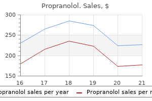
Propranolol dosages: 80 mg, 40 mg, 20 mg
Propranolol packs: 60 pills, 90 pills, 120 pills, 180 pills, 270 pills, 360 pills

Propranolol 20 mg discount amex
Because the ulnar nerve is near 5 arteries bypass buy propranolol 20 mg on line the hook o the hamate quivering arteries propranolol 20 mg discount, the nerve may be injured, inflicting decreased grip strength o the hand. The head o the bone rotates over the distal end o the shat, producing a fexion deormity. Because o the extremely developed sensation within the e ngers, these injuries are extremely painul. Fractures o the proximal and center phalanges are often the end result o crushing or hyperextension accidents. Because o the shut e relationship o phalangeal ractures to the fexor tendons, the bone ragments should be careully realigned to restore regular unction o the ngers. Fracture o Metacarpals the metacarpals (except the 1st) are closely certain collectively; hence, isolated ractures are inclined to be stable. The upper limb is composed o our increasingly cellular segments: the proximal three (shoulder, arm, and orearm) serve primarily to position the ourth section (hand), which is used or greedy, manipulation, and contact. The clavicle serves as a movable crane-like strut (extended support) rom which the scapula and ree limb are suspended at a distance rom the trunk that permits reedom o motion. Shocks acquired by the upper limb (especially the shoulder) are transmitted by way of the clavicle, leading to a racture that mostly happens between its middle and lateral thirds. This triangular at bone is curved to conorm to the thoracic wall and offers giant surace areas and edges or attachment o muscles. These muscular tissues (1) transfer the scapula on the thoracic wall on the physiological scapulothoracic joint and (2) extend to the proximal humerus maintaining the integrity o-and producing movement at-the glenohumeral joint. The coracoid process o the scapula is the site o attachment or the coracoclavicular ligament, which passively helps the higher limb, and a site or muscular (tendon) attachment. Humerus: the lengthy, strong humerus is a mobile strut- the frst in a series o two-used to position the hand at a top (level) and distance rom the trunk to maximize its efciency. The spherical head o the humerus enables a great vary o movement on the mobile scapular base; the trochlea and capitulum at its distal end acilitate the hinge movements o the elbow and, on the similar time, the pivoting o the radius. The long shat o the humerus allows reaching and makes it an eective lever or power in liting, as well as offering surace area or attachment o muscles that act primarily at the elbow. Added surace area or attachment o exors and extensors o the wrist is offered continued on next page 158 Chapter 3 Upper Limb the Bottom Line (continued) by the epicondyles, the medial and lateral extensions o the distal end o the humerus. Because the orearm unit is ormed by two parallel bones, and the radius is ready to pivot about the ulna, supination and pronation o the hand are possible throughout elbow exion. Proximally, the bigger medial ulna orms the primary articulation with the humerus, whereas distally, the shorter lateral radius orms the primary articulation with the hand via the wrist. Hand: Each phase o the upper limb will increase the unctionality o the tip unit, the hand. The carpal bones are organized into two rows o our bones every and, as a group, articulate with the radius proximally and the metacarpals distally. The extremely exible, elongated digits-extending rom a semirigid base (the palm)-enable the flexibility to grip, manipulate, or perorm complicated tasks involving multiple and simultaneous particular person motions. Surace anatomy: the higher limb presents multiple palpable bony eatures that are useul (1) when diagnosing ractures, dislocations, or malormations; (2) or approximating the position o deeper buildings; and (3) or exactly describing the location o incisions and websites or therapeutic puncture, or areas o pathology or harm. The pectoral ascia invests the pectoralis main and is continuous ineriorly with the ascia o the anterior abdominal wall. The pectoral ascia leaves the lateral border o the pectoralis main and becomes the axillary ascia, which orms the foor o the axilla (compartment deep to skin o the armpit). Deep to the pectoral ascia and pectoralis main, another ascial layer, the clavipectoral ascia, descends rom the clavicle, enclosing the subclavius after which pectoralis minor, becoming continuous ineriorly with the axillary ascia. The half o the clavipectoral ascia between the pectoralis minor and subclavius, the costocoracoid membrane, is pierced by the lateral pectoral nerve, which primarily supplies the pectoralis major. The half o the clavipectoral ascia inerior to the pectoralis minor, the suspensory ligament o the axilla, helps the axillary ascia and pulls it and the overlying skin upward during abduction o the arm, orming the axillary ossa (armpit). The scapulohumeral muscles that cover the scapula, and orm the majority o the shoulder, are additionally ensheathed by deep ascia. The deltoid ascia descends over the supercial surace o the deltoid rom the clavicle, acromion, and scapular backbone. From the deep surace o the deltoid ascia, quite a few septa (connective tissue partitions) penetrate between the ascicles (bundles) o the muscle. Ineriorly, the deltoid ascia is continuous with the pectoral ascia anteriorly and the dense inraspinous ascia posteriorly. The muscle tissue that cover the anterior and posterior suraces o the scapula are lined supercially with deep ascia, which is hooked up to the margins o the scapula and posteriorly to the backbone o the scapula. This arrangement creates osseobrous subscapular, supraspinous, and inraspinous compartments; the muscular tissues in each compartment connect to (originate rom) the deep surace o the overlying ascia in part, permitting the muscular tissues to have higher bulk (mass) than could be the case i solely bony attachments occurred. The supraspinous and inraspinous ascia overlying the supraspinatus and inraspinatus muscular tissues, respectively, on the posterior side o the scapula are so dense and opaque that they should be removed throughout dissection to view the muscles. The brachial ascia, a sheath o deep ascia, encloses the arm like a comfortable sleeve deep to the pores and skin and subcutaneous tissue. It is steady superiorly with the deltoid, pectoral, axillary, and inraspinous ascias. The brachial ascia is connected ineriorly to the epicondyles o the humerus and the olecranon o the ulna. This ascia is continuous with the antebrachial ascia, the deep ascia o the orearm. Two intermuscular septa-the medial and lateral intermuscular septa-extend rom the deep surace o the brachial ascia to the central shat and medial and lateral supra-epicondylar ridges o the humerus. Axillary ascia orms the oor o the axilla and is continuous with the pectoral ascia. The pectoral ascia surrounds the pectoralis major, orming the anterior layer o the anterior axillary wall. The clavipectoral ascia extends between the coracoid process o the scapula, the clavicle, and the axillary ascia. The ascial compartments o the higher limb are essential clinically because additionally they include and direct the unfold o inection or hemorrhage within the limb. In the orearm, related ascial compartments are surrounded by the antebrachial ascia and are separated by the interosseous membrane connecting the radius and ulna. The antebrachial ascia thickens posteriorly over the distal ends o the radius and ulna to orm a transverse band, the extensor retinaculum, which retains the extensor tendons in position. The antebrachial ascia also orms an anterior thickening, which is continuous with the extensor retinaculum however is ocially unnamed; some authors identiy it as the palmar carpal ligament. Immediately distal and at a deeper stage to the latter, the antebrachial ascia is also continued as the exor retinaculum (transverse carpal ligament). The deep ascia o the upper limb continues beyond the extensor and fexor retinacula because the palmar ascia.
Fever Grass (Lemongrass). Propranolol.
- How does Lemongrass work?
- Are there safety concerns?
- Stomach and intestinal spasms, stomach ache, high blood pressure, convulsions, pain, vomiting, cough, rheumatism, fever, common cold, exhaustion, headache, use as an antiseptic and astringent, and other uses.
- What is Lemongrass?
- Dosing considerations for Lemongrass.
Source: http://www.rxlist.com/script/main/art.asp?articlekey=96704
Propranolol 20 mg cheap free shipping
Alternately arteries disease in legs buy 20 mg propranolol overnight delivery, when the orearm is pronated kansas cardiovascular center 40 mg propranolol cheap fast delivery, the biceps is the primary (most powerul) supinator o the orearm. The biceps barely operates as a fexor when the orearm is pronated, even in opposition to resistance. Arising rom the supraglenoid tubercle o the scapula, and crossing the pinnacle o the humerus throughout the cavity o the glenohumeral joint, the rounded tendon o the long head o the biceps continues to be surrounded by synovial membrane because it descends within the intertubercular sulcus o the humerus. A broad band, the transverse humeral ligament, passes rom the lesser to the higher tubercle o the humerus and converts the intertubercular groove right into a canal. Distally, the main attachment o the biceps is to the radial tuberosity by way of the biceps tendon. However, a triangular membranous band, the bicipital aponeurosis, runs rom (continued on p. In this dissection o the best arm, the veins have been removed, besides or the proximal part o the axillary vein. Note the courses o the musculocutaneous, median, and ulnar nerves and the brachial artery along the medial (protected) facet o the arm. Their programs typically parallel the medial intermuscular septum that separates the anterior and posterior compartments in the distal two thirds o the arm. In this transverse section o the right arm, the three heads o the triceps and the radial nerve and its companion vessels (in contact with the humerus) lie within the posterior compartment. The pectoralis main and minor muscles are reected superolaterally, and the lateral and medial cords o the brachial plexus are reected superomedially. All major vessels and nerves arising rom the medial and lateral cords o the brachial plexus (except or the musculocutaneous nerve arising rom a phase o the lateral cord) are removed. The posterior cord, ormed by the merging o the posterior divisions o all three trunks o the brachial plexus, is demonstrated. It offers rise to fve peripheral nerves, our o which provide the muscle tissue o the posterior wall o the axilla and posterior compartments o the higher limb. It attaches indirectly by means o the ascia to the subcutaneous border o the ulna. The proximal part o the aponeurosis may be simply elt the place it passes obliquely over the brachial artery and median nerve. The aponeurosis aords safety or these and other buildings within the cubital ossa. It additionally helps lessen the stress o the biceps tendon on the radial tuberosity during pronation and supination o the orearm. To test the biceps brachii, the elbow joint is fexed towards resistance when the orearm is supinated. It acts throughout both slow and quick actions and within the presence or absence o resistance. Unlike the biceps, the brachialis fexes the orearm in all the coracobrachialis is an elongated muscle in the superomedial part o the arm. For example, the musculocutaneous nerve pierces it, and the distal part o its attachment signifies the location o the nutrient oramen o the humerus. The coracobrachialis helps fex and adduct the arm and stabilize the glenohumeral joint. Because its long head crosses the glenohumeral joint, the triceps helps stabilize the adducted glenohumeral joint by serving as a shunt muscle, resisting inerior displacement o the top o the humerus. The medial head is the workhorse o orearm extension, energetic in any respect speeds and within the presence or absence o resistance. The lateral head is the strongest but is it recruited into activity primarily towards resistance (Hamill and Knutzen, 2014). Just proximal to the distal attachment o the triceps is a riction-reducing subtendinous olecranon bursa, between the triceps tendon and the olecranon. Its strength ought to be comparable with the contralateral muscle, given consideration or lateral dominance (right or let handedness). The lateral head o the triceps brachii is split and displaced to present the buildings traversing the quadrangular space and the radial nerve and prounda brachii artery. The exposed bone o the radial groove, which is devoid o muscular attachment, separates the humeral attachments o the lateral and medial heads o the triceps. The anconeus assists the triceps in extending the orearm and tenses the capsule o the elbow joint, stopping its being pinched during extension. It can additionally be stated to exert an abducting orce on the ulna during pronation o the orearm. Brachial Artery the brachial artery offers the main arterial supply to the arm and is the continuation o the axillary artery. The brachial artery, comparatively supercial and palpable throughout its course, lies anterior to the triceps and brachialis. At rst, it lies medial to the humerus where its pulsations are palpable in the medial bicipital groove. It then passes anterior to the medial supra-epicondylar ridge and trochlea o the humerus. As it passes inerolaterally, the brachial artery accompanies the median nerve, which crosses anterior to the artery. During its course via the arm, the brachial artery provides rise to many unnamed muscular branches, and the humeral nutrient artery. The main named branches o the brachial artery arising rom its medial side are the prounda brachii artery and the superior and inerior ulnar collateral arteries. The collateral arteries assist orm the peri-articular arterial anastomoses o the elbow area. Other arteries involved are recurrent branches, sometimes double, rom the radial, ulnar, and interosseous arteries, which run superiorly anterior and posterior to the elbow joint. These arteries anastomose with descending articular branches o the deep artery o the arm and the ulnar collateral arteries. The prounda brachii accompanies the radial nerve alongside the radial groove as it passes posteriorly across the shat o the humerus. The prounda brachii terminates by dividing into center and radial collateral arteries, which participate within the periarticular arterial anastomoses across the elbow. Here, it anastomoses with the posterior ulnar recurrent and inerior ulnar collateral arteries, participating within the peri-articular arterial anastomoses o the elbow. It then passes ineromedially anterior to the medial epicondyle o the humerus and joins the peri-articular arterial anastomoses o the elbow region by anastomosing with the anterior ulnar recurrent artery. Veins o Arm Two sets o veins o the arm, supercial and deep, anastomose reely with each other. The supercial veins are in the subcutaneous tissue, and the deep veins accompany the arteries. The ensuing collateral circulation permits blood to reach the orearm when exion o the elbow compromises ow via the terminal part o the brachial artery. In this deep dissection, half o the biceps is excised and the cubital ossa is opened extensively by retracting the orearm extensor muscular tissues laterally and the exor muscle tissue medially.
Propranolol 20 mg discount visa
Between the fexor tendons and the ascia covering the deep palmar muscle tissue are two potential spaces cardiovascular system glucose cheap propranolol 80 mg, the thenar area and the midpalmar area arteries 2014 ryerson propranolol 80 mg buy line. The spaces are bounded by brous septa passing rom the edges o the palmar aponeurosis to the metacarpals. Between the two spaces is the especially sturdy lateral brous septum, which is attached to the third metacarpal. Although most ascial compartments finish at the joints, the midpalmar area is continuous with the anterior compartment o the orearm via the carpal tunnel. Thenar muscle tissue within the thenar compartment: abductor pollicis brevis, fexor pollicis brevis, and opponens pollicis. Hypothenar muscle tissue within the hypothenar compartment: abductor digiti minimi, fexor digiti minimi brevis, and opponens digiti minimi. Short muscles o the hand, the lumbricals, in the central compartment with the lengthy fexor tendons. The proximal end or apex o the triangular palmar aponeurosis is steady with the fexor retinaculum and the palmaris longus tendon. When the palmaris longus is present, the palmar aponeurosis is the expanded tendon o the palmaris longus. Distal to the apex, the palmar aponeurosis orms our longitudinal digital bands or rays that radiate rom the apex and connect distally to the bases o the proximal phalanges and become continuous with the brous digital sheaths. The high degree o reedom o the actions outcomes rom the 1st metacarpal being unbiased, with cell joints at both ends. Transverse section via the middle o the palm illustrating the ascial compartments o the hand. The midpalmar space underlies the central compartment o the palm and is said distally to the synovial tendon sheaths o the 3rd�5th digits and proximally to the frequent exor sheath as it emerges rom the carpal tunnel. The thenar space underlies the thenar compartment and is expounded distally to the synovial tendon sheath o the index fnger and proximally to the widespread exor sheath distal to the carpal tunnel. This movement happens on the carpometacarpal joint and ends in a "cupping" o the palm. Bringing the tip o the thumb into contact with the fifth nger, or any o the opposite ngers, involves significantly extra motion than may be produced by the opponens pollicis alone. The rst our movements o the thumb happen at the carpometacarpal and metacarpophalangeal joints. Opposition, a posh motion, begins with the thumb within the prolonged place and initially entails abduction and medial rotation o the first metacarpal (cupping the palm) produced by the motion o the opponens pollicis on the carpometacarpal joint and then fexion on the metacarpophalangeal joint. In pulp-to-pulp opposition, actions o the nger opposing the thumb are additionally involved. This may be confrmed by noting the course the nail o the thumb aces in contrast with the nails o the other fngers. Thus, abduction and adduction occur in a sagittal airplane and exion and extension happen in a coronal plane. Opposition, the motion bringing the tip o the thumb in touch with the pulps o the opposite fngers. The elements o opposition are abduction and medial rotation on the carpometacarpal joint and exion o the metacarpophalangeal joint. The pores and skin and subcutaneous tissue have been removed, as have most o the palmar aponeurosis and the thenar and hypothenar asciae. The superfcial palmar arch is situated instantly deep to the palmar aponeurosis, superfcial to the lengthy exor tendons. Three thenar and three hypothenar muscular tissues attach to the exor retinaculum and to the our marginal carpal bones united by the retinaculum. It fexes and rotates the first metacarpal medially on the carpometacarpal joint during opposition; this motion occurs when choosing up an object. The an-shaped muscle has two heads o origin, which are separated by the radial artery as it enters the palm to orm the deep palmar arch. Deep dissection o the palm revealing the anastomosis o the palmar carpal branch o the radial artery with the palmar carpal branch o the ulnar artery to orm the palmar carpal arch and deep palmar arch. The abductor digiti minimi is probably the most supercial o the three muscles orming the hypothenar eminence. The abductor digiti minimi abducts the fifth nger and helps fex its proximal phalanx. The exor digiti minimi brevis is variable in dimension; it lies lateral to the abductor digiti minimi. The fexor digiti minimi brevis fexes the proximal phalanx o the fifth nger on the metacarpophalangeal joint. The opponens digiti minimi is a quadrangular muscle that lies deep to the abductor and lexor muscles o the fifth inger. The opponens digiti minimi attracts the 5th metacarpal anteriorly and rotates it laterally, thereby deepening the hole o the palm and bringing the 5th inger into opposition with the thumb. Like the opponens pollicis, the opponens digiti minimi acts exclusively at the carpometacarpal joint. The palmaris brevis is a small, thin muscle in the subcutaneous tissue o the hypothenar eminence. The palmaris brevis wrinkles the skin o the hypothenar eminence and deepens the hollow o the palm, thereby aiding the palmar grip. It is attached proximally to the medial border o the palmar aponeurosis and to the pores and skin on the medial border o the hand. The lumbricals fex the ngers at the metacarpophalangeal joints and extend the interphalangeal joints. The examiner uses one nger to apply resistance along the palmar surace o the proximal phalanx o digits 2�5 individually. The our dorsal interosseous muscular tissues (dorsal interossei) are situated between the metacarpals; the three palmar interosseous muscles (palmar interossei) are on the palmar suraces o the metacarpals within the interosseous compartment o the hand. The 1st dorsal interosseous muscle is easy to palpate; oppose the thumb rmly in opposition to the index nger and it can be easily elt. The our dorsal interossei abduct the ngers, and the three palmar interossei adduct them. Acting collectively, the dorsal and palmar interossei and the lumbricals produce lexion on the metacarpophalangeal joints and extension o the interphalangeal joints (the socalled Z-movement). This happens because o their attachment to the lateral bands o the extensor expansions. The individual is asked to "maintain the ngers together" to stop the paper rom being pulled away by the examiner. To take a look at the dorsal interossei, the examiner holds adjacent prolonged and adducted ngers between thumb and middle nger, offering resistance as the person attempts to abduct the ngers (the person is requested to "unfold the ngers aside").
20 mg propranolol order overnight delivery
The size o the bile duct varies rom 5 to 15 cm coronary heart anatomy order propranolol 20 mg otc, depending on where the cystic duct joins the widespread hepatic duct blood vessels in spanish propranolol 40 mg discount with amex. The bile duct descends posterior to the superior part o the duodenum and lies in a groove on the posterior surace o the head o the pancreas. On the let side o the descending part o the duodenum, the bile duct comes into contact with the principle pancreatic duct. These ducts run obliquely by way of the wall o this part o the duodenum, where they unite, orming a dilation, the hepatopancreatic ampulla. The distal end o the ampulla opens into the duodenum via the main duodenal papilla. The circular muscle around the distal end o the bile duct is thickened to orm the sphincter o the bile duct (L. The veins o the gallbladder neck talk with cystic veins along the cystic and biliary ducts. Small cystic veins move rom the adherent portion o the gallbladder into the sinusoids o the liver. The relationship o the gallbladder to the duodenum is so intimate that the superior part o the duodenum is normally stained with bile in the cadaver. Because the liver and gallbladder must be retracted superiorly to expose the gallbladder. Magnetic resonance cholangiopancreatography o the gallbladder, bile passages, pancreatic duct and descending part o duodenum. Peritoneum fully surrounds the undus o the gallbladder and binds its body and neck to the liver. The hepatic surace o the gallbladder attaches to the liver by connective tissue o the brous capsule o the liver. Body: main portion that contacts the visceral surace o the liver, transverse colon, and superior part o the duodenum. Neck: narrow, tapering end, opposite the undus and directed toward the porta hepatis; it typically makes an S-shaped bend and joins the cystic duct. The cystic duct (3�4 cm long) connects the neck o the gallbladder to the common hepatic duct. The spiral old helps maintain the cystic duct open; thus, bile can easily be diverted into the gallbladder when the distal finish o the bile duct is closed by the sphincter o the bile duct and/or hepatopancreatic sphincter, or bile can move to the duodenum because the gallbladder contracts. The spiral old additionally oers additional resistance to sudden dumping o bile when the sphincters are closed, and intra-abdominal pressure is suddenly increased, as throughout a sneeze or cough. The cystic duct passes between the layers o the lesser omentum, usually parallel to the widespread hepatic duct, which it joins to orm the bile duct. The cystic artery generally arises rom the best hepatic artery within the triangle between the widespread hepatic duct, cystic duct, and visceral surace o the liver, the cystohepatic triangle (o Calot). Right hepatic department and duct the venous drainage rom the neck o the gallbladder and cystic duct fows through the cystic veins. These small and usually a number of veins enter the liver immediately or drain through the hepatic portal vein to the liver, ater becoming a member of the veins draining the hepatic ducts and proximal bile duct. The veins rom the undus and body o the gallbladder cross immediately into the visceral surace o the liver and drain into the hepatic sinusoids. Because that is drainage rom one capillary (sinusoidal) bed to one other, it constitutes an additional (parallel) portal system. The proper phrenic nerve (somatic aerent bers) could carry ache caused by gallbladder infammation. Parasympathetic stimulation causes contractions o the gallbladder and rest o the sphincters on the hepatopancreatic ampulla. In most people, Right hepatic branch and duct Left hepatic branch and duct Right hepatic branch and duct Left hepatic department and duct Left hepatic branch and duct Common hepatic duct Cystic artery Cystic duct Bile duct Gastroduodenal artery (A) 75. The cystic artery normally arises rom the right hepatic artery within the cystohepatic triangle (o Calot), bounded by the cystic duct, frequent hepatic duct, and visceral surace o the right liver. Anastomoses present a collateral circulation in instances o obstruction within the liver or portal vein. Here, the portal tributaries are darker blue and systemic tributaries are lighter blue. A is between the submucosal esophageal veins draining into either the azygos vein (systemic) or the let gastric vein (portal); when dilated, these are esophageal varices. B is between the inerior and center rectal veins draining into the inerior vena cava (systemic) and the superior rectal vein, persevering with as the inerior mesenteric vein (portal). The submucosal veins concerned are normally dilated (varicose in appearance), even in newborns. D is on the posterior aspects (bare areas) o secondarily retroperitoneal viscera, or the liver, the place twigs o visceral veins-or instance, the colic vein, splenic veins, or the portal vein itsel (portal system)-anastomose with retroperitoneal veins o the posterior abdominal wall or diaphragm (systemic system). As it approaches the porta hepatis, the hepatic portal vein divides into right and let branches. The hepatic portal vein collects blood with reduced oxygenation but wealthy in nutrients rom the abdominal part o the alimentary system, together with the gallbladder and pancreas, in addition to the spleen, and carries it to the liver. Within the liver, its branches are distributed in a segmental pattern (see "Blood Vessels o Liver") and finish in expanded capillaries, the venous sinusoids o the liver. Portal�systemic anastomoses, by which the portal venous system communicates with the systemic venous system, are ormed in the submucosa o the inerior esophagus, in the submucosa o the anal canal, in the peri-umbilical area, and on the posterior features (bare areas) o secondarily retroperitoneal viscera, or the liver. However, the amount o blood orced by way of the collateral routes may be extreme, leading to probably atal varices (abnormally dilated veins) (see the Clinical Box "Portal Hypertension," p. Blunt trauma to the let aspect or to other areas o the abdomen that trigger a sudden, marked enhance in intra-abdominal stress. The shut relationship o the spleen to the ribs that usually shield it might be detrimental when there are rib ractures. Severe blows on the let side might racture a number of o these ribs, and rupture the underlying spleen, or sharp bone ragments might lacerate the spleen. When the spleen is diseased, resulting rom, or example, granulocytic leukemia (high leukocyte and white blood cell count), it may enlarge to 10 or extra occasions its regular measurement and weight (splenomegaly). Generally, i its decrease edge could be detected when palpating below the let costal margin at the end o inspiration. Accessory Spleen(s) and Splenosis One or extra small accent spleens may develop prenatally near the splenic hilum. They could also be e embedded partly or wholly within the tail o the pancreas, between the layers o the gastrosplenic ligament, in n the inracolic compartment, in the mesentery, or in shut proximity to an ovary or testis. Accessory spleens are relatively widespread, are normally small (approximately 1 cm in diameter, and range rom 0. Awareness o the potential presence o an accessory spleen is necessary as a outcome of i not eliminated throughout a splenectomy, the signs that indicated elimination o the spleen. Splenosis-generalized autoimplantation o ectopic splenic tissue into the peritoneum, omentum, or mesenteries- typically ollows splenic rupture. This potential space descends to the extent o the 10th rib in the midaxillary line. Its existence have to be kept in thoughts when doing a splenic needle biopsy, or when injecting radiopaque material into the spleen or visualization o the hepatic portal vein (splenoportography). Blockage o Hepatopancreatic Ampulla and Pancreatitis Because the principle pancreatic duct joins the bile duct to orm the hepatopancreatic ampulla and pierces the duodenal wall, a gallstone passing along the extrahepatic bile passages might lodge in the constricted distal finish o the ampulla, where it opens at the summit o the major duodenal papilla.
Propranolol 20 mg buy with mastercard
As well as allowing motion between adjoining vertebrae blood vessels in 1 pound of fat generic 20 mg propranolol visa, their resilient deormability allows them to serve as shock absorbers arteries used to count pulse rate 20 mg propranolol order fast delivery. The anuli insert into the graceful, rounded epiphysial rims on the articular suraces o the vertebral bodies ormed by the used anular epiphyses. The bers orming every lamella run obliquely rom one vertebra to another, about 30 or extra levels rom vertical. The bers o adjacent lamellae cross each other obliquely in opposite instructions at angles o greater than 60�. The anulus becomes decreasingly vascularized centrally, and only the outer third o the anulus receives sensory innervation. At birth, these pulpy nuclei are about 88% water and are initially more cartilaginous than brous. The nuclei turn into broader when compressed and thinner when tensed or stretched (as when hanging or suspended). Compression and tension occur concurrently in the identical disc throughout anterior and lateral fexion and extension o the vertebral column. During these movements, as properly as during rotation, the turgid nucleus acts as a semifuid ulcrum. The superfcial layers o the anulus have been minimize and spread apart to present the direction o the fbers. The fbrogelatinous nucleus pulposus occupies the middle o the disc and acts as a cushion and shock-absorbing mechanism. The pulpy nucleus attens and the anulus bulges when weight is utilized, as occurs throughout standing and more so throughout liting. The anulus is concurrently positioned underneath compression on one aspect and rigidity on the opposite. The nucleus pulposus is avascular; it receives its nourishment by diusion rom blood vessels at the periphery o the anulus brosus and vertebral physique. However, their thickness relative to the dimensions o the our bodies they join is most clearly associated to the range o motion, and relative thickness is best in the cervical and lumbar areas. The discs are thicker anteriorly within the cervical and lumbar regions, their various shapes producing the secondary curvatures o the vertebral column. Uncovertebral "joints" or clets (o Luschka) commonly develop between the unci o the bodies o C3 or C4� C6 or C7 vertebrae and the beveled inerolateral suraces o the vertebral our bodies superior to them ater 10 years o age. The articulating suraces o these joint-like buildings are lined with cartilage moistened by fuid contained inside an interposed potential space, or "capsule. The uncovertebral "joints" are requent websites o bone spur ormation in later years, which can trigger neck pain. These small, synovial joint-like structures are between the unci o the bodies o the lower vertebrae and the beveled suraces o the vertebral our bodies superior to them. The inerior thoracic (T9�T12) and superior lumbar (L1�L2) vertebrae, with related discs and ligaments, are shown. The pedicles o the T9�T11 vertebrae have been sawn through and their our bodies and intervening discs eliminated to present an anterior view o the posterior wall o the vertebral canal. This ligament prevents hyperextension o the vertebral column, maintaining stability o the joints between the vertebral our bodies. The posterior longitudinal ligament is a much narrower, somewhat weaker band than the anterior longitudinal ligament. The posterior longitudinal ligament runs within the vertebral canal along the posterior facet o the vertebral our bodies. This ligament weakly resists hyperfexion o the vertebral column and helps stop or redirect posterior herniation o the nucleus pulposus. The joint capsule is connected to the margins o the articular suraces o the articular processes o adjoining vertebrae. Accessory ligaments unite the laminae, transverse processes, and spinous processes and assist stabilize the joints. The zygapophysial joints allow gliding actions between the articular processes; the shape and disposition o the articular suraces decide the kinds o motion attainable. The zygapophysial joints are innervated by articular branches that come up rom the medial branches o the posterior rami o spinal nerves. As these nerves move postero-ineriorly, they lie in grooves on the posterior suraces o the medial elements o the transverse processes. Each articular department provides two adjoining joints; thereore, each joint is equipped by two nerves. These articulations are aircraft synovial joints between the superior and inerior articular processes (G. Those in the cervical area are particularly skinny and loose, the laminae o adjacent vertebral arches are joined by broad, pale yellow bands o elastic tissue, the ligamenta ava (L. Superior to the distinguished spinous course of o C7 (vertebra prominens), the spinous processes are deeply positioned and connected to an overlying nuchal ligament. The pedicles o the superior two vertebrae have been sawn via and the vertebral arches eliminated to reveal the posterior longitudinal ligament. Intertransverse, supraspinous, and interspinous ligaments are demonstrated in association with the vertebrae with intact vertebral arches. The intertransverse ligaments, connecting adjoining transverse processes, consist o scattered bers within the cervical region and brous cords within the thoracic area. The medial department offers rise to articular branches which might be distributed to the zygapophysial joint at that degree and to the joint one degree inerior to its exit. Thus, every zygapophysial joint receives articular rami rom the medial department o the posterior rami o two adjacent spinal nerves. The medial branches o each posterior rami need to be ablated to denervate a zygapophysial joint. The faval ligaments bind the lamina o the adjoining vertebrae collectively, orming alternating sections o the posterior wall o the vertebral canal. The ligamenta fava are lengthy, thin, and broad in the cervical region, thicker in the thoracic region, and thickest within the lumbar region. The strong, elastic yellow ligaments assist preserve the normal curvatures o the vertebral column and assist with straightening o the column ater fexing. Adjoining spinous processes are united by weak, oten membranous interspinous ligaments and robust brous supraspinous ligaments. The skinny interspinous ligaments connect adjoining spinous processes, attaching rom the foundation to the apex o each process. The cordlike band orming the supraspinous ligaments connects the tips o the spinous processes rom C7 to the sacrum and merge superiorly with the nuchal ligament on the back o the neck (Fr. It extends as a median band rom the external occipital protuberance and posterior border o the oramen magnum to the spinous processes o the cervical vertebrae. Because o the shortness and depth o the C3�C5 spinous processes, the nuchal ligament provides There are two sets o craniovertebral joints, the atlantooccipital joints, ormed between the atlas (C1 vertebra) and the occipital bone o the cranium, and the atlanto-axial joints, ormed between the atlas and axis (C2 vertebra). Their design provides a wider vary o movement than in the rest o the vertebral column.
Propranolol 20 mg buy low price
Pronation rotates the radius medially in order that the palm o the hand aces posteriorly and its dorsum aces anteriorly 403 cardiovascular disease project propranolol 40 mg purchase with amex. When the elbow joint is fexed cardiovascular location buy discount propranolol 20 mg on line, pronation strikes the hand so that the palm aces ineriorly. Supination is the opposite rotational motion, rotating the radius laterally and uncrossing it rom the ulna, returning the pronated orearm to the anatomical position. When the elbow joint is fexed, supination moves the hand so that the palm aces superiorly. Inversion moves the only o the oot towards the median airplane (acing the sole medially). Pronation o the oot actually reers to a mix o eversion and abduction that ends in reducing o the medial margin o the oot (the eet o an individual with fats eet are pronated), and supination o the oot usually implies movements resulting in raising the medial margin o the oot, a combination o inversion and adduction. Opposition is the movement by which the pad o the 1st digit (thumb) is brought to one other digit pad. Reposition describes the motion o the 1st digit rom the place o opposition back to its anatomical position. Protrusion is a movement anteriorly (orward) as in protruding the mandible (chin), lips, or tongue. Retrusion is a movement posteriorly (backward), as in retruding the mandible, lips, or tongue. The related phrases protraction and retraction are used mostly or anterolateral and posteromedial movements o the scapula on the thoracic wall, inflicting the shoulder region to transfer anteriorly and posteriorly. Elevation raises or moves a part superiorly, as in elevating the shoulders when shrugging, the upper eyelid when opening the attention, or the tongue when pushing it up against the palate (roo o mouth). Depression lowers or strikes an element ineriorly, as in depressing the shoulders when standing comfy, the upper eyelid when closing the eye, or pulling the tongue away rom the palate. Anatomical variations are oten found throughout imaging or surgical procedures, at autopsy, or during anatomical research in individuals who had no awareness o or adverse eects rom the variation. A congenital anomaly or birth deect is a variation oten evident at birth or soon aterward as a end result of aberrant orm or unction. It is necessary to understand how such variations and anomalies may infuence physical examinations, prognosis, and therapy. However, sometimes a specific structure demonstrates a lot variation inside the regular vary that the commonest pattern is ound lower than hal the time! Oten, college students ignore the variations or inadvertently damage them by trying to produce conormity. In a random group o folks, individuals obviously dier supercially rom each other in physical look. The bones o the skeleton vary not solely in dimension but extra subtly in their basic form and in lesser details o surace structure. Similarly, appreciable variation exists in the patterns o branching o neurovascular structures (veins, arteries, and nerves). However, the requency o variation oten diers among human groups, and variations collected in a single inhabitants may not apply to members o another population. Some variations, similar to these occurring within the origin and course o the cystic artery to the gallbladder, are clinically signiicant (see Chapter 5, Abdomen). Being aware o these variations is essential in medical apply, particularly surgical procedure. Clinically signicant variations are described in scientific correlation (blue) packing containers identied with an Anatomical Variation icon (at let). Humans exhibit considerable genetic variation beyond sexual and racial dierences, similar to polydactyly (extra digits). Discovering anatomical variations in cadavers is actually one o the numerous benets o rsthand dissection, as a end result of it permits students to develop an consciousness o the incidence o variations and a way o their requency. The pores and skin offers: Protection o the body rom environmental eects, similar to abrasions, fuid loss, harmul substances, ultraviolet radiation, and invading microorganisms. Thermal regulation via the evaporation o sweat and/ or the dilation or constriction o supercial blood vessels. The dermis is a keratinized epithelium-that is, it has a tricky, sexy supercial layer that provides a protective outer surace overlying its regenerative and pigmented deep or basal layer. The dermis is supplied by arteries that enter its deep surace to orm a cutaneous plexus o anastomosing arteries. The pores and skin can be provided with aerent nerve endings which are sensitive to contact, irritation (pain), and temperature. Vascular and lymphatic capillary beds in superficial dermis Afferent nerve endings Small arteriole feeding vascular capillary bed Collagen and elastin fibers Arrector muscle of hair Sebaceous gland Hair follicle Fat Cutaneous nerve Lymphatic vessel Superficial blood vessels Hair Basal (regenerating) layer of epidermis Epidermis Dermis Subcutaneous tissue (superficial fascia) Deep fascia Skeletal muscle Skin ligament (L. Integumentary System thirteen the dermis is a dense layer o interlacing collagen and elastic bers. Although the bundles o collagen bers in the dermis run in all instructions to produce a tough eltlike tissue, in any specic location most bers run in the identical direction. The predominant sample o collagen bers determines the characteristic tension and wrinkle lines in the skin. The tension traces (also referred to as cleavage strains or Langer lines) are inclined to spiral longitudinally within the limbs and run transversely in the neck and trunk. Tension lines at the elbows, knees, ankles, and wrists are parallel to the transverse creases that appear when the limbs are fexed. The deep layer o the dermis contains hair ollicles, with associated easy arrector muscle tissue and sebaceous glands. Hair ollicles are typically slanted to one aspect, and a variety of other sebaceous glands lie on the facet the hair is directed toward ("points to") because it emerges rom the skin. Thus, contraction o the arrector muscles causes the hairs to rise up straighter, thereby compressing the sebaceous glands and serving to them secrete their oily product onto the skin surace. The evaporation o the watery secretion (sweat) o the sweat glands rom the skin provides a thermoregulatory mechanism or heat loss (cooling). Also concerned in the loss or retention o body warmth are the small arteries (arterioles) within the dermis. They dilate to ll supercial capillary beds to radiate heat (skin seems red) or constrict to reduce surace heat loss (skin, especially o the lips and ngertips, seems blue). Other skin buildings or derivatives include the nails (ngernails, toenails), the mammary glands, and the enamel o tooth. Located between the overlying pores and skin (dermis) and underlying deep ascia, the subcutaneous tissue (supercial ascia) consists principally o loose connective tissue and saved at and incorporates sweat glands, supercial blood vessels, lymphatic vessels, and cutaneous nerves. The neurovascular buildings o the integument (cutaneous nerves, supercial vessels) course within the subcutaneous tissue, distributing only their terminal branches to the pores and skin. In addition, the distribution o subcutaneous tissue varies significantly in dierent websites in the same 1 An incision made throughout the cleavage lines is more likely to gape, growing therapeutic time, and end in elevated scar tissue.
Syndromes
- Lose weight
- Washing of the skin (irrigation) -- perhaps every few hours for several days
- Norpramin
- Slowed respirations
- Nosebleeds
- Take drugs to lower your cholesterol, if needed.
- Rocky Mountain spotted fever
- A lower jaw that is short compared to the upper jaw (retrognathia)
- Chromosome study to look for changes (mutations) in the ABCD1 gene
Propranolol 80 mg generic visa
Tibial ractures in children are extra critical i they contain the epiphysial plates as a result of continued regular growth o the bone could additionally be jeopardized blood vessels yahoo propranolol 40 mg purchase mastercard. The tibial tuberosity often orms by inerior bone growth rom the superior epiphysial heart at roughly 10 years o age cardiovascular disease journal generic 80 mg propranolol with mastercard, but a separate middle or the tibial tuberosity may appear at roughly 12 years o age. Disruption o the epiphysial plate on the tibial tuberosity might trigger infammation o the tuberosity and continual recurring ache during adolescence (Osgood-Schlatter disease), especially in younger athletes. Eversion (A) Tibial and fibular fractures Fibular Fractures Fibular ractures generally happen 2�6 cm proximal to the distal end o the lateral malleolus and are oten associated with racture�dislocations o the ankle joint, that are combined with tibial ractures. When a person slips and the oot is orced into an excessively inverted position, the ankle ligaments tear, orcibly tilting the talus against the lateral malleolus, and should shear it o. Even ater a segment o the shat has been removed, walking, running, and jumping can be normal. Awareness o the situation o the nutrient oramen in the bula is essential when perorming ree vascularized bular transers. Because the nutrient oramen is situated in the center third o the bula generally. The needle is inserted into the fats space o bone approximately 2 cm distal and barely medial rom the tibial tuberosity. Special needles designed or handbook insertion are used; battery-powered or impact-driven devices also can be found to assist assist insertion. Calcaneal Fractures A hard all onto the heel, rom a ladder, or example, might racture the calcaneus into several pieces, producing a comminuted racture. A calcaneal racture is usually disabling because it disrupts the subtalar (talocalcaneal) joint, where the talus articulates with the calcaneus. It is used primarily in cases o traumatic shock and in children with circulatory collapse. Metatarsal ractures are additionally common in dancers, particularly emale ballet dancers who use the demi-pointe approach. These ractures, often transverse, end result rom repeated stress on the metatarsals. When the oot is abruptly and violently inverted, the tuberosity o the 5th metatarsal could also be avulsed (torn away) by the tendon o the bularis brevis muscle. This injury produces pain and edema on the base o the 5th metatarsal and could additionally be related to a extreme ankle sprain. This ailure could additionally be attributable to utilized stress (orceul plantarfexion) in the course of the early teenagers. Occasionally, a partly and even ully ossied center could racture and progress to nonunion. Either occasion could lead to a bone (accessory ossicle) known as an os trigonum, which occurs in 14�25% o adults, extra commonly bilaterally. Each hip bone is specialized to receive hal the weight o the upper physique when standing and all o it periodically during walking. Thin parts o the bone present a broad surace or attachment o powerul muscle tissue that move the emur. The pelvic girdle encircles and protects the pelvic viscera, particularly the reproductive organs. Femur: Through development, our largest bone, the emur, has developed a bend (angle o inclination) and has twisted (medial rotation and torsion in order that the knee and all joints inerior to it ex posteriorly) to accommodate our erect posture and to enable bipedal strolling and working. The angle o inclination and attachment o the abductors and rotators to the higher trochanter enable elevated leverage, superior placement o the abductors, and oblique orientation o the emur in the thigh. Combined with the torsion angle, indirect rotatory movements on the hip joint are transformed into actions o exion�extension and abduction�adduction (in the sagittal and coronal planes, respectively) in addition to o rotation. Patella: the patella is a triangular bone that articulates posteriorly with the distal emur. It is a sesamoid bone in the tendon o the quadriceps emoris muscle, providing the muscle with mechanical benefit in extending the knee. Tibia and bula: Our second largest bone, the tibia, is a vertical column bearing the burden o all superior to it. Through development, the 2 bones have turn into permanently pronated to present or a steady stance and acilitate locomotion. Bones o oot: the various bones o the oot orm a unctional unit that allows weight to be distributed to a wide platorm to keep stability when standing, allow conormation and adjustment to terrain variations, and perorm shock absorption. They also transer weight rom the heel to the oreoot as required in walking and running. The subcutaneous tissue o the hip and thigh is steady with that o the inerior part o the anterolateral belly wall and buttocks. At the knee, the subcutaneous tissue loses its at and blends with the deep ascia, however at is again present distal to the knee in the subcutaneous tissue o the leg. The deep ascia o the decrease limb is very sturdy, investing the limb like an elastic stocking. This ascia limits outward enlargement o contracting muscle tissue, making muscular contraction extra ecient in compressing veins to push blood towards the heart. The medial margin o the opening is easy but its superior, lateral, and inerior margins orm a pointy crescentic edge, the alciorm margin. The connective tissue is pierced by quite a few openings (thus its name) or the passage o eerent lymphatic vessels rom the supercial inguinal lymph nodes and by the great saphenous vein and its tributaries. Ater passing through the saphenous opening and cribriorm ascia, the good saphenous vein enters the emoral vein. Superiorly, the ascia lata attaches to and is steady with the inguinal ligament, pubic arch, physique o pubis, and pubic tubercle anteriorly. Ineriorly, the ascia lata attaches to and is steady with uncovered parts o bones across the knee. This broad band o bers is the shared aponeurosis o the tensor asciae latae and gluteus maximus muscle tissue. The iliotibial tract extends rom the iliac tubercle to the anterolateral tubercle o the tibia (Gerdy tubercle). The partitions o these compartments are ormed by the ascia lata and three ascial intermuscular septa that arise rom its deep side and connect to the linea aspera o the emur. The lateral intermuscular septum extends deeply rom the iliotibial tract to the lateral lip o the linea aspera and lateral supracondylar line o the emur. The deep ascia o the leg is thick in the proximal part o the anterior side o the leg, where it orms half o the proximal attachments o the underlying muscle tissue. Although thinner distally, the deep ascia o the leg orms thickened bands each superior and anterior to the ankle joint, the extensor retinacula. Anterior and posterior intermuscular septa cross rom the deep surace o the lateral deep ascia o the leg and connect to the corresponding margins o the bula.
Propranolol 80 mg buy without a prescription
It normally decreases substantially within the first days of life with a unbroken necessary decline over the next 4�6 weeks and a slow gradual decline beyond that for a number of months cardiovascular system gas exchange propranolol 80 mg buy cheap on-line. Over the first 4�6 weeks of life coronary heart causes purchase propranolol 80 mg without prescription, an more and more loud murmur might develop and is often accompanied by the onset of symptoms of congestive coronary heart failure. Thus the kid will turn out to be increasingly tachypneic, notably associated to feeding. Physical examination normally demonstrates hepatomegaly in addition to tachycardia, a hyperactive precordium and pansystolic murmur. It is usually potential to manage the signs of congestive coronary heart failure with acceptable medical therapy, together with digoxin and lasix. This tissue will lower the scale of the defect and will ultimately result in its full closure. Because of the massive quantity of blood returning to the left atrium, the foramen ovale could turn out to be "stretched" and permit a left to proper shunt at the atrial level along with the ventricular degree shunt. With additional progression of vascular illness, pulmonary arterioles become fibrosed and even occluded with thrombus. It is tough to predict which children will develop an early and accelerated type of pulmonary vascular disease. Over the final decade, quite a few innovative units have been developed that are designed to keep away from injury to these structures. The greater pressure within the left ventricle serves to seal the device against the ventricular septum. In the past, a big sheath measurement was required which necessitated the kid being no much less than 8�10 kg in weight. Since this subsequently requires the kid to undergo a surgical procedure for removal of the pulmonary artery band and reconstruction of the principle pulmonary artery, our most well-liked strategy within the small symptomatic toddler is to proceed with one-stage surgical closure (see below). This avoids the risk of late distortion of the pulmonary arteries consequent to the band, as nicely as the extra costs of a tool and additional process. The pulmonary arteries are outstanding and the lung fields are congested and plethoric. This is in contrast to the scenario with cineangiography within the catheterization laboratory the place generally a restricted number of dye injections are made. Very small VsD within the teenager or younger aDult There is ongoing controversy regarding the need to shut very small defects. On the opposite hand, others argue that the continuing threat of bacterial endocarditis, in addition to the need for regular surveillance for monitoring of the aortic valve, argue in favor of surgical closure of the very small defect by the time a child reaches midteenage years. Symptoms not uncommonly embody failure to thrive with the kid falling progressively off the growth curve, in addition to frequent respiratory infections. Often a defect that leads to greater than 50% systemic stress within the pulmonary arteries is clearly a large defect which in all probability has a low chance of spontaneous closure. If imaging demonstrates accumulation of fibrous tissue significantly as a windsock or aneurysm within the membranous area, then there could additionally be the next probability of spontaneous closure. Rather remarkably for the time, five of the primary eight sufferers were infants and three of those survived. Barratt-Boyes was capable of demonstrate that preliminary floor cooling adopted by brief cooling on cardiopulmonary bypass, then a period of circulatory arrest with subsequent rewarming using a mixture of cardiopulmonary bypass and floor warming could probably be undertaken with a remarkably low mortality. Device closure within the catheterization laboratory was described by Lock and colleagues in 1987. Therefore, the kid must be closely monitored with common examination together with echocardiography to observe the competence and anatomy of the aortic valve. Hopefully, there will be evidence of progressive closure of the defect which is able to encourage an strategy of ongoing conservative remedy quite than proceeding to surgery. Caval cannulation with thin-walled plastic proper angle cannulas is usually handy, though straight cannulas inserted via the atrium may additionally be used. The pH stat technique should be employed and the hematocrit ought to be maintained above 25�30%. We usually cool to 28�C to scale back the inflammatory results of bypass, enhance myocardial safety, and to present an added margin of safety, though others use mild hypothermia, corresponding to 32�34�C. When bypass move has been stabilized, the ascending aorta is cross-clamped and cardioplegic resolution is infused into the root of the aorta. However, open arch surgery is frequently carried out today with no apparent deleterious results as lengthy as air is displaced at the finish of the process. It can be best to keep away from division of the crista terminalis which can additionally contribute to sinus node blood provide and stability. An appropriate retractor is positioned in the atrial incision and passes although the tricuspid annulus. Sutures are positioned sequentially working in a clockwise path with great care within the region of the conduction bundle at the posterior and inferior angle of the defect. As sutures method the tricuspid annulus, they should be positioned at least 2 or three mm inferior to the inferior margin of the defect. Two pledgets are used for the transition stitch, one lies towards the muscle of the ventricular septum, while the second lies on the proper atrial facet of the septal leaflet of the tricuspid valve. An extra two or three sutures are placed backhand by way of the septal leaflet close to however not within the annulus of the tricuspid valve with the pledgets additionally mendacity on the proper atrial aspect of the tricuspid valve. The most superior of these atrial sutures will cross through the leaflet very near the annulus and emerge in conal septal muscle. If the suture have been to be handed by way of into the left ventricular aspect of the septum there would be a threat of injuring the aortic valve. This typically demonstrates that the aortic valve is instantly adjacent to this most cephalad suture. A transition sew (pledgets 4a and 4b) is helpful in laying the patch directly over the bundle. Under these circumstances, autologous pericardium handled with glutaraldehyde is commonly preferred, notably when the fabric should adopt a fancy three-dimensional shape. Under these circumstances, Dacron might buckle or kink inwards regardless of the upper stress on its left ventricular facet. The transition stitch is completed by passing the suture again by way of the septal leaflet of the tricuspid valve in order that one pledget lies under the leaflet, while one lies on the atrial side of the septal leaflet. Traction on suture 6 as nicely as essentially the most just lately positioned suture aids publicity of the superior margin of the defect. This can be achieved by tensioning the suture away from probably the most lately tied suture. The "noose" is then tightened to set the appropriate pressure after which the suture is locked by throwing throws in opposite directions. Teflon-coated polyester sutures require at least 5 - 6 throws to be securely locked into place. De-Airing Left heart return is commonly minimal within the child who has had a large left to right shunt. For this reason, it may be necessary to fill the left coronary heart with saline and to simultaneously vent air through the cardioplegia infusion web site.
Propranolol 40 mg purchase with amex
Partial gastrectomy (removal o part o the stomach) may be perormed to take away a region o the stomach concerned by a carcinoma cardiovascular system simplified generic 80 mg propranolol, or instance arteries with plaque safe 20 mg propranolol. Because the anastomoses o the arteries supplying the stomach provide good collateral circulation, one or more arteries could additionally be ligated during this procedure with out significantly aecting the blood provide to the half o the stomach remaining in place. Truncal (A), selective gastric (B), and selective proximal (C) vagotomy are proven. The mind interprets the pain as if the irritation occurred in the skin o the epigastric area, which can additionally be supplied by the same sensory ganglia and spinal twine segments. Pain arising rom the parietal peritoneum is o the somatic kind and is normally severe. The anatomical foundation or this localization o pain is that the parietal peritoneum is equipped by somatic sensory bers via thoracic nerves, whereas a viscus such as the appendix is provided by visceral aerent bers within the lesser splanchnic nerve. When digital pressure is utilized to the anterolateral stomach wall over the location o infammation, the parietal peritoneum is stretched. When the ngers are abruptly removed, excessive localized pain is usually elt, often recognized as rebound tenderness. Most (65%) duodenal ulcers occur in the posterior wall o the superior half o the duodenum inside 3 cm o the pylorus. Occasionally, an ulcer (especially one situated anteriorly) perorates the duodenal wall, allowing the contents to enter the peritoneal cavity and causing peritonitis. Because the superior part o the duodenum carefully relates to the liver, gallbladder, and pancreas, any o these structures might turn into adherent to the infamed duodenum. They can also turn out to be ulcerated as the lesion continues to erode the tissue that surrounds it. Visceral Reerred Pain Pain is an disagreeable sensation associated with precise or potential tissue damage and mediated by specic nerve bers to the mind, where its acutely aware appreciation may be modied. Organic ache arising rom an organ such as the stomach varies rom boring to severe; nonetheless, the pain is poorly localized. It radiates to the dermatome level, which receives visceral aerent bers rom the organ concerned. Gallbladder Liver Abdominal Viscera 481 Developmental Changes in Mesoduodenum During the early etal interval, the complete duodenum has a mesentery; however, most o it uses with the posterior abdominal wall because o pressure rom the overlying transverse colon. Paraduodenal Hernias There are two or three inconstant olds and ossae (recesses) across the duodenojejunal fexure. The paraduodenal old and ossa are giant and lie to the let o the ascending half o the duodenum. Brie Review o Embryological Rotation o Midgut An understanding o the rotation o the midgut claries the grownup arrangement o the intestines. Pain arising rom oregut derivatives- esophagus, abdomen, pancreas, duodenum, liver, and biliary ducts-localizes in the epigastric area. Inferior duodenal fossa Inferior mesenteric vessels midgut derivatives-the small gut distal to bile duct, cecum, appendix, ascending colon, and most o the transverse colon-localizes in the peri-umbilical region. Pain arising rom hindgut derivatives-the distal half o the transverse colon, descending colon, sigmoid colon, and rectum- localizes in the hypogastric region (see Table 5. It is connected to the umbilical vesicle (yolk sac) by the omphalo-enteric duct (yolk stalk). As the relative size o the liver and kidneys decreases, the midgut returns to the abdominal cavity as increased area turns into available. As the parts o the gut reach their denitive positions, their mesenteric attachments undergo modication. Malrotation o the midgut (intestine) leads to a quantity of congenital anomalies corresponding to volvulus (twisting) o the intestine (Moore et al. Place your arms on both sides o the intestine and its mesentery after which ollow the mesentery with your ngers to its root (its attachment to the posterior belly wall), untwisting the loop o gut as necessary. Once the mesentery and intestine are straightened to match the course o the foundation, the cranial finish should be the orad end, and the caudal finish the aborad finish. Ileus is accompanied by a extreme colicky pain, along with abdominal distension, vomiting, and oten ever and dehydration. Abdominal Viscera 483 Ileal Diverticulum An ileal diverticulum (or Meckel diverticulum) is a congenital anomaly that occurs in 1�2% o the population. A remnant o the proximal part o the embryonic omphalo-enteric duct (yolk stalk), the diverticulum often appears as a nger-like pouch. It is all the time at the site o attachment o the omphalo-enteric duct on the antimesenteric border (border reverse the mesenteric attachment) o the ileum. The diverticulum is normally located 30�60 cm rom the ileocecal junction in inants and 50 cm in adults. Although its mucosa is generally ileal in type, it may additionally embrace areas o acid-producing gastric tissue, pancreatic tissue, or jejunal or colonic mucosa. An ileal diverticulum could turn out to be infamed and produce pain mimicking that produced by appendicitis. Position o Appendix A retrocecal appendix extends superiorly toward the right colic fexure and is often reely cellular. When it lies beneath the peritoneal overlaying o the cecum, it could become used to the cecum or the posterior stomach wall. An infamed appendix on this position is extra dicult to take away, especially laparoscopically. The anatomical position o the appendix determines the symptoms and the site o muscular spasm and tenderness when the appendix is infamed. Appendicitis Acute infammation o the appendix, appendicitis, is a typical trigger o an acute stomach (severe belly pain arising suddenly). Usually, digital strain over the McBurney point registers most abdominal tenderness. Appendicitis in younger folks is often attributable to hyperplasia o lymphatic ollicles within the appendix that occludes the lumen. In older people, the obstruction normally results rom a ecalith (coprolith), a concretion that orms round a center o ecal matter. The pain o appendicitis often commences as a imprecise ache in the peri-umbilical region as a outcome of aerent pain bers enter the spinal twine on the T10 level. Later, extreme pain in the best decrease quadrant outcomes rom irritation o the parietal peritoneum lining the posterior stomach wall (usually ormed by the psoas and iliacus muscle tissue within the area o the appendix). Acute inection o the appendix might lead to thrombosis (clotting o blood) in the appendicular artery, which oten leads to ischemia, gangrene (death o tissue), and peroration o an infamed appendix. Rupture o the appendix results in inection o the peritoneum (peritonitis), increased stomach pain, nausea and/or vomiting, and stomach rigidity (stiness o abdominal muscles). Flexion o the best thigh ameliorates the ache as a end result of it causes rest o the right psoas muscle, a fexor o the thigh. Ileal diverticulum 5 Ilium Antimesenteric border (A) Ileal diverticulum Umbilicus Ileum and diverticulum opened (B) Appendectomy Surgical removal o the appendix (appendectomy) may be perormed via a transverse or gridiron (muscle-splitting) incision centered at the McBurney level in the best lower quadrant (see the Clinical Box "Abdominal Surgical Incisions," p. Traditionally, a gridiron incision is made perpendicular to the spino-umbilical line, but a transverse incision can additionally be generally used.
Order propranolol 80 mg fast delivery
The posterolateral elements o the 5th�7th intercostal spaces are necessary sites or posterior thoracotomy incisions cardiovascular hospitals of america propranolol 20 mg cheap free shipping. In general 50th cardiovascular disease epidemiology and prevention discount propranolol 40 mg with amex, a lateral strategy is most satisactory or entry through the thoracic cage. This elevates and laterally rotates the inerior angle o scapula, allowing entry as high because the 4th intercostal area. Most commonly, rib retraction permits procedures to be perormed by way of a single intercostal house ollowing rib retraction, with care to avoid the superior neurovascular bundle. I wider publicity is required, surgeons use an H-shaped incision to incise the supercial facet o the periosteum that ensheathes the rib, strip the periosteum rom the rib, after which excise a large segment o the rib to gain better access, as may be required to enter the thoracic cavity and take away a lung (pneumonectomy), or example. Ater the operation, the lacking pieces o ribs regenerate rom the intact periosteum, though imperectly. In many instances, intrathoracic surgery may be perormed using a minimally invasive endoscopic strategy (see the Clinical Box "Thoracoscopy" on this chapter). Ossifed Xiphoid Process People in their early 40s might abruptly become aware o their partly ossied xiphoid process and seek the advice of their doctor about the hard lump within the "pit o their stomach" (epigastric ossa). Such injuries o the cartilage may end up in heterotropic ossication o the upper part o the incision. The installation and use o air bags in automobiles has reduced the quantity o sternal ractures. A racture o the sternal body is often a comminuted racture (a break resulting in a number of pieces). Displacement o the bone ragments is rare as a result of the sternum is invested by deep ascia (brous continuities o radiate sternocostal ligaments;. The most typical site o sternal racture in aged folks is on the sternal angle, the place the manubriosternal joint has used. The mortality (death rate) related to sternal ractures is 25�45%, largely owing to these underlying injuries. Patients with sternal contusion ought to be evaluated or underlying visceral harm (Marx et al. Resection could also be required to relieve stress on these constructions, which can be perormed through a transaxillary approach (incision in axillary ossa or armpit). Supernumerary (extra) ribs even have clinical signicance in that they might conuse the identication o vertebral levels in radiographs and different diagnostic pictures. Median Sternotomy To achieve entry to the thoracic cavity or surgical operations within the mediastinum, the sternum is divided (split) in the median plane and retracted, or instance, or coronary artery bypass grating. The fexibility o ribs and costal cartilages enables spreading o the halves o the sternum throughout procedures requiring median sternotomy. Such "sternal splitting" additionally provides good publicity or removing o tumors within the superior lobes o the lungs. Recovery is much less painul than when a muscle-splitting thoracotomy incision is used (see previous Clinical Box, "Thoracotomy, Intercostal Space Incisions, and Rib Excision"). Protective Function and Aging o Costal Cartilages Costal cartilages present resilience to the thoracic cage, stopping many blows rom racturing the sternum and/or ribs. In elderly folks, the costal cartilages lose some o their elasticity and become brittle; they might endure calcication, making them radiopaque. Consequently, 304 Chapter 4 Thorax Sternal Biopsy the sternal physique is oten used or bone marrow needle biopsy because o its breadth and subcutaneous position. The needle rst pierces the thin cortical bone and then enters the vascular spongy bone. Sternal biopsy is usually used to obtain specimens o marrow or transplantation and or detection o metastatic most cancers and blood dyscrasias (abnormalities). Rib dislocations are widespread in body contact sports activities; complications could outcome rom pressure on or injury to close by nerves, vessels, and muscles. Displacement o interchondral joints often happens unilaterally and entails ribs 8, 9, and 10. Trauma sucient to displace these joints oten injures underlying buildings, such as the diaphragm and/or liver, inflicting extreme pain, particularly during deep inspiratory movements. Sternal Anomalies the sternum develops through the usion o bilateral, vertical condensations o precartilaginous tissue, sternal bands or bars. Complete sternal clet is an unusual anomaly by way of which the heart could protrude (ectopia cordis). Partial clets involving the manubrium and superior hal o the body are V- or U-shaped and could be repaired during inancy by direct apposition and ixation o the sternal halves. Sometimes a peroration (sternal oramen) stays within the sternal body as a outcome of o incomplete usion. The xiphoid process is commonly perorated in aged persons as a outcome of o age-related changes; this peroration is also not clinically signicant. Separation o Ribs "Rib separation" reers to dislocation o the costochondral junction between the rib and its costal cartilage. In separations o the 3rd�10th ribs, tearing o the perichondrium and periosteum usually occurs. One can detect paralysis o the diaphragm radiographically by noting its paradoxical movement. Instead o descending because it normally does during inspiration owing to diaphragmatic contraction. Instead o ascending throughout expiration, the paralyzed dome descends in response to the optimistic strain in the lungs. Inspiration Resting (normal expiration) Thoracic Outlet Syndrome Anatomists reer to the superior thoracic aperture because the thoracic inlet as a end result of noncirculating substances (air and ood) could enter the thorax solely through this aperture. The domed form o the thoracic cage provides it energy, and its osteocartilaginous parts and joints give it exibility. Laterally and anteriorly, the cage consists o 12 ribs which would possibly be continued anteriorly by costal cartilages. The superior thoracic aperture is a small passageway or the transmittal o structures to and rom the neck and upper limbs. The massive inerior thoracic aperture provides a rim to which the diaphragm is connected. Joints o thoracic wall: the joints allow and decide movements o the thoracic wall. Posteriorly, ribs articulate with the semiexible thoracic vertebral column via costovertebral joints. These include joints o heads o ribs and costotransverse joints, both strongly supported by multiple ligaments. Costal cartilages 1�7 articulate directly and costal cartilages 8�10 indirectly with the sternum through the synchondrosis o the 1st rib, synovial sternocostal joints, and interchondral joints. Movements o thoracic wall: the movements o most ribs occur around a typically transverse axis that passes via the head, neck, and tubercle o the rib.