

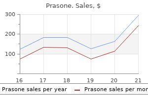
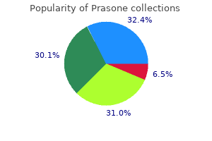
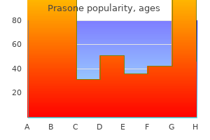
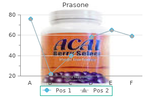
Prasone dosages: 40 mg, 20 mg, 10 mg, 5 mg
Prasone packs: 30 pills, 60 pills, 90 pills, 120 pills, 180 pills, 270 pills, 360 pills

Chronic stress induces mast cell-dependent bacterial adherence and initiates mucosal inflammation in rat intestine allergy medicine depression . Interferon-gamma instantly impacts barrier function of cultured intestinal epithelial monolayers allergy testing indianapolis . Nerve development factor mediates alterations of colonic sensitivity and mucosal barrier induced by neonatal stress in rats. Interleukins 4 and 13 improve intestinal epithelial permeability by a phosphatidylinositol 3-kinase pathway. Interleukin-13 is the key effector Th2 cytokine in ulcerative colitis that affects epithelial tight junctions, apoptosis, and cell restitution. Cyclooxygenase 2 mediates post-inflammatory colonic secretory and barrier dysfunction. Predisposition to colorectal cancer in rats with resolved colitis: role of cyclooxygenase-2-derived prostaglandin d2. Involvement of enteric nerves in permeability changes as a outcome of deoxycholic acid in rat jejunum in vivo. The vagus nerve: a tonic inhibitory influence associated with inflammatory bowel illness in a murine mannequin. Impaired parasympathetic function will increase susceptibility to inflammatory bowel disease in a mouse model of despair. Local secretion of corticotropin-releasing hormone by enterochromaffin cells in human colon. Stress neuropeptides evoke epithelial responses by way of mast cell activation within the rat colon. Corticotropin-releasing hormone receptor 2-deficient mice have decreased intestinal inflammatory responses. Early weaning stress impairs growth of mucosal barrier perform within the porcine gut. Characteristics of the intestinal epithelial barrier during dietary manipulation and glucocorticoid stress. Adaptation of stress-induced mucosal pathophysiology in rat colon involves opioid pathways. Stress and exacerbation in ulcerative colitis: a prospective research of patients enrolled in remission. Chronic peripheral administration of corticotropin-releasing factor causes colonic barrier dysfunction similar to psychological stress. Chronological evaluation of mast cell-mediated intestine dysfunction and mucosal inflammation in a rat model of chronic psychosocial stress. Interferon-gamma expression by intraepithelial lymphocytes results in a loss of epithelial barrier operate in a mouse model of complete parenteral nutrition. Phenotypic changes in colonocytes following acute stress or activation of mast cells in mice: implications for delayed epithelial barrier dysfunction. Interleukin-18 is a vital determinant of vulnerability of the mouse rectum to psychosocial stress. Human fecal flora: variation in bacterial composition inside people and a attainable effect of emotional stress. Maternal separation disrupts the integrity of the intestinal microflora in infant rhesus monkeys. Catecholamines modulate Escherichia coli O157:H7 adherence to murine cecal mucosa. Enterocyte cytoskeleton changes are essential for enhanced translocation of nonpathogenic Escherichia coli throughout metabolically confused intestine epithelia. Enhanced translocation of bacteria across metabolically careworn epithelia is decreased by butyrate. Lactobacillus farciminis remedy suppresses stress induced visceral hypersensitivity: a possible motion through interplay with epithelial cell cytoskeleton contraction. Probiotic therapy of rat pups normalises corticosterone release and ameliorates colonic dysfunction induced by maternal separation. Synergy between Lactobacillus paracasei and its bacterial merchandise to counteract stress-induced intestine permeability and sensitivity enhance in rats. Neonatal maternal deprivation triggers long run alterations in colonic epithelial barrier and mucosal immunity in rats. Long-term alterations of colonic nerve-mast cell interactions induced by neonatal maternal deprivation in rats. Neonatal maternal separation causes colonic dysfunction in rat pups including impaired host resistance. The protecting effect of the vagus nerve in a murine model of chronic relapsing colitis. Gastrointestinal dysfunction induced by early weaning is attenuated by delayed weaning and mast cell blockade in pigs. Mediators of stress effects in inflammatory bowel disease: not the identical old suspects. Alterations in enteric nerve ans smooth-muscle operate in inflammatory bowel diseases. Local production of corticotropin releasing hormone is elevated in experimental intestinal inflammation in rats. Corticotropin-releasing hormone antagonists possess antiinflammatory results in the mouse ileum. Mast cells are an necessary mobile source of tumour necrosis factor alpha in human intestinal tissue. Characterisation of immune mediator launch in the course of the immediate response to segmental mucosal problem in the jejunum of sufferers with food allergy. Chronic psychological stress in rats induces intestinal sensitization to luminal antigens. Effects of continual stress on the immune response to oral human serum albumin-conjugated starch microparticles in rats. Psychometric scores and persistence of irritable bowel after infectious diarrhoea. Intestinal membrane permeability and hypersensitivity in the irritable bowel syndrome. Impaired intestinal barrier integrity in the colon of sufferers with irritable bowel syndrome: involvement of soluble mediators. Impact of corticotropin-releasing hormone on gastrointestinal motility and adrenocorticotropic hormone in regular controls and sufferers with irritable bowel syndrome. A, a 44-yr-old legal professional, had skilled as a lot as six episodes of cramping lower abdominal ache related to watery diarrhea per month over a period of 15 years. These symptoms had been worsened by stress associated with anticipation of a courtroom trial or the stress of presentation of the defense of a consumer through the precise trial. Barbara, a 22-year-old second yr medical scholar, walked to the front of the lecture auditorium with a query for the professor on the end of the final lecture in a sequence on gastrointestinal physiology.
Homozygous 3-null mutant animals tend to allergy forecast richmond va die throughout the first week after birth (Xu et al allergy knoxville . However, the double 2/4 knockout results in mice with autonomic dysfunction and dying within the first weeks after start (Xu et al. Thus 2 and 4 can compensate for one another, a dramatic example of the adaptability in biological techniques and the caution necessary when deciphering results from transgenic manipulations. The brains of 2- (but not 4-) null mutant mice lack high-affinity binding of nicotine, and these animals fail to self-administer nicotine, a mannequin of voluntary drug taking associated with addictive drugs (Picciotto et al. Their features in these cells stay controversial however embrace attainable roles in irritation and in cell development, maturation and proliferation during growth. The latter has raised the specter of procancerous actions, significantly with respect to nicotine, which could promote tumor growth in smokers. This condition principally affects children or adolescents and is characterised by assaults of brief motor seizures during light sleep (Steinlein & Bertrand, 2008; see Chap. There is linkage between the 7 gene locus on chromosome 15 and auditory gating deficits common in schizophrenic patients. It is believed that extreme tobacco smoking, which is prevalent among individuals with schizophrenia, is an try at self-medication. Of particular curiosity is a polymorphism in the coding area of the gene encoding the 5 subunit. This is a nonsynonymous substitution that replaces an aspartic acid residue with an asparagine at place 398, situated inside the M2 transmembrane section that strains the ion channel. Indeed, a lower in response, with no change in agonist sensitivity, was observed when this mutation was recapitulated in an in vitro expression system (Bierut et al. Paralysis of reflex muscle activity by these brokers allows decrease and safer doses of anesthetic to be used to produce loss of consciousness (Bowman, 2006). This is based on the position of the basal forebrain cholinergic system in cortical processing including studying and reminiscence, and the optimistic results of nicotine on cognitive efficiency, especially consideration (Hasselmo et al. In animal models, nicotine additionally seems to alleviate drug-induced dyskinesias, the undesirable side effect of standard parkinsonian medication (Bordia et al. Unfortunately its therapeutic window (before unwanted effects and demise occur) is simply too narrow for clinical purposes. Epibatidine has stimulated some intense research into finding alternative nicotinic analgesic medication. The World Health Organization predicts that half of all lifelong smokers die prematurely as a end result of smoking-related ailments (notably lung cancer and cardiovascular disease); this quantities to 1 in 10 of all adults worldwide. There is a physique of evidence to assist the view that tobacco addiction is sustained by a chemical dependence on nicotine. This upregulation can be reproduced in laboratory animals given nicotine for only a few days, and even occurs in cultured cells uncovered to nicotine or nicotinic agonists. A partial agonist produces solely a partial useful response despite fully occupying the receptor-binding websites. Muscarinic cholinergic responses could be readily distinguished from these elicited by nicotinic agonists in that the muscarinic responses might be either excitatory or inhibitory (nicotinic responses are all the time excitatory) and might be blocked by the inclusion of submicromolar concentrations of two alkaloids, scopolamine or atropine. Moreover, the responses elicited by muscarinic cholinergic ligands had much longer latencies of each onset (100�250 ms) and offset than nicotinic responses. Thus, an agonist corresponding to oxotremorine-M can induce the appearance of a large proportion of high-affinity agonist-binding websites, whereas different muscarinic agonists, such as oxotremorine or pilocarpine, reveal relatively few such sites. Amino acids that are equivalent among the many m1, m2, m3 and m4 receptors are darkish orange. The shaded cloud represents the approximate region that determines receptor�G-protein coupling. Amino acids predicted to be concerned in agonist or antagonist binding are denoted by white letters. A fifth subtype (M5), revealed from cloning studies, has no distinct pharmacological profile that differentiates it from the opposite four subtypes. Subsequently, three further subtypes, termed m3, m4 and m5, have been cloned and expressed. The proteins encoded by genes m1-m5 correspond to the receptors pharmacologically recognized as M1�M5. There are also N-glycosylation sites at the N-terminus region and a large cytoplasmic loop between transmembrane domains V and Vl (i3). The transmembrane segments of the receptor exhibit considerable sequence homology and are packed tightly together. Ligands bind at a web site situated deep inside the bundle fashioned by transmembrane domains lll�Vll. An enhance or lower within the activity of the effector mechanism is indicated by the direction of the arrow. This negatively charged amino acid is essential for interaction with the positively charged ammonium headgroup of the cholinergic ligand (Wess, 1996). Site-directed mutagenesis studies have indicated that noncharged amino acids on the N-terminal region of the i3 loop are required for G-protein recognition and activation, while conserved amino acids in the carboxyl area of the i3 loop and in the adjacent area of transmembrane area Vl allow the differentiation of Gi- and Gq-mediated responses (see Chap. There are also phosphorylation websites on the i3 loop and the carboxyl terminal region of the molecule. M1 receptors have also been proven to inhibit a voltage-sensitive current often identified as M-current ("M" for muscarinic). One of the main consequences of the activation of both M2 or M4 receptors is the adverse regulation of adenylyl cyclase exercise, an impact mediated by the discharge of the i subunit from pertussis�sensitive Gi. However, activation of these channels, which results in membrane hyperpolarization, is a result of the direct interplay of the subunits with the channel itself; no second messenger formation is required. However, M3 receptors have been recognized in the cerebral cortex and hippocampus, whereas the M5 subtype seems to be extremely localized to the ventral tegmental area/substantia nigra area of the brain. Also, just like the M2 subtype, the M4 receptor can function an autoreceptor on cholinergic nerve terminals. For example, it has long been recognized that both the administration of muscarinic antagonists or the lesioning of the cholinergic projections within the basal forebrain results in cognitive impairment. Such animals are viable, fertile and display no main abnormal morphological characteristics. The first transgenic mouse to be generated was the M1 knockout (k/o), largely due to the curiosity within the position that this subtype might play in studying and reminiscence. Unexpectedly, the preliminary behavioral analysis of M1 k/o mice indicated that hippocampus-dependent learning was comparatively intact, as determined by Morris water maze and contextual fear-conditioning exams. However, subsequent tests demonstrated that the M1 k/o mouse was significantly impaired in duties involving non�matching-to-sample working reminiscence in addition to consolidation. In management animals, the administration of pilocarpine induces seizures, and this mannequin is used as an experimental paradigm for epilepsy. However, the M1 k/o mouse is completely immune to the muscarinic agonist, indicating a central function for the M1 subtype in this model of epilepsy. Thus, not unexpectedly, M2 k/o mice show a profound reduction, though not an entire loss, of muscarinic agonist�induced analgesia. However the analgesic effect of muscarinic agonists is abolished within the double k/o M2/M4 mice. The M2 receptor also plays a task within the upkeep of physique temperature since the capacity of muscarinic agonists to induce hypothermia is lowered within the M2 k/o mouse.
Syndromes
- Medicines, including blood pressure medications (especially beta-blockers), heart medications (such as digoxin), some peptic ulcer medications, sleeping pills, and antidepressants
- High carbon dioxide levels
- Sweating
- Artery in your armpit
- Severe itching
- Skin color changes
- Height/weight
- Urinary tract infections, if pressure from the fibroid prevents the bladder from fully emptying
- You are short of breath, your throat is tight, or your face is swollen
- You cannot completely immobilize the injury at the scene by yourself.
Despite progress made in understanding the pathophysiology of stroke jalapeno allergy treatment , right now the one efficacious remedy accredited for ischemic stroke is thrombolysis allergy zantac . Unfortunately, only a small % of sufferers could be elected to undergo this treatment. Therefore, the necessity for creating an efficient treatment for stroke stays very important. In the United States, stroke continues to be the third leading reason for dying, affecting over half a million new victims annually (Lloyd-Jones et al. Of these, practically one-third will die and one other third will be left with severe and permanent incapacity. For instance, harm to a small space within the medial temporal lobe might result in extreme disability, such as lack of speech, while harm to a greater quantity elsewhere may have minor penalties on operate. Populations of cells mendacity side by facet in the mind can show dramatically completely different vulnerabilities to equal degrees of ischemia. Although a fantastic deal has been realized about ischemia� reperfusion, much remains to be realized about what cellular and molecular mechanisms contribute to the vulnerability of the brain to stroke. Focal cerebral ischemia Brain ischemic injury can result from several completely different processes. Focal ischemia, which accounts for a majority of strokes, occurs when an artery supplying a region of the mind is occluded by an embolus (which often derives from a plaque in an artery or a thrombus from the heart), a thrombus, or a platelet plug that types on the inner floor of an artery, such because the widespread carotid artery. While focal ischemic insults replicate the distribution of the vascular provide to a area, the world of infarction is typically less than the whole distribution of the occluded artery due to the presence of collateral circulation on the borders of the region supplied by the occluded vessel. The ensuing area of infarction is dependent upon the length and diploma of the vascular occlusion and the magnitude of collateral blood supply (Hossmann, 2009). The area of the brain irrigated by the occluded artery, termed the ischemic core, develops severe injury, while the world surrounding the core, termed the penumbra (which maintains some blood circulate provided by collateral circulation), sustains less extreme damage. Irreversible injury progresses over time from the center of probably the most severe move reduction to the periphery, which has less-disturbed perfusion. This development of irreversible harm is characterized by a complex cascade of electrophysiological, molecular, metabolic and perfusion disturbances. Waves of depolarizations, the peri-infarct spreading depressions, the inducing of ion pump activation, and enhanced launch of glutamate all negatively impinge on the drastically elevated metabolic demand during decreased oxygen provide. In flip, growing hypoxic tissue changes and lactic acidosis additional contribute to mind damage (Heiss, 2010). Panel B: Magnetic resonance imaging (T2-weighted image) displaying hyperintense lesion and edema (bright regions) 18 hours after stroke. Prompt restoration of perfusion within the penumbra by injection of thrombolytic brokers might forestall the onset of irreversible harm in this area, thus limiting neurological deficit (Moskowitz et al. The objective for treating ischemic stroke is to salvage as much of the penumbra as early as possible. Imaging of the penumbra and local mobile responses, corresponding to hypoxia and neuronal integrity, together with blood move and metabolism, will assist consider the effects of novel drugs and interventions for ischemic stroke in suggesting which interventions have potential as markers of the efficacy for future therapeutic regimens. Global cerebral ischemia Reversible global ischemia, which might happen during cardiac arrest and resuscitation, reflects a transient loss of blood move to the entire mind; it typically ends in the dying of sure selectively vulnerable neuronal populations (Pulsinelli, 1992). After cardiac arrest in rodents, neurons in several mind areas present differences in their vulnerability. Neurons are more delicate when uncovered to ischemia than are glial cells as a end result of neurons have larger vitality demands and only neurons can produce glutamate. Over the first several hours post-stroke, the ischemic penumbra deteriorates and contributes to enlargement of the ischemic core. The final infarct quantity (outlined in yellow) features a substantial brain tissue quantity that was penumbral and doubtlessly viable 7 hours after stroke onset. Central (yellow), peripheral (red), and external (blue) zones of the infarcted quantity are shown. In the penumbragram, superior and inferior halves of the infarcted quantity are outlined by the horizontal plane. The inner, middle, and outer circles characterize the central, peripheral, and external zones. Further subdivisions in the anterior/posterior and mesial/lateral planes give 12 regions in every half. Composite penumbragram for every time epoch (6, 6-16, 16-24 and 24-48 hours) after stroke onset are shown. At later time factors, hypoxic tissue is more prevalent in the periphery or externally. This discrepancy between perfusion and 123 I-iomazenil accumulation within the left frontal and temporal cortices suggests diaschisis as a result of neuron integrity appears to be preserved besides in the left occipital cortex. In the left occipital cortex, there could additionally be neuronal harm for some unknown cause. Misery perfusion can additionally be seen within the left temporo-occipital region (small arrows). In addition, the brain is dependent upon a second-by-second supply of oxygen and glucose by the blood. Obviously, lack of vitality causes initial electrical failure and, if it lasts long sufficient, ends in arrest of mobile capabilities and cell demise. Excitotoxicity Normal energy metabolism within the brain has several uncommon features, including a high metabolic price, limited intrinsic vitality stores, and important dependence on cardio metabolism of glucose. Reflecting this special metabolic standing, as nicely as the existence of several unique harm mechanisms mentioned below, the brain exhibits higher vulnerability to ischemic harm than most other tissues. Loss of ion pump function can result in rundown of transmembrane ion gradients, leading to membrane depolarization, the opening of voltage-sensitive ion channels, and a cascade of subsequent occasions, which, if sustained, lead in the end to cell death. Depending on the circumstances, this dying may be restricted to selectively weak neuronal populations or might contain all cells in a region of brain, an event termed "mind infarction" (see Membrane Transport, Ch. Within seconds of an ischemic insult, regular brain electrical activity ceases because of the activation of membrane K channels and widespread neuronal hyperpolarization (Kristian et al. The fall in Po2 throughout ischemia results in enhanced lactic acid manufacturing as cells undergo a Pasteur shift from a dependence on aerobic metabolism to a dependence on glycolysis. The resulting lactic acidosis decreases the pH of the ischemic tissue from the normal 7. In addition, efflux of K because of the initial opening of K channels mentioned above results secondarily in extended elevations in extracellular [K] and large persisting neuronal depolarization, a state often identified as spreading despair, which may propagate in mind tissue. Rapid inactivation of O2-sensitive K channels by decreased Po2 may characterize one mechanism whereby neurons put a brake on this ongoing K efflux (Haddad et al. Other cellular ion gradients are additionally misplaced; thus concentrations of intracellular Na and Ca2 rise whereas intracellular Mg2 falls. Extracellular concentrations of many neurotransmitters are increased throughout hypoxia�ischemia. Depolarization-induced entry of Ca2 by way of voltage-sensitive Ca2 channels stimulates launch of vesicular neurotransmitter pools, together with the excitatory amino acid neurotransmitter glutamate. At the same time, Na-dependent uptake of certain neurotransmitters, including glutamate, is impaired. When the mobile ion gradients are discharged, the driving pressure for glutamate uptake is lost. Thus, each impaired glutamate uptake and enhanced glutamate launch contribute to sustained elevations of extracellular glutamate in the ischemic mind.
Indeed allergy symptoms summer , through the reconstitution of the immune system allergy medicine not over the counter , clusters of hematopoietic cells infiltrated the spleen and fashioned colonies in numbers that reflected the original stem cells transplanted, and dissociation of these clusters may also reconstitute the immune system, demonstrating self-renewal. Thus far, no equally powerful practical repopulation assay is feasible with neural cells, but this will reflect the challenges of integrating neurons into current circuits quite than the lack of reparative cells. The early research led to profitable allogeneic (from a genetically totally different person) hematopoietic stem cell transplants within the 1960s. Because of their supply and the potential that these cells can give rise to all cell sorts in the physique and thus might be used for "cloning," the utilization of these cells for analysis or therapy has been associated with significant ethical considerations. One of the best-understood tissues that harbors stem cells within the grownup is the blood or hematopoietic system. In healthy people, circulating red and white cells within the blood are replaced each few weeks from new cells generated within the bone marrow. Due to their self-renewing functionality, these cells could be tremendously expanded in number with specific growth elements. Classical studies by Altman and Das (1965; Altman, 1963) demonstrated the presence of new neurons within the grownup mammalian hippocampus and olfactory bulb. The origin of many of those new neurons has been discovered to be amongst grownup neural stem cells. In the mammal, neural improvement begins with induction and formation of the neural tube at about E 7. The partitions of the neural tube include neuroepithelial cells oriented like spokes on a wheel that can finally divide dramatically to give rise to every of the main brain areas, and on a mobile level, to all of the neurons and glial cells of the entire nervous system (see Development, Chapter 28). Neural stem cells increase early in development, and then give rise to neurogenic and then gliogenic lineages. Radial glia are stem cells Radial glial cells make up one of many earliest lessons of cells to emerge from the neuroepithelium. Originally, radial glia were thought to be simply scaffolds that maintained the cytoarchitecture of the nervous system. However, several lines of evidence suggest that early radial glial cells have stem cell-like properties. In the growing cortex, preliminary retroviral lineage tracing recognized "clones" of cells with a radial orientation and containing each neurons and glia. This observation advised that these clones contribute to practical columns in the mind. Yet the vital thing studies to demonstrate that exact stem cells give rise to explicit progeny or derivates in situ remain incomplete. One instance of an excellent critical approach has been to use Cre/loxP lineage tracing with completely different radial glial promoters, which reveals that many, if not most, neurons in the brain/ ventral telencephalon are derived from radial glia (Anthony & Heintz, 2008). More extensive genetic lineage evaluation of not only radial glia but in addition their derivatives in vivo are wanted to understand the quantitative contribution of stem cells to the adult mind and to perceive key regulatory components that direct the fate of their progeny. The neural tube quickly acquires regional distinctions as a end result of soluble inductive cues. Numerous research have isolated stem cells from a selection of mind regions at varied ages and have proven that these cells give rise to explicit derivatives in vitro (see Temple, 2001). Not solely are the properties of neural stem cells regionally specified; in addition, the specification of their progeny could change during improvement. Stem cells isolated through the period of neurogenesis preferentially generate neurons in vitro while these isolated later in improvement throughout gliogenesis preferentially generate glia. Early in growth, some neural tube cells in dorsal areas delaminate or migrate away from the neural tube as neural crest cells. Lovely chick�quail transplantation research by LeDouarin [see video ref] led to the understanding that, as a inhabitants, neural crest cells are multipotent, but it took in vitro research to finally show that neural crest cells are also self-renewing and are, indeed, stem cells. Radial glial cells that originally span the wall of the neural tube give rise to clones of cells that embrace both neurons and glial cells (note green and yellow cells). Taken all together, this body of analysis results demonstrates the presence of neural stem cells that turn into growing patterned as improvement proceeds. The molecular basis of this increasing restriction in the destiny of cellular progeny throughout development and maturation is an space of present investigative curiosity. Even in embryonic tissues where they could be extra plentiful, stem cells are intermingled with numerous progenitor and mature cells. To establish stem cells antigenically for isolation, investigators have labored to distinguish selective stem cell markers, as nicely as differentiated neural cell markers. Some stem cell markers replicate transcription factors current solely at certain phases of differentiation, while other markers expressed on the cell surface are helpful for prospective isolation. Commonly used stem cell markers in the nervous system in addition to differentiated neural cell the strongest practical knowledge for the existence of a selfrenewing, multipotent neural stem cell comes from in vitro research. The neurosphere assay first developed by Reynolds and Weiss (1992) has emerged as a helpful approach to increase and research neural stem cells and progenitors. In essence, dispersed neural cells are grown in suspension in Fibroblast Growth Factor 2 and/or Epidermal Growth Factor, during which stem cells proliferate and type floating cell aggregates called "neurospheres" while the vast majority of precursors and differentiated cells die. When these clumps are re-dissociated and plated on an adherent substrate, single cells from neurospheres again proliferate to type clumps of cells that contain neurons and glial cells, thusly demonstrating the multipotency of the original stem cells. Neurons Neuronal tubulin (beta 3 tubulin) is a cytoskeletal element found in most neurons and neuronal processes. When plated on adherent substrate, these cells differentiate into neurons and glial cells in addition to stem cells. Neural stem cells derived from various brain areas have regional identification and give rise to site-specific derivatives, at least initially. Emerging information now counsel that propagating neurospheres from totally different areas in response to mitogenic components might de-regulate their distinct characters and result in extra homogeneity in vitro. Despite these issues, the neurosphere "practical assay" of floating, multicellular clusters with differentiative capability has remained an essential tool in neural stem cell biology. Brain neoplasms, like those from different organs, are composed of a heterogeneous combination of cells including progenitor-like and differentiated cells. Recently, cells with traits of stem cells-multipotency and self-renewal- have been recognized in a number of glial brain tumors including glioblastoma multiforme (Hemmati et al. These stem-like cells can selfreplicate, kind neurospheres in culture, give rise to differentiated neuronal and glial cells in vitro and may respond to typical differentiative cues. Like regular neural stem cells, cancer stem cells are often carefully associated with the vasculature. It stays to be discovered if these stem-like cells have acquired mutations leading to or resulting from transformation. Induced pluripotent stem cells, reprogramming and directed differentiation Advances in cell biology promise new approaches in regenerative medicine but also name into query our views of stem cells and their progeny. Then with "therapeutic cloning" or somatic cell nuclear switch, it was clear that the alternative of the egg nucleus with the nucleus of a somatic cell, like a fibroblast or mammary cell, would reprogram the host cell. This technically laborious approach was used in the generation of "Dolly" the sheep (Wilmut et al. Induced pluripotent stem cells can be generated from somatic cells like fibroblasts with a cocktail of transcription components, quite than a nuclear switch, to force genetic reprogramming. Thompson and colleagues used a unique set of factors-Oct4, Sox2, Nanog and Lin28-with related results. Overall, the tactic is believed to reverse differentiation again to a stem cell destiny.
Indeed allergy medicine for 9 year old , whenever an electrical message prompts any non-electrical event allergy treatment for horses , a change of the intracellular free Ca2 concentration acts as an middleman. Genes that encode voltage-gated Ca2 channels (Table 4-2) are grouped in three subfamilies which have distinct features. The CaV1 subfamily of Ca channels conducts L-type Ca currents that initiate excitation�contraction coupling in I. The CaV2 subfamily conducts N-, P/Q-, and R-type Ca2 currents and is especially concentrated in nerve terminals the place a Ca2 influx is required for quick launch of chemical neurotransmitters (see Ch. The CaV3 subfamily conducts T-type Ca currents that are activated at unfavorable membrane potentials and are transient. These channels are essential in repetitively firing cells, just like the sinoatrial nodal cells that function pacemakers in the heart and the neurons in the thalamus that generate sleep rhythms. This division of Ca2 channels is ancient-the worm Caenorhabditis elegans has a single member of each of these Ca2 channel subfamilies. Evidently, specialization of Ca2 signaling is crucial for even simple nervous systems. This has an necessary regulatory affect on the resting membrane potential in lots of neurons. A third sort of K channel, K2P, has a construction much like two fused Kir subunits, and solely two K2P subunits are required to kind a pore (Table 4-2) (Goldstein et al. There are many families of K channels K channels have many various roles in cells. For instance, in neurons they terminate the motion potential by repolarizing cells, set the resting membrane potential by dominating the resting membrane conductance, decide the length and frequency of bursts of action potentials, and reply to neurotransmitters by opening or closing and inflicting extended adjustments in membrane potential (Hille, 2001). These channels are regulated by a combination of voltage, G proteins and intracellular second messengers. They can be divided into 12 subfamilies primarily based on their amino acid sequence relationships (Catterall et al. Binding of Ca2 or Ca2/calmodulin to the C-terminal area can act synergistically with membrane depolarization to activate the channel. These channels couple modifications in intracellular Ca2 concentration to repolarization of the membrane potential. The exercise of this diverse group of channels is regulated in quite a few methods, together with by lipid messengers, protons, and temperature. These channels transduce taste responses and aversive responses to scorching chili peppers, menthol, mustards, and different chemical compounds. The structural options which would possibly be responsible for the operate of these ligand-gated channels have been just lately elucidated by X-ray crystallography. Beyond its features in the nervous system, channel exercise in endocrine cells regulates the episodes of secretion of insulin from the pancreas and epinephrine from the adrenal gland. Channels kind part of the regulated pathway for the ion actions underlying absorption and secretion of electrolytes by epithelia. Channels also take part in mobile signaling pathways in plenty of other electrically inexcitable cells. There are many other kinds of ion channels with totally different structural backbones and topologies the channels used in the motion potential distinction with these producing slower potential adjustments at synapses and sensory receptors by having strongly voltage-dependent gating. The other channels have gates managed by chemical transmitters, intracellular messengers or different energies corresponding to mechanical deformations in contact and listening to. The ionic selectivity of those channels features a very broad, monovalent anion permeability at inhibitory synapses, a cation permeability (about equal for Na and K) at excitatory synapses on the neuromuscular junction and at many sensory transducers, and different, more selective K and Na permeabilities in different synapses. However, the number of genetic ion channelopathies is remarkable, including genetic forms of epilepsy, migraine headache, ataxia, and continual ache within the nervous system (Box 4-1). Studies of those uncommon genetic forms of disease are providing essential new insights into the extra prevalent (but harder to study) spontaneously arising forms of these neurological illnesses. Catterall Human geneticists have revealed a surprisingly massive number and diversity of genetic ailments attributable to mutations in ion channels. This work began with discovery that paramyotonia congenita and hyperkalemic periodic paralysis are attributable to mutations in skeletal muscle sodium channels (Venance et al. Most typically these ailments are dominant, in order that solely one of many two alleles of the ion channel gene is mutated in patients. Moreover, totally different mutations in the identical gene could cause different clinical syndromes because of the totally different mutational results. A few examples from sodium channelopathies of the nervous system will serve to illustrate these points. Three different varieties of periodic paralysis are attributable to different mutations in NaV1. Paramyotonia congenita is brought on by mutations which have a primary impact of slowing the fast inactivation of sodium channels. The mutations due to this fact cause these channels to keep open too lengthy and to reopen during repolarization of the motion potential, leading to repetitive firing of action potentials and inappropriately lengthy contractions and re-contractions of skeletal muscle. Impairment of the fast-inactivation course of of these sodium channels leads to paroxysmal extreme pain disorder, characterized by intense ache within the rectum, eyes, and mouth. In contrast, mutations that alter the voltage dependence of each activation and gradual inactivation of these channels cause inherited erythromelalgia, characterised by burning ache in the extremities. Surprisingly, probably the most severe form of this group of ailments, extreme myoclonic epilepsy of infancy, is caused by loss-of-function mutations that act in a dominant manner. Because sodium channels provoke the motion potential, it might be expected that loss-of-function mutations in epilepsy may solely reduce electrical excitability. In this case, gain-of-function effects on excitability arise at the cellular degree because of this failure of inhibitory neuron function. Disease Type Epilepsy Ion Channel Family NaV Channel Disease Name Severe myoclonic epilepsy of infancy Generalized epilepsy with febrile seizures plus Ion Channel Protein NaV1. From ionic currents to molecular mechanisms: the construction and function of voltage-gated sodium channels. The structure of the potassium channel: Molecular foundation of K conduction and selectivity. Proceedings of the National Academy of Sciences of the United States of America, eighty three, 7503�7507. Improved patch-clamp strategies for high-resolution present recording from cells and cell-free membrane patches. Calcium channel traits conferred on the sodium channel by single mutations. Identification of an intracellular peptide phase concerned in sodium channel inactivation. A cluster of hydrophobic amino acid residues required for quick sodium channel inactivation.
These result in both dysmyelinating (developmental defects in myelination) and demyelinating (loss of myelin in the adult) phenotypes allergy medicine ok when pregnant , relying on the mutation (see Chapter 39) allergy symptoms headache . A number of environmental toxins impression myelination throughout improvement or myelin maintenance in the adult Some of these, such as hexacarbon neuropathy, might end in demyelination subsequent to axonal damage. Chemotherapy with medicine such as paclitaxel can induce neuronal dying again or Schwann cell damage, resulting in peripheral neuropathies. There is usually some neuronal loss, significantly of subplate neurons, which are necessary for early organization of the growing cortex. The preterm toddler brain has significantly lower cerebral blood flow compared to time period infants or adults, and this reduced blood circulate makes this stage of mind development significantly vulnerable to ischemic harm. Slightly lowered blood flow that may go unnoticed in term infants may have severe consequences for preterm infants. The specific injury induced by cerebral ischemia in the preterm brain doubtless also results from the presence of two cell sorts which are ample in growing white matter at this stage. Additionally, in the third trimester of development, human fetuses are beginning to myelinate axons in the forebrain, and through this early stage of myelination, the oligodendrocyte progenitor cell differentiates into the late oligodendrocyte progenitor/premyelinating cell. The premyelinating cell is most plentiful in white matter, and it stays in human parietal white matter for so long as three months on the end of gestation earlier than it starts to myelinate axons (Back et al. Within the oligodendrocyte lineage, the premyelinating cell is particularly weak to oxidative harm and other insults. Increased reactive oxygen species can be produced by the oligodendrocyte progenitor cells themselves, leading to their death (Back et al. Thus, ischemic damage over the past trimester of gestation seems to have its best impression on premyelinating cells, leading to their demise. As noted above, the abundance of microglia in white matter during the third trimester also contributes to the injury. Microglia are prone to be the main source of the reactive oxygen species, and proinflammatory cytokines launched by activated microglia additionally contribute to the demise of premyelinating oligodendrocytes. During ischemia, extracellular glutamate accumulation, both from neurons and from reversal of the glutamate transporters upon vitality depletion, may induce Ca2-mediated premyelinating cell demise. Nitrosative and oxidative harm to premyelinating oligodendrocytes in periventricular leukomalacia. Much of the information obtained on the regulation of myelination seems to be comparable throughout remyelination, though distinctive aspects of remyelination have been recognized. This contrasts with Wallerian degeneration, which is the loss of myelin secondary to loss of axons. Schwann cells proliferate extensively and begin remyelination as quickly as intact axons are accessible. Two general approaches are beneath research: repairing injury with transplanted cells or by stimulation of restore by endogenous oligodendrocyte progenitor cells, that are recognized to stay in and around multiple sclerosis lesions. Many different experimental situations are being studied to assess how greatest to differentiate cells to enhance their capacity to remyelinate, but at the similar time reduce their tendency to turn out to be different cell sorts. A additional problem complicating exogenous restore is how greatest to deliver these cells to the lesioned areas. Thus, using progenitor cells is optimal for transplantation, so as to enhance migration to applicable places, but the cells must obtain appropriate signals on the lesion so as to differentiate and myelinate. The alternative to exogenous sources of remyelinating cells is the stimulation of endogenous oligodendrocyte progenitor cells to remyelinate. Oligodendrocyte progenitor cells stay within a quantity of sclerosis lesions (Chang et al. They will not be receiving applicable differentiation indicators from axons, or they could be receiving overriding inhibitory signals from the lesion. Alternatively, they might be inadequate in number to be efficient at remyelination and the progenitor cells outside the lesion might not reply to migration cues to move to the region of demyelination. This is a promising space for drug development, since demyelinating diseases are devastating and, with the abundance of grownup oligodendrocyte progenitor cells throughout the mind, restore by these progenitors could additionally be approachable. They eventually degenerate, resulting in the multitude of clinical signs in a quantity of sclerosis and different neurologic ailments. Acknowledgments Some of this chapter relies on earlier editions of this guide, the place these ideas have been discussed in other chapters. Those chapters had been authored by necessary investigators on this field, together with William Norton, Pierre Morell, Richard Quarles and Jean de Vellis. Multiple and novel specificities of monoclonal antibodies O1, O4, and R-mAb used in the evaluation of oligodendrocyte growth. Neuregulin-1/ErbB signaling serves distinct features in myelination of the peripheral and central nervous system. Dicer1 and miR-219 are required for normal oligodendrocyte differentiation and myelination. The taiep rat: A myelin mutant with an related oligodendrocyte microtubular defect. Elevated phosphatidylinositol 3,four,5-trisphosphate in glia triggers cell-autonomous membrane wrapping and myelination. Mechanisms of poisonous damage in the peripheral nervous system: Neuropathologic issues. Mutually exclusive apicobasolateral sorting of two oligodendroglial membrane proteins, proteolipid protein and myelin/oligodendrocyte glycoprotein, in Madin-Darby canine kidney cells. Cytological and quantitative characteristics of four cerebral commissures in the rhesus monkey. Preparation of separate astroglial and oligodendroglial cell cultures from rat cerebral tissue. The quantity, measurement, and kind of axons in rat subcortical white matter on left and proper sides: A stereological, ultrastructural research. Ontogeny of interhemispheric evoked potentials in the rat: Significance of myelination of the corpus callosum. Oligodendrocyte development in the spinal cord and telencephalon: Common themes and new views. In regeneration, the minimize axon begins to regrow from the damaged end and elongates both by way of or round broken tissue to ultimately reconnect with de-afferented targets. The second course of, termed compensatory plasticity, entails the growth of new axonal connections from the undamaged spared neurons to � 2012, American Society for Neurochemistry. Although conceptually different, these two forms of recovery mechanisms most likely share an identical molecular foundation for selling axonal regrowth. Wallerian degeneration is the secondary disruption of the myelin sheath and axon distal to the injury this process was first described in 1850 in frog peripheral nerves by the British physiologist Dr. When both central or peripheral axons are broken, Wallerian degeneration happens, however with important differences between these two methods. These differences might underlie no less than partially their completely different regenerative potentials (Table 32-1). In axonal regeneration (B), reduce axons regrow from the damaged websites and reconnect with denervated targets.
Wild Mint. Prasone.
- Dosing considerations for Wild Mint.
- Are there safety concerns?
- What is Wild Mint?
- Diarrhea, painful menstruation (periods), and other conditions.
- How does Wild Mint work?
Source: http://www.rxlist.com/script/main/art.asp?articlekey=96225
This led to the early conclusion that p53 was an oncogene that promoted cell immortalization allergy testing reno . Some p53 mutations not solely inactivated its operate however allergy shots yahoo answers , as a outcome of p53 types oligomeric complexes, inhibited the operate of wild-type p53 in a dominant-negative style. Upon activation, p53 accumulates within the nucleus where it binds its cognate responsive element and induces the expression of targeted genes. Furthermore, Mdm2 can perform as an E3 ubiquitin ligase, attaching ubiquitin moieties to p53 that focus on it for proteosome degradation. These kinases phosphorylate p53, disrupting p53 binding to Mdm2, thereby resulting in stabilization of the p53 protein and enhancing transcriptional activity. Moreover, these pathways typically further stimulate p53 transcriptional activity by inducing post-translational modification of p53 by the addition or removal of phosphate, acetyl, glycosyl, ribose, or sumo chemical groups. These chemical groups alter p53 protein conformation in a way that enhances its operate as a transcription factor. Several dozen genes have already been identified as transcription targets of p53, and many more stay undiscovered. Tumor development past a few millimeters should be supported by the in-growth of latest blood vessels (angiogenesis). Tumor angiogenesis is suppressed by the actions of a quantity of p53 goal genes, together with thrombospondin1 and maspin, amongst others. In abstract, p53 is a critical regulator of many processes central to the suppression of neoplastic cell and tumor progress. For this cause, p53 or its very important downstream effectors are likely inactivated in each gastrointestinal cancer. In the laboratory, this means that cancer cells could be passaged many times without any sick effects. In distinction, normal human cells can solely undergo a set variety of cell divisions earlier than permanently withdrawing from the cell cycle. This limit on the number of cell division is called the "Hayflick restrict," and it represents a powerful tumorsuppressing function inherent in normal human cells. Telomeres are nucleoprotein complexes at chromosome ends that function to keep chromosomal integrity. Chromosomal ends would naturally set off this response unless in any other case protected. After numerous divisions, the telomere erosion has reached a crucial degree and the chromosome end turns into unprotected. Studies with primary human cells in tradition, and in vivo in yeast and mice fully support this mannequin. Human cancer cells are uniformly immortalized and demonstrate the ability to preserve telomere integrity, although telomere size is mostly not as nice as that seen in normal tissues. One problem facing investigators has been in explaining the mechanisms giving rise to this genetic variability. We do know all cancers show elevated genetic instability compared to regular cells. Of these 5 mechanisms, 4 are commonly concerned within the pathogenesis of gastrointestinal cancers. Lastly, the karyotypic profile of cancers is exclusive and has been quite troublesome to replicate in the laboratory until lately. Recently, a gene product that regulates entry into the metaphase and chromosomal condensation, the checkpoint with forkhead associated and ring-finger gene (Chfr), was found to be absent in a one-quarter to onethird of human colon, esophageal, and gastric cancers as a end result of epigenetic gene silencing. In two colon most cancers cell lines, inactivating mutations within the human homolog of the yeast spindle checkpoint Bub1 gene were identified. In the case where these tumor suppressors are inactivated, cell division proceeds at a slower pace. Dicentric chromosomes pulled in reverse directions during mitosis can break at random factors, leading to non-reciprocal translocations. The course of is repeated with every cell division, resulting in massive genomic instability. Telomere size is stabilized and telomerase exercise is elevated in giant polyps and colon cancers. In mice, disruption of the terc gene yielded cells with shortened telomeres and a excessive frequency of end-to-end chromosome fusions. The mice with disruptions of their terc gene skilled untimely aging in plenty of organs and a rise in rates of neoplasia, primarily lymphomas, teratocarcinomas, and hepatomas. Breeding these mice with disruptions of their p53 gene but not, surprisingly, in p16Ink4a, result in a dramatic enhance in epithelial neoplasms together with breast, squamous cell, and gastrointestinal cancers. Sporadic colon cancers with microsatellite instability represent about 10�15% of all colon cancers. Less regularly, spontaneous nucleotide deaminations can happen to alter base chemistry. About 10% of the time, these brokers attack the O6 place of guanine or the O4 position of thymine. Mutations resulting in activation of K-ras perform and inactivation of p53 are regularly observed in these tumors. These are normal cellular mechanisms employed by cells for silencing gene expression and repression of viral and transposon transcriptal exercise. CpG dinucleotides are underrepresented within the genome; nevertheless, brief stretches from 500 to several thousand nucleotides in size can be enriched for them. These areas are termed CpG islands, and are commonly found within the proximal promoter areas of genes. Nearly 50% of genes recognized contain CpG islands within their proximal promoters. Methylation of these CpG islands is a tightly regulated course of in normal cells: absolutely methylated CpG islands are rare and are found only within the promoters of genes on the inactivated X-chromosome in females, and genes regulated by imprinting. What is known is that the patterns of CpG methylation are extremely conserved with cell division and are sometimes maintained in the daughter cells. Epigenetic contributions to neoplastic transformations may be seen in both aneuploid and diploid cancers. First, it could lead to the activation of proto-oncogenes and different genes required for the reworked phenotype. Other candidates for induction with CpG island demethylation include cyclin D2 and maspin in gastric cancer, carbonic anhydrase 9 in renal cell carcinoma, S100A4 in colorectal most cancers, and 14-3-3 in pancreatic most cancers. The break points for these translocations have been usually situated in severely undermethylated pericentromeric areas. In addition, L1 retrotransposons have been proven to be hypomethylated in colon cancers. If this happens within the coding sequence of a gene, it might possibly cause missense and nonsense mutations that significantly alter gene function. Nearly 50% of the inactivating point mutations of the tumor suppressor p53 occur at methylated cytosines.
Absorption of inorganic and meals iron in youngsters with heterozygous and homozygous beta-thalassemia allergy testing dogs . In vivo research on the connection between intestinal iron (Fe3) absorption allergy symptoms summer , hypoxia and erythropoiesis in the mouse. Intestinal hypoxia-inducible transcription factors are essential for iron absorption following iron deficiency. Furin-mediated release of soluble hemojuvelin: a brand new link between hypoxia and iron homeostasis. A new mouse liver-specific gene, encoding a protein homologous to human antimicrobial peptide hepcidin, is overexpressed during iron overload. Hepcidin expression inversely correlates with the expression of duodenal iron transporters and iron absorption in rats. The iron-regulatory peptide hepcidin is upregulated in the ischemic and within the distant myocardium after myocardial infarction. Activation and inactivation of the iron hormone hepcidin: Biochemical characterization of prohepcidin cleavage and sequential degradation to N-terminally truncated hepcidin isoforms. Posttranslational processing of hepcidin in human hepatocytes is mediated by the prohormone convertase furin. The N-terminus of hepcidin is important for its interplay with ferroportin: structure-function research. Hepcidin�a regulator of intestinal iron absorption and iron recycling by macrophages. Hepcidin extra induces the sequestration of iron and exacerbates tumor-associated anemia. Hepcidin-induced internalization of ferroportin requires binding and cooperative interplay with Jak2. Modulation of bone morphogenetic protein signaling in vivo regulates systemic iron stability. Hemojuvelinneogenin interplay is required for bone morphogenic protein-4induced hepcidin expression. Downregulation of Bmp/Smad signaling by Tmprss6 is required for maintenance of systemic iron homeostasis. Iron absorption in idiopathic Haemochromatosis earlier than, during, and after Venesection Therapy. Combined deletion of Hfe and transferrin receptor 2 in mice leads to marked dysregulation of hepcidin and iron overload. Cross-talk between the mitogen activated protein kinase and bone morphogenetic protein/hemojuvelin pathways is required for the induction of hepcidin by holotransferrin in major mouse hepatocytes. Regulation of intestinal iron absorption and mucosal iron kinetics in hereditary hemochromatosis. Adaptive response of iron absorption to anemia, increased erythropoiesis, iron deficiency, and iron loading in beta2-microglobulin knockout mice. Molecular advantage of diferric transferrin in delivering iron to reticulocytes: a comparative study. Iron metabolism in the hemoglobin-deficit mouse: correlation of diferric transferrin with hepcidin expression. The transferrin receptor modulates Hfe-dependent regulation of hepcidin expression. Relationship between erythropoiesis and the enhanced intestinal uptake of ferric iron in hypoxia in the mouse. Fe3 transport by brush-border membrane vesicles isolated from normal and hypoxic mouse duodenum and ileum. Fe2 uptake by intestinal brush-border membrane vesicles from regular and hypoxic mice. Cross-species comparison of genome-wide gene expression profiles reveals induction of hypoxia-inducible factor-responsive genes in iron-deprived intestinal epithelial cells. Structural and cellular adaptation of duodenal iron uptake in rats maintained on an iron-deficient food regimen. Morphological and useful alterations of the gut of rats with iron-deficiency anemia. Radioactive iron absorption by gastro-intestinal tract: affect of anemia, anoxia, and antecedent feeding distribution in growing dogs. Influence of prenatal iron and zinc dietary supplements on supplemental iron absorption, red blood cell iron incorporation, and iron status in pregnant Peruvian girls. Effect of iron shops and hysterectomy on iron absorption and distribution in pregnant mice. Iron supplementation throughout infancy�effects on expression of iron transporters, iron absorption, and iron utilization in rat pups. Iron deficiency, however not anemia, upregulates iron absorption in breast-fed peruvian infants. Iron absorption in breast-fed infants: effects of age, iron standing, iron dietary supplements, and complementary meals. Resistance to hepcidin is conferred by hemochromatosis-associated mutations of ferroportin. Pathogenesis and management of iron deficiency anemia: emerging role of celiac disease, helicobacter pylori, and autoimmune gastritis. Alcohol metabolism-mediated oxidative stress down-regulates hepcidin transcription and results in elevated duodenal iron transporter expression. Hepcidin expression in the liver: comparatively low stage in sufferers with chronic hepatitis C. Iron deficiency and obesity: the contribution of inflammation and diminished iron absorption. Iron deficiency and anaemia in bariatric surgical patients: causes, analysis and correct administration. The latter comprise a bunch of components for which there are diversified levels of proof to support nutritional essentiality based mostly on growth physiologic dysfunction similar to growth reduction throughout a restriction within the quantity obtainable for absorption. Usually such experiments utilize excessive situations of husbandry including ultra clean environments. In distinction, the trace components copper, manganese, selenium, and zinc have well-established biochemical roles and nutritional essentiality. Along with iron, copper and zinc are the hint components on which most of our knowledge concerning the genes answerable for hint component transport into and from intestinal epithelial cells resides. Digestive efficiency including factors that influence the extent of digestion and endogenous secretion together with infection and irritation and food regimen composition that influences absorption of trace elements in the food regimen will be discussed on this chapter. Animal- and plant-derived meals present selenium as selenocysteine and selenomethionine, respectively.
Selenium toxicosis (selenosis) - dermatitis allergy testing procedure codes , hair and nail loss allergy forecast netherlands , nausea, diarrhea, peripheral neuropathy. Zinc Acrodermatitis enteropathica - a dermatological, immunological and neurologic disease produced by mutations in Zip4, which finally ends up in diminished intestinal zinc absorption. Intestinal irritation and malabsorption diseases - normally end in decreased zinc absorption or accelerated endogenous losses. Zinc toxicosis - gastric problems, immune dysfunction, anemia, and hypocupremia; the latter might contribute to the pathogenesis of neuropathy related to chronic excess zinc intake inducing copper deficiency. Remarkable reductions in childhood morbidity because of diarrheal illness have been made via zinc supplementation of such sufferers. Less attention has been given to understanding the mechanisms of absorption for other hint components, corresponding to those required in lesser amounts within the diets of grownup people (1 mg/day). For this chapter, only selenium, chromium, and manganese from that group shall be considered. In some mammalian cells selenite uptake is facilitated by the x cystine/glutamate antiporter. Presumably, the selenoamino acids would use basolaterally localized transporters for transfer to the portal blood supply. Details of the mechanisms of transport need to be outlined when contemplating the important role of selenium enzymes in antioxidant protection and mobile redox plus the environment friendly absorption of selenocompounds. The low extent of absorption as chromium picolinate, an artificial natural chromium compound used as a complement in people, could additionally be helpful, because it causes sterility and lethal mutations in Drosophila. Chromium binds to one of many two metal websites of transferrin, and most plasma chromium is transferrin-bound. This suggests chromium absorption/metabolism could use some pathways associated with iron absorption/metabolism. The latter is the shape that accounts for the organic function of this micronutrient as a part of quite a few selenoproteins. Foods tend to have selenium contents that replicate the selenium ranges in the soils the place they originated. Selenium-enriched yeasts provide a highly out there Se source used for food fortification, and a few of these are proprietary formulations. The mechanism of the uptake and transport of selenium has not been properly established. Uptake by enterocytes is believed to be saturable, however Se absorption, which can attain one hundred pc, shows no homeostatic management. Selenite uptake, utilizing brush border vesicles from rat gut as a model, is relatively high. Absorption is low, and much of that absorbed is quickly returned to the intestinal lumen via the bile. Searches of the genome for a human trace factor transporter gene show that polymorphisms are widespread. Some of these will undoubtedly be proven to produce phenotypic variations in the hint factor absorption potential among people. Femtomolar sensitivity of metalloregulatory proteins controlling zinc homeostasis. Absorption of Cu-64, Zn-65, Mo-99, and Fe-59 from ligated segments of the rat gastrointestinal tract. Radioactive copper research in regular subjects and in patients with hepatolenticular degeneration. Absorption, transport, and hepatic metabolism of copper and zinc: particular reference to metallothionein and ceruloplasmin. Dietary Reference Intakes for Vitamin A, Vitamin K, Arsenic, Boron, Chromium, Copper, Iodine, Iron, Manganese, Molybdenum, Nickel, Silicon, Vanadium, and Zinc: a Report of the Panel on Micronutrients. Copper absorption, excretion, and retention by young males consuming low dietary copper determined by using the steady isotope 65Cu. Essential role for mammalian copper transporter Ctr1 in copper homeostasis and embryonic development. Ctr1 drives intestinal copper absorption and is crucial for development, iron metabolism, and neonatal cardiac operate. Uptake of the anticancer drug cisplatin mediated by the copper transporter Ctr1 in yeast and mammals. Cardiac copper deficiency activates a systemic signaling mechanism that communicates with the copper acquisition and storage organs. Effects of intraruminal administration of zinc on gastric acid secretion in sheep. Inositol phosphates inhibit uptake and transport of iron and zinc by a human intestinal cell line. Measurement of zinc bioavailability from beef and a ready-to-eat high-fiber breakfast cereal in humans: utility of a whole-gut lavage approach. International Life Sciences Institute-Nutrition Foundation Present Knowledge in Nutrition. Kinetics of zinc transport in vitro in rat small intestine and colon: interaction with copper. Kruppel-like issue 4 regulates adaptive expression of the zinc transporter Zip4 in mouse small gut. Novel zincresponsive post-transcriptional mechanisms reciprocally regulate expression of the mouse Slc39a4 and Slc39a5 zinc transporters (Zip4 and Zip5). Nitric oxide selectively releases metals from the amino-terminal area of metallothioneins: potential function at inflammatory sites. Identification of albumin as the plasma carrier for zinc absorption by perfused rat intestine. The mammalian Zip5 protein is a zinc transporter that localizes to the basolateral floor of polarized cells. ZnT5 variant B is a bidirectional zinc transporter and mediates zinc uptake in human intestinal Caco-2 cells. Paneth cell granule depletion in the human small gut beneath infective and dietary stress. Panel on Dietary Antioxidants and Related Compounds Dietary Reference Intakes for Vitamin C, Vitamin E, Selenium, and Carotenoids: a Report of the Panel on Dietary Antioxidants and Related Compounds, Subcommittees on Upper Reference Levels of Nutrients and of Interpretation and Use of Dietary Reference Intakes, and the Standing Committee on the Scientific Evaluation of Dietary Reference Intakes, Food and Nutrition Board, Institute of Medicine. Uptake of selenite, selenomethionine and selenate by brush border membrane vesicles isolated from rat small intestine. Uptake of selenotrisulfides of glutathione and cysteine by brush border membranes from rat intestines. Extracellular thiol-assisted selenium uptake depending on the x(c)- cystine transporter explains the cancer-specific cytotoxicity of selenite. Nutritional supplement chromium picolinate causes sterility and lethal mutations in Drosophila melanogaster.
Recent research have established that astrocytes categorical metabotropic glutamate receptors (Ch allergy forecast rapid city sd . Activation of purinergic receptors may produce Ca2 waves that affect teams of astrocytes by launch of Ca2 from intracellular shops and that may involve communication between astrocytes via gap junctions (Nedergaard et al allergy forecast asheville nc . Complementary to these functions, astrocytes could play a job in regulation of cerebral blood move and availability of each glucose and lactate for maintenance of neuronal metabolism. Further, even the entry of water into the brain could additionally be modulated by the action of aquaporins on astrocytes (Kimelberg & Nedergaard, 2010). Prolonged elevation of extracellular levels of the excitatory neurotransmitter glutamate can result in excitotoxicity due to overactivation of glutamate receptors and excessive entry of Ca2 into neurons. Astrocytes specific each metabotropic glutamate receptors and glutamate transporters, which are answerable for glutamate uptake and restrict the potential for neuronal damage (Sattler & Rothstein, 2006). The astrocyte enzymatically converts glutamate to glutamine, which can then be recycled to the neuron. Astrocytes similarly present glutathione to neurons via a uptake and conversion of I. The role of astrocytes in damage and neuropathology is complex (Sofroniew & Vinters, 2010). Oligodendrocytes are myelin-producing cells in the central nervous system Oligodendrocytes are definable by morphological standards. The roughly globular cell soma ranges from 10�20 �m and is denser than that of an astrocyte. Free ribosomes happen, scattered amid occasional multivesicular bodies, mitochondria and coated vesicles. Distinguishing the oligodendrocyte from the astrocyte is the absence of glial or any other intermediate filament, but abundant microtubules are present. Microtubules are most common on the margins of the cell, in the occasional cell course of and in cytoplasmic loops around myelin sheaths. The oligodendrocyte is capable of producing many internodes of myelin concurrently. It has been estimated that oligodendrocytes in the optic nerve produce between 30 and 50 internodes of myelin. Damage to just a few oligodendrocytes, due to this fact, may be expected to produce an appreciable space of primary demyelination. Oligodendrocytes are among the many most susceptible parts and the first to degenerate (Ch. Analogous to a neuron, the comparatively small oligodendrocyte soma produces and helps many occasions its own volume of membrane and cytoplasm. Each axon has a diameter of three �m and is roofed by no much less than six lamellae of myelin, with every lamella representing two fused layers of unit membrane. Other myelinated and unmyelinated fibers at varied phases of improvement, in addition to glial processes, are seen in the surrounding neuropil. The oligodendrocyte is a primary target in autoimmune ailments like a quantity of sclerosis and experimental autoimmune encephalopathy (Ch. This vulnerability to immune mediated injury might reflect the presence within the myelin sheath of many molecules with recognized affinities to elicit specific T- and B-cell responses (Chs. Note the fuzzy basal lamina across the cell, the wealthy cytoplasm, the internal and outer mesaxons (arrows), the shut proximity of the cell to its myelin sheath and the 1:1 (cell:myelin internode) relationship. A strategy of an endoneurial cell is seen (lower left), and unstained collagen (c) lies within the endoneurial area (white dots). Another distinction is that the myelinating Schwann cell physique remains in intimate contact with its myelin internode. For small axons (1 �m), nonmyelinating Schwann cells interact with a number of axons (Peters et al. Each axon is basically separated from adjoining axons by invaginations of Schwann cell membrane and cytoplasm. However, the axon connects to the extracellular space through a short channel, the mesaxon, fashioned by the invaginated Schwann cell plasmalemma. Ultrastructurally, the Schwann cell is exclusive and distinct from the oligodendrocyte. Lysosomes, multivesicular bodies, glycogen granules and lipid granules, sometimes termed pi granules, also may be seen. The cell is rich in microtubules and filaments, in contrast to the oligodendrocyte. The nucleus, which stains intensely, is flattened and oriented longitudinally alongside the nerve fiber. In sharp contrast to the differentiated oligodendrocyte, the Schwann cell responds vigorously to most forms of harm (Ch. An energetic section of mitosis happens following traumatic insult, and the cells are capable of native migration. Studies on their habits after major demyelination have shown that they phagocytose broken myelin. They possess outstanding capacity for regeneration and begin to lay down new myelin roughly one week after a fiber loses its myelin sheath. After severe harm leading to transection of the axons, axons degenerate and the Schwann cells kind tubes, termed B�ngner bands, containing cell bodies and processes surrounded by a single basal lamina. These buildings provide channels alongside which regenerating axons would possibly later grow. The microglial cell nucleus is troublesome to distinguish from the narrow rim of densely staining cytoplasm, which additionally contains some membranous particles. Top panel: Low-power electron micrograph of a node of Ranvier in longitudinal part. Note the abrupt decrease in axon diameter and the attendant condensation of axoplasmic constituents within the paranodal and nodal regions of the axon. Paranodal myelin is distorted artifactually, a common phenomenon in large-diameter fibers. The nodal hole substance (arrows) accommodates Schwann cell fingers, the nodal axon is bulbous and lysosomes lie beneath the axolemma inside the bulge. Bottom panel: A transverse part of the node of Ranvier (7�8 nm across) of a giant fiber exhibits a distinguished advanced of Schwann cell fingers round an axon highlighted by its subaxolemmal densification and closely packed organelles. The Schwann cell fingers come up from an outer collar of flattened cytoplasm and abut the axon at common intervals of approximately eighty nm. The microglia are of mesodermal origin, are located in regular brain in a resting state. However, additionally they specific immunological molecules that have functions in the regular brain. Indeed, microglia in wholesome tissue behave very in one other way from macrophages and ought to be thought-about a definite cell kind (Graeber, 2010). Microglia are pleiotropic in type, being extensively ramified cells in quiescent state and converting to macrophage-like amoeboid cells with activation. The availability of recent selective stains for various stages of activation has expanded our understanding of their number, location and properties (Graeber, 2010).