

Methotrexate dosages: 10 mg, 5 mg, 2.5 mg
Methotrexate packs: 10 pills, 20 pills, 30 pills, 60 pills, 90 pills, 120 pills, 180 pills, 270 pills, 360 pills

Buy 5 mg methotrexate otc
Mother (A and B) and daughter (C and D) symptoms kidney cancer generic methotrexate 10 mg otc, both with the 3q29 microdeletion syndrome symptoms 5 weeks into pregnancy discount 2.5 mg methotrexate with visa. Seven-year-old boy with an elongated face, prominent nasal bridge, and large ears. In common, the severity of the behavioral and motor deficiencies increases over time, and the deficiencies become more obvious after adolescence. Gradual loss of previously discovered motor and communication expertise, a progressive immobility, and, ultimately, inflexible flexion of the arms and arms and a decline in motivational and efficiency features occur. Four deaths-three that occurred at lower than 1 12 months of age, secondary to respiratory failure or apnea-have been reported. This was confirmed by the identification of point mutations within the gene in sufferers who had the typical phenotype but who lacked the microdeletion. The virtual lack of detection of larger terminal deletions in stay births is assumed to replicate lethality. In addition, three familial cases have been reported during which the deletion was current within the moms in a mosaic pattern. Prenatal onset overgrowth with respect to weight (9%�20%), short stature (13%�39%), chubby (20%�30%), weight problems. Developmental delay, delicate to extreme intellectual incapacity, hypotonia, speech delay, sleep disturbance. In older kids, outbursts of anger; delinquent, compulsive, and self-stimulating behaviors; and stereotypic movements. Defects in 50% to 61%, including dilated ventricles, white matter anomalies, corpus callosum hypoplasia or agenesis, cerebellar hypoplasia. Microcephaly (>50%), brachycephaly, hypertelorism, midface hypoplasia; synophrys, with distinguished broad arched eyebrows; short nostril with anteverted nares; open mouth with protruding tongue; thin higher lip with downturned corners of mouth; full everted decrease lip; prognathism; pointed chin; malformed ears. Abnormalities in 30% to 60%, together with cryptorchidism, hypospadias, and micropenis. Cormier-Daire V, et al: Cryptic terminal deletion of chromosome 9q34: A novel reason for syndromic obesity in childhood, J Med Genet forty:300, 2003. Willemsen M, et al: Familial Kleefstra syndrome because of maternal somatic mosaicism for interstitial 9q34. Facial options of nine kids with 9q34 microdeletion syndrome from 15 months to 15 years of age. Note the synophrys, arched eyebrows, quick anteverted nose, thin tented upper lip, and macroglossia in a single case. Midface hypoplasia, quick nostril with anteverted nares, facial phenotype similar to that of sufferers with a microdeletion. More than 30 individuals have been reported since then, and the estimated incidence is 1 in 42,000. Connective tissue laxity is clear in plenty of sufferers, with unfastened joints, hernias, and scoliosis. Nearly half of the sufferers have a historical past of recurrent infections, suggesting some form of immunodeficiency is current, not but outlined. Several adults have been reported, with variable cognitive and behavioral impairment and no main extra health points. The precise measurement and breakpoints of the deletion differ among sufferers, with most deletions occurring due to nonallelic homologous recombination between segmental duplication blocks (low copy repeats). Duplications distal to the deletion crucial region also appear to have related phenotypic penalties, with important behavioral and cognitive features. Mild to severe mental incapacity (100%), scarce to absent speech, hypotonia, autistic conduct, food seeking and obsessive compulsive behaviors, poor sleep. Cortical atrophy, neuronal heterotopia, abnormal corpus callosum with cysts, enlarged ventricles, hypoplastic olfactory bulbs, enlarged cisterna magna. Microcephaly (20%); long, slim, triangular face; facial asymmetry; high anterior hairline; excessive forehead; deep-set eyes; epicanthal folds; hypertelorism; downslanting palpebral fissures; sparse, broad medial eyebrows that taper laterally; low nasal bridge; broad nasal base with notched flaring alae nasi; long, easy philtrum; full decrease lip; small mouth; small pointed chin; abnormal ears (large, protuberant, cup-shaped, thick anteverted lobes). Ocular abnormalities in 60%, particularly strabismus and nystagmus but also iris and chorioretinal coloboma, anisocoria, and hypermetropia. Small hands, quick fifth fingers, brachydactyly of fourth and fifth metacarpals, hypoplastic and proximally implanted thumbs, camptodactyly of toes, overriding toes, hypoplastic fifth toes, cutaneous syndactyly of fingers or toes, sandal gap. Hypospadias (40%), micropenis, cryptorchidism in males, labial adhesions in females. Congenital heart defects, joint laxity, scoliosis, kyphosis, hernias, recurrent infections. Van Esch H, et al: Congenital diaphragmatic hernia is part of the model new 15q24 microdeletion syndrome, Eur J Med Genet fifty two:153, 2009. Patient with the 15q24 microdeletion at 9 months (A), 2 years (B), and three years (C) of age. Note facial asymmetry, high anterior hairline, high forehead, deep-set eyes, epicanthal folds, hypertelorism, strabismus, downslanting palpebral fissures, low nasal bridge, lengthy distinguished philtrum, small pointed chin, and protuberant ears. A, Note low nasal bridge, gentle hypertelorism, sparse medial eyebrows that taper laterally, and broad nasal base with notched alae nasi. Frontal (A) and lateral (B) view of the face of a 2-year-old with the 15q24 microdeletion. Note distinguished forehead, mild hypertelorism, strabismus, low nasal bridge, broad nasal base, and small pointed chin. The telomeric breakpoint appears to be the identical in all patients, whereas the proximal breakpoint has been variable. However, recurrent microdeletion and reciprocal microduplication at a contiguous area on 16p11. Growth has been regular in 4 patients, below the third percentile in two sufferers. Hypotonia, unsteady gait, intellectual disability, extreme expressive language disorder, hyperactivity. Long, narrow flat face; deep-set eyes; downslanting palpebral fissures; low-set, malformed, posteriorly rotated ears. Tetralogy of Fallot, pulmonary atresia, bicuspid aortic valve, tricuspid regurgitation. Speech development is a particular problem, as is hyperactivity with brief consideration span and impairment of nice motor expertise. The deletions References Hernando C, et al: Comparative genomic hybridization reveals a partial de novo deletion 16p11. Battaglia A, et al: Further characterization of the new microdeletion syndrome of 16p11. Note the deep-set eyes; low-set, posteriorly rotated ears; mildly pointed chin; and thin upper lip. More than 70 circumstances have now been reported, with an estimated prevalence of 1 in 16,000. It has lately been proven that haploinsufficiency for a single gene in the interval is the cause for the general features of this microdeletion. In infancy, hypotonia of the face, with an open mouth appearance and a protruding tongue, is characteristic.
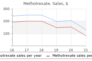
Methotrexate 10 mg order mastercard
In such cases medications xanax buy methotrexate 5 mg, a Z-plasty of the skin could additionally be done to relieve the partial constriction medicine 72 hours best methotrexate 5 mg. If there has been persistent amnion leakage, the neonate might show features of the oligohydramnios deformation sequence, including incomplete growth of the lung, with respiratory insufficiency. Every try must be made to oxygenate and assist such an infant, since, with continued lung morphogenesis, the prognosis could be glorious. Hence, the features evident by surface examination are normally the only abnormalities. Those uncommon exceptions are identified or presumed to be attributable to trauma and embrace two examples of tried early termination of pregnancy by using a coat hanger and one incident of a girl falling from a horse whereas pregnant. It has typically been a sporadic occasion in an in any other case normal household, and therefore the recurrence danger is normally stated as being negligible. Before that point, the amnion and chorion are utterly separate membranes and, as such, it has been advised that the amnion is susceptible to rupture. Torpin R: Amniochorionic mesoblastic fibrous strings and amniotic bands: Associated constricting fetal malformations of fetal dying, Am J Obstet Gynecol ninety one:sixty five, 1965. Torpin R: Fetal Malformations Caused by Amnion Rupture during Gestation, Springfield, Ill, 1968, Charles C Thomas. Moerman P, et al: Constrictive amniotic bands, amniotic adhesions, and limb-body wall complicated: Discrete disruption sequences with pathogenetic overlap, Am J Med Genet forty two:470, 1992. Jamsheer A, et al: Comparative examine of medical traits of amniotic rupture sequence with and without physique wall defect: Further proof for separation, Birth Defects Res A Clin Mol Teratol 85:211, 2009. Note the amnion that has stripped off the left aspect of the fetal surface of the placenta and is rolled up at the base of the umbilical cord. A�D, Bands constricting the ankle, resulting in deformational defects and amputation. The vast majority of circumstances are spontaneously aborted; the remainder are stillborn. Thoraco-abdominoschisis involves an anterolateral body wall defect with evisceration of thoracic or stomach organs into a persistent extraembryonic coelom. Failure of the ventral physique wall to fuse because of harm to part of the physique wall or failure of regular ventral folding of the embryo leads to a persistence of the extraembryonic coelom. The amnion is continuous with the pores and skin at the edge of the defect and the umbilical cord is short and partially devoid of its regular amniotic membrane overlaying. Limb defects just like those seen in the amnion rupture sequence, similar to amputations secondary to ring constrictions and pseudosyndactyly, happen often. The encephaloceles are normally anterior, typically multiple, and sometimes attached to the amnion. The developmental pathogenesis, as nicely as the etiology of limb�body wall advanced, is controversial. Adhesion of the amnion to these necrotic areas may lead secondarily to amniotic adhesive bands. Failure of the ventral body wall to close because of vascular compromise may lead to persistence of the extraembryonic coelom. In 2011, Hunter et al concluded that this complicated originates at the embryonic disc stage. They instructed that a defect or deficiency of the ectoderm of the embryonic disc was answerable for many of the malformations seen on this complicated. Russo R, et al: Limb body wall complex: A crucial evaluate and a nosological proposal, Am J Med Genet 47:893, 1993. A�E, Affected fetuses with multiple involvement of limbs, body wall, and craniofacial space. The occurrence of epibulbar dermoid with this sample of anomaly, especially when accompanied by vertebral anomalies, was designated as the Gold enhar syndrome, and the predominantly unilateral occurrence was designated as hemifacial microso mia. However, the occurrence of assorted combina tions and gradations of this pattern of anomalies, both unilateral and bilateral, with or with out epi bulbar dermoid, and with or with out vertebral anomalies, has advised that hemifacial microso mia and the Goldenhar syndrome could simply rep resent variable manifestations of a similar error in morphogenesis. Ventricular and atrial septal defects, patent ductus arteriosus, tetralogy of Fallot, conotrun cal defects, and coarctation of aorta, in decreas ing order of frequency. Ectopic or fused kidneys, renal agen esis, vesicoureteral reflux, ureteropelvic junc tion obstruction, ureteral duplication, and multicystic dysplastic kidney. Estimated recurrence in first diploma family members is approximately 2%, although minor options of this dysfunction could also be more com monly famous in relatives. Based on research using an animal model, Poswillo concluded that this disorder was as a result of interference with vascular provide and focal hemorrhage within the developing first and second branchial arch. Hypoplasia of malar, maxillary, or mandibular area, especially ramus and condyle of man dible and temporomandibular joint; lateral cleftlike extension of the nook of the mouth (macrostomia); hypoplasia of facial muscula ture; hypoplasia of depressor anguli oris. Microtia, accent preauricular tags or pits, mostly in a line from the tragus to the nook of the mouth; center ear anomaly with variable deafness. Diminished to absent parotid secretion, anomalies in function or structure of tongue, malfunction of soppy palate. Hemivertebrae or hypoplasia of verte brae, mostly cervical but may also be thoracic or lumbar. Hydrocephalus, Arnold Chiari malformation, occipital encephalocele, agenesis of corpus callosum, calcification of falx cerebri, hypoplasia of septum pellucidum, enlarged ventricles, intracranial dermoid cyst, lipoma in corpus callosum, polymicrogyria. Pashayan H, et al: Hemifacial microsomiaoculoauriculo vertebral dysplasia: A patient with overlapping fea tures, J Med Genet 7:185, 1970. Poswillo D: the pathogenesis of the primary and second bran chial arch syndrome, Oral Surg 35:302, 1973. Nijhawan N, et al: Caruncle abnormalities in the oculo auriculovertebral spectrum, Am J Med Genet 113:320, 2002. Wang R, et al: Infants of diabetic moms are at elevated threat for the oculoauriculovertebral sequence: A case based mostly and casecontrol method, J Pediatr 141:611, 2002. Str�mland K, et al: Oculoauriculovertebral spectrum: Associated anomalies, useful deficits and attainable developmental danger components, Am J Med Genet 143:1317, 2007. Wieczorek D, et al: Reproduction abnormalities and twin pregnancies in parents of sporadic sufferers with oculoauriculovertebral spectrum/Goldenhar syn drome, Hum Genet 121:369, 2007. Note the variable features including the lateral cleft-like extension of the mouth (A), preauricular tags (B), and microtia (C). Kaplan and colleagues empha sized a "community" or spectrum of problems and instructed widespread parts in modes of develop mental pathogenesis. The presumed vascular downside is more more probably to happen in distal areas, such as the distal limbs, tongue, and sometimes parts of the mind. Chorionic villus sampling, par ticularly when performed between 56 and 66 days of gestation, has been related to this dysfunction, as has using misoprostol as an abortifacient, giving further credence to a disruptive vascular pathogenesis. Small mouth, micrognathia, hypo glossia, variable clefting or aberrant connect ments of tongue; mandibular hypodontia; complete bony fusion of the maxilla and man dible, choanal atresia, cleft palate; cranial nerve palsies, together with Moebius sequence; broad nose; telecanthus; decrease eyelid defect; facial asymmetry. Hypoplasia of various levels, to point of adactyly; syndactyly, angelshaped phalanx. Brain defects, particularly of cranial nerve nuclei, inflicting Moebius sequence; splenogo nadal fusion, hypoplasia of atlas with cranio cervical junction malformation, gastroschisis. Serious problems with hyper thermia can happen in kids with fourlimb amputation. The speculation that the References Rosenthal R: Aglossia congenita: A report of the situation mixed with other congenital malformations, Am J Dis Child 44:383, 1932.
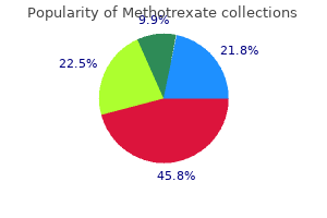
Methotrexate 5 mg generic with mastercard
Occasionally treatment keratosis pilaris 5 mg methotrexate with visa, females can have brief stature medicine wheel images methotrexate 2.5 mg discount line, developmental delay, facial dysmorphism, and gonadal dysgenesis. Sanlaville D, Schluth-Bolard C, Turleau C: Distal Xq duplication and practical Xq disomy, Orphanet J Rare Dis 4:four, 2009. Severe neonatal encephalopathy resulting in demise earlier than age 2 years is most frequent in affected males. References Sanlaville D, et al: Functional disomy of the Xq28 chromosome region, Eur J Hum Genet thirteen:579, 2005. B, Hypertelorism, epicanthal folds, depressed nasal bridge, upturned nares, small and open mouth, thin tented upper lip. As the molecular etiology of this condition has been elucidated, each a classical and a milder phenotype are recognized. Hirsutism (78%), cutis marmorata and perioral pale "cyanosis" (56%), hypoplastic nipples and umbilicus (50%), low posterior hairline (92%) Limbs. Micromelia (93%), phocomelia and oligodactyly (27%), clinodactyly of fifth fingers (74%), single transverse palmar crease (51%), proximal implantation of thumbs (72%), flexion contracture of elbows (64%), syndactyly of second and third toes (86%), chilly extremities Genitalia. Hypoplasia in males (57%), undescended testes (73%), hypospadias (33%), hypoplastic labia majora Gastrointestinal. Mandibular spur present up to three months of age, dislocated/hypoplastic radial head, hypoplastic first metacarpal and fifth center phalanx, brief sternum with precocious fusion and 13 ribs, enlarged cerebral ventricles, white matter atrophy. Prenatal onset development deficiency with size and weight less than tenth percentile. Mean adult top 156 cm (males) and 131 cm (females), Retarded osseous maturation (100%). Initial hypertonicity (100%) Low-pitched, weak, growling (74%) cry in infancy High pain tolerance, extreme (23%) speech and language delays, autism spectrum problems, seizures Broad-based gait Behavior. Hyperactivity, short consideration span, aggression, self-injurious behavior, excessive shyness, anxiousness, depression, obsessivecompulsive behavior, perseveration, sleep disturbance, circadian rhythm issues. Microbrachycephaly (93%); bushy eyebrows and synophrys (98%), long, thick, curly eyelashes (99%), arched eyebrows (98%); ptosis; high myopia; peripapillary pigmentation; microcornea; tear duct malformation; depressed nasal bridge (83%); anteverted nares (85%); long philtrum, skinny upper lip, and downturned angles of mouth (94%); high-arched palate (86%); late eruption of widely spaced enamel (86%); thick dysplastic posteriorly rotated ears; micrognathia (84%); outstanding symphysis (66%) 118 Brachmann�De Lange Syndrome 119 swallowing difficulties, typically continue well past 6 months. Although a excessive proportion of affected kids have severe mental incapacity, a major quantity have a a lot larger potential relative to efficiency than earlier studies have instructed, particularly amongst those much less classically affected. Puberty happens on the normal time although it might be incomplete with irregular menses being widespread. Episodes of aspiration in infancy, apnea, issues related to bowel obstruction, diaphragmatic hernia, and cardiac defects seem to constitute the main hazards for survival in these sufferers. This gene is inherited in an X-linked method and accounts for many of the familial, in addition to lots of the milder, cases observed. It is anticipated that mutations in different cohesin-related genes will account for the not quite 50% of instances in which no mutation has yet been identified. C References Brachmann W: Ein Fall von symmetrischer Monodaktylie durch ulnadefekt mit symmetrischer Flughautbildung in den Ellenbeugen, sowie anderen Abnormitaten (Zwerghaftigheit, Halsrippen, Behaarung), Jahrb Kinderheilk 84:225�235, 1916. Oliver C, et al: Cornelia de Lange syndrome: extending the physical and psychological phenotype, Am J Med Genet 152A:1127�1135, 2010. Note the synophrys, thin downturned higher lip, lengthy philtrum, hirsutism, small palms and toes, and extreme limb defects. A and B, Note the fifth finger clinodactyly and proximal implantation of the thumb. This dysfunction is uncommon, occurring with an estimated frequency of 1 in one hundred,000 to 1 in 125,000 newborns. Defects, most frequent of which are patent ductus arteriosus, ventricular septal defect, and atrial septal defect, occur in roughly one third of circumstances. Global developmental delay is universal, with most patients testing within the severe to moderate range of intellectual disability. Recurrent ear infections with hearing loss and dental problems primarily related to overcrowding of the enamel occur incessantly. Hand and/or foot surgical procedure frequently improves grasp, oppositional perform, and luxury. Unusual reactions to anesthesia (respiratory misery and cardiac arrhythmias) have been reported, in addition to tracheal collapse after muscle relaxants. More than 90% of affected people survive into adulthood and most obtain some independence in self-care and communication. Postnatal onset of progress deficiency; in adults, average peak of 153 cm in males and 147 cm in females; average weight of 48 kg in males and 55 kg in females. Social and pleasant in childhood with short consideration span, attention-deficit/hyperactivity disorder, motor stereotypies. Microcephaly (35%), large anterior fontanel (41%), delayed closure of fontanel (24%), frontal bossing (33%), frontal hair upsweep (20%), low anterior hairline (24%), low posterior hairline (42%), downslanting palpebral fissures (88%; nevertheless, only half of youngsters younger than age 5 will manifest this), maxillary hypoplasia with slim palate (100%), small mouth (56%), outstanding or beaked nostril with or without nasal septum extending beneath alae nasi and brief columella (90%), deviated nasal septum (71%), micrognathia (49%), low-set and/or malformed ears (84%), heavy eyebrows (76%), extremely arched eyebrows (73%), long eyelashes (87%), nasolacrimal duct stenosis (43%), ptosis (36%), epicanthal folds (55%), strabismus (69%), enophthalmos (22%) Limbs. Broad thumbs with radial angulation (87%), broad great toes (100%), other fingers broad (87%), fifth finger clinodactyly (62%), persistent fetal fingertip pads (31%), deep plantar crease between first and second toes (33%), flat toes (72%) Other skeletal. Cryptorchidism (78% of males), renal anomalies (52%) Rubinstein-Taybi Syndrome a hundred twenty five and keloids are frequent issues for adults. Affected individuals have an increased danger for a selection of benign and malignant tumors although no screening protocol has been recommended. Translocations, inversions, and enormous deletions involving contiguous genes at 16p13. Tetralogy of Fallot, atrial septal defect, vertebral fusions, and different anomalies have been noted. Young infant (A and B); 21-month-old youngster (C); and 10-year-old youngster (D and E) with Rubinstein-Taybi syndrome. Note the hirsutism, downslanting palpebral fissures, maxillary hypoplasia, outstanding nostril with nasal septum extending below alae nasi, and low posteriorly rotated ears. Tendency towards extra sweating, particularly on the head and upper trunk, during infancy; liability to fasting hypoglycemia from about age 10 months to age 2 to 3 years. Feeding difficulties are seen in 86% and embody weak suck, absence of hunger, gastroesophageal reflux, and food aversion. There tends to be a gradual enchancment in progress in weight and look throughout childhood and especially during adolescence. Small, triangular facies with frontal prominence, late closure of the anterior fontanel, downturned corners of mouth; facial asymmetry, possible bluish sclerae in early infancy, low-set posteriorly rotated ears, and micrognathia; eye findings embrace visible refractive errors, strabismus, and tortuous retinal vessels. Russell-Silver Syndrome 129 As a outcome, the adult usually seems more regular than the toddler with this disorder. Because of the small facies, the higher head could appear massive, although head circumference is well within the normal range. Somewhat frequent feedings and sufficient glucose intake during illness should be ensured from age 6 months until age 3 years, the period of enhanced liability to fasting hypoglycemia. A small number of patients with submicroscopic deletions or duplications in these imprinted regions have additionally been described. Several instances with maternally inherited microduplications or translocations have been familial. Russell-Silver syndrome is likely one of the imprinting issues that has been related to the utilization of assisted reproductive technologies, although the explanation for this apparent relationship stays obscure.
10 mg methotrexate order mastercard
B: Aspirate smear exhibits several big hypogranular bands (arrows) and a dysplastic erythroid precursor with asymmetric binucleation is located slightly below the centrally located hypogranular band treatment lung cancer methotrexate 2.5 mg purchase with amex. C: Aspirate smear reveals increased blasts and two dysplastic micromegakaryocytes (arrows) medicine and health 5 mg methotrexate generic otc. B: Low-power magnification of aspirate smear demonstrates hypercellular marrow fragments consisting exclusively of monotonous sheets of primitive 386 mononuclear cells. C: High-power view of aspirate smear discloses nucleolated undifferentiated blasts. B: Aspirate smear demonstrates immature myeloid precursors, some of which show the presence of quite a few main granules in the cytoplasm. A: Blood smear shows a spectrum mainly immature and uncommon mature myeloid precursors. A: Aspirate smear reveals a leukemic blast population composed largely of promyelocytes that could possibly be confused with a case of acute promyelocytic leukemia. B: Myeloperoxidase stains show robust diffuse staining that obscures nuclear morphology. C: A hypercellular clot section consists virtually solely of enormous blasts with "open" chromatin pattern (vesicular nuclei), two to three small nucleoli, and indented nuclear contours. B: An aspirate smear demonstrates the identical dimorphic blast population composed of monocytes and myeloblasts (open and closed arrows, respectively). A: Blood smear exhibits a particular dimorphic blast population consisting of immature nucleolated myeloblasts and mature monocytes. B: Aspirate smear stained with butyrate esterase displays optimistic staining in only the larger sized monocytic blast population. A: Blood smear exhibits a dual inhabitants of small and large blasts, myeloblasts and monoblasts, respectively. On the left facet are three mature monocytic blasts characterised by massive dimension, low nuclear-to-cytoplasmic (N:C) ratios with somewhat condensed chromatin, absent nucleoli, irregularly folded nuclear contours, and vacuolated, finely granular, neutral-staining cytoplasm. The right higher corner of the slide contains three myeloblasts characterised by medium-size, larger N:C ratios, open chromatin, and scant cytoplasm. A small mature lymphocyte with closed chromatin is present in the proper decrease corner. B: Myeloperoxidase staining demonstrates robust granular cytoplasmic staining in a subpopulation of blasts. Undifferentiated monoblasts with excessive N:C ratios, open chromatin, nucleoli, and basophilic-staining cytoplasm are current within the higher panels, promonocytes within the center panels, and differentiated monocytic blasts with closed chromatin sample and lobulated nuclei are proven within the decrease panels. B: Bone marrow aspirate options promonocytes characterized by giant size with low nuclear-to-cytoplasmic (N:C) ratios, nuclei with reasonably open chromatin, folded nuclear contours (some kidney bean-shaped), absent nucleoli, and agranular, slightly basophilic-staining cytoplasm. For comparability, an arrow factors to a small mature lymphocyte with larger N:C ratio and condensed chromatin. A: this peripheral smear reveals leukocytosis consisting of huge undifferentiated blasts with low to average N:C ratios and basophilic-staining cytoplasm. C: Butyrate esterase stains are adverse on this explicit case of acute monoblastic leukemia. A benign histiocyte situated near the underside of the slide serves as inner constructive management. B: Butyrate esterase stains show localized positivity restricted to the perinuclear granules, confirming the monocytic origin for this case of acute monoblastic leukemia with closely granulated blasts. A: Increased cellularity and architectural distortion, corresponding to swirling and lining up of individual marrow cells (so-called Indian filing), recommend the presence of serious bone marrow fibrosis. B: Large, monotonous-appearing, wide-spaced, immature hematopoietic cells that replace the complete marrow cavity. E and F: Immunohistochemistry for lysozyme stains with equal intensity in both the benign histiocytes and leukemic monoblasts. B: A bone marrow aspirate smear exhibits monotonous sheets of monoblasts displaying open chromatin with a quantity of outstanding nucleoli and vacuolated, basophilic-staining cytoplasm. A: Monoblasts encompass a big, benign histiocyte with lengthy tentacles of cytoplasmic projections full of dark-staining pigment (likely iron) on this case of acute monoblastic leukemia. B: In the same case, the 2 decrease cells are monocytic leukemic blasts and the larger cell in the higher area is a benign histiocyte. Histiocytes, often "innocent" benign bystanders in marrow illnesses, are characterised by oval-shaped nuclei, smooth nuclear contours, "bland" chromatin patterns, and ample neutralstaining cytoplasm. C: Positive butyrate esterase staining is seen within the quite a few leukemic monoblasts (on the right) and a benign histiocyte (long arrow). A peripheral blood smear displays two circulating promonocytes and a small mature lymphocyte. The two promonocytes are characterized by large dimension, low N:C ratios, distinctly lobulated nuclear contours, and ample "agranular" neutral-staining cytoplasm. B: A high-power view of a bone marrow aspirate smear reveals monoblasts with bilobed nuclei harking back to the microgranular variant of acute promyelocytic leukemia. C: Butyrate esterase histochemistry demonstrates adverse staining in the promonocytes (a benign histiocyte serves as optimistic control). An aspirate smear shows a pleomorphic population of immature monocytic precursors. A: High-magnification view of a blood smear shows 4 large mature monocytes which have low N:C ratios, lobulated nuclei with reasonably closed chromatin pattern and neutral staining, and finely granular, vacuolated cytoplasm. B: Aspirate smear reveals clusters of monocytic blasts which have a more immature appearance than do the circulating blasts. A and B: Blood smears show peripheral monocytosis consisting of mature monocytes that have a closed chromatin sample, lobulated nuclei, and ample granular, vacuolated cytoplasm. C: Bone marrow biopsy demonstrates hypercellular marrow consisting almost exclusively of monocytoid precursors. A: Bone marrow aspirate smear consists completely of a heterogeneous mixture of dysplastic erythroid blasts, many displaying cytoplasmic vacuolation. A: Aspirate smear reveals a mixture of dysplastic erythroid precursors and immature myeloid cells, together with quite a few blasts. B: All the erythroid cells from the sector in A had been digitally eliminated, clearly demonstrating a nonerythroid blast depend exceeding 20% (blasts shown by arrows). A: Aspirate smear shows erythroid hyperplasia, elevated numbers of blasts, and hemophagocytosis. In the middle of the determine, a benign-appearing histiocyte engulfs mobile debris. Hemophagocytosis can occur in both benign and malignant bone marrow problems, including acute erythroid leukemia. B: Bone marrow aspirate smear reveals only scattered megaloblastic erythroid precursors. C: Biopsy at medium magnification shows a hypercellular marrow comprising a heterogeneous mononuclear cell inhabitants. D: Biopsy at greater magnification reveals substitute of the bone marrow cavity by a blended erythroid population. E: Immunohistochemistry for the erythroid lineage marker glycophorin C demonstrates constructive staining in the leukemic cells, and negative-staining megakaryocytes function controls. A: Bone marrow aspirate smear exhibits erythroid hyperplasia and numerous dysplastic erythroid blasts with vacuolated cytoplasm.
Buy 2.5 mg methotrexate amex
Microcephaly treatment 02 binh methotrexate 2.5 mg generic without prescription, sloping forehead symptoms bladder cancer methotrexate 10 mg cheap fast delivery, broad nasal bridge, small facies, shallow supraorbital ridge with nasal bridge at about identical level as brow, broad nasal tip, quick palpebral fissures with telecanthus and appearance of hypertelorism, variable ptosis and blepharophimosis, epicanthal folds, prominent or mildly dysplastic ears, micrognathia, delayed dental eruption, lacking tooth, caries. Eczema-like skin disorder on face and flexural areas, sparseness of lateral eyebrows and scalp hair. Brachyclinodactyly of fifth fingers; syndactyly of second and third toes; cryptorchidism; seizures; ocular abnormalities including strabismus, microphthalmia, hyperopia, megalocornea, hypoplasia of iris, and coloboma; abnormalities of the ocular fundus embody abnormal veins, tapetoretinal degeneration, and ocular albinism. A�C, Note the quick palpebral fissures, asymmetric ptosis, shallow supraorbital ridges, and mild micrognathia. Feeding issues are frequent throughout infancy, likely secondary to severe gastroesophageal reflux. The facial erythema may be very seldom present at birth, normally appearing throughout infancy following exposure to sunlight; it might excoriate, however improves after childhood. Although studying disabilities happen, the overwhelming majority of sufferers are throughout the regular range for intelligence. Malignancy has been the major known cause of dying and develops in 50% of sufferers. The cancers that develop in Bloom syndrome are similar in kind and distribution to those seen within the common population; nonetheless, malignancy develops at a much younger age. An increased rate of chromosomal breakage and sister chromatid trade is found in cultured leukocytes and fibroblasts from all patients studied, but not reliably so within the heterozygotes. The frequency of the gene carrier within the Ashkenazi Jewish inhabitants is estimated at 1: a hundred. Several people have developed myelodysplastic problems following remedy for most cancers. Prenatal onset of growth deficiency imply birth weight of males and females are 1760 and 1754 gm respectively; common adult male top, 149 cm, and adult female height, 138 cm; decreased adipose tissue leads to wasted look. Mild microcephaly with dolichocephaly; malar hypoplasia, with or with out small nostril; loss of decrease lashes; fissure of lower lip. Facial telangiectatic erythema includes the butterfly midface area, is exacerbated by sunlight, and normally develops through the first year. Bloom D: the syndrome of congenital telangiectatic erythema and stunted growth, J Pediatr sixty eight:103�113, 1966. Sawitsky A, Bloom D, German J: Chromosomal breakage and acute leukemia in congenital telangiectatic erythema and stunted growth, Ann Intern Med 65:487� 495, 1966. Photograph of elevated sister chromatin exchange in a baby with Bloom syndrome on the left in comparison with the control on the right. This syndrome incorporates elements of ectodermal dysplasia with endocrine and exocrine insufficiency plus development and mental disability. Improvement in growth fee could happen when the patient is treated with thyroid substitute, pancreatic enzymes, and fat-soluble vitamins. Diabetes and lack of glucagon response to hypoglycemia develop in adolescence and adults as a end result of ongoing destruction of the pancreas. The protein product is an E3 ubiquitin ligase of the N-end rule pathway, which targets proteins to proteasomal degradation based mostly on the identity of their N-terminal residue. Missense mutations within the gene trigger a milder phenotype than those who abolish gene perform. Intellectual incapacity, generally extreme (67%), sensorineural deafness (75%), hypotonia (80%) Craniofacial. Mild to average microcephaly (50%), distinguished brow; frontal upsweep of hair pattern; midline scalp defect most sometimes posterior however could be anterior or over vertex (87%), variable sparse hair with frontal upsweep (96%), upslanting palpebral fissures; hypoplastic to aplastic alae nasi (100%), nasolacrimal duct cutaneous fistulae (66%), absence of superior or inferior puncta, hypoplastic deciduous tooth, absent permanent enamel (90%). Imperforate or anteriorly placed anus (40%), rectoureteral or rectovaginal fistula (18%) Genitourinary. Caliectasis to hydronephrosis; vesicoureteral reflux; defects occurring in 25% embrace vagina septate or double, cryptorchidism, micropenis, hypospadias, or single urogenital orifice Endocrine. Johanson A, Blizzard R: A syndrome of congenital aplasia of the alae nasi, deafness, hypothyroidism, dwarfism, absent permanent teeth, and malabsorption, J Pediatr seventy nine:982�987, 1971. Rezaei N, et al: Eponym: Johanson-Blizzard syndrome, Eur J Pediatr 170:179�183, 2011. A, 2-month-old affected toddler with hypoplastic ala nasi and in B marked frontal hair upsweep and a mild diploma of aplasia cutis noted in insert. Large areola, a number of caf� au lait spots, freckling, patches of hyper- and hypopigmentation. Gracile long bones, dysharmonic bone maturation with delayed epiphyseal ossification, extreme coxa vara, metaphyseal flaring, excessive slim ilia, flat acetabular angles, delicate platyspondyly, eleven ribs, pseudoepiphyses of metacarpals, small facial bones, massive sella, abnormal brain myelination, ventriculomegaly, hypoplasia/cyst of corpus callosum. Vascular tortuosities and aneurysm resembling Moya-moya disease predispose to vascular accidents, stroke, and cognitive decline. Severe prenatal growth deficiency with proportionate microcephaly, delivery weight 1500 g, start size 40 cm; progressive postnatal microcephaly; progressive disproportion with shortening of middle and distal limbs; ultimate grownup top < 100 cm. Normal intelligence to mild-moderate intellectual disability; short attention span; hyperactivity; sociable outgoing persona; excessive, squeaky, nasal voice; sleep disturbance. Microcephaly with secondary craniosynostosis; relatively regular brow; distinguished nostril; elevated broad nasal root and bridge; hypoplastic ala nasi; micrognathia; outstanding cheeks; low-set, easy ears with lack of lobule; comparatively large eyes with downslanting palpebral fissures; shallow orbits; strabismus; small teeth; enamel hypoplasia; oligodontia. Moderate prenatal onset development deficiency with extreme microcephaly; postnatal development deficiency with progressive disproportionate microcephaly with out skeletal dysplasia or physique disproportion. Moderate to extreme mental disability; hyperactive, aggressive habits; seizures. Sloping forehead, upslanting or downslanting palpebral fissures, prominent nose, high nasal bridge, relatively massive ears, micrognathia, abnormal enamel. Severe pre- and postnatal growth deficiency with brachymelic physique proportions; microcephaly. Platyspondyly; cleft vertebral arches; delayed epiphyseal ossification; low, broad, dysplastic pelvis; poor acetabular formation; quick, broad, bowed humeri and femora; unremarkable metaphyses; agenesis of corpus callosum; colpocephaly; marked lissencephaly; heterotopias and different neuromigrational defects; vermis agenesis; arachnoid cyst. Majewski F, Goecke T: Studies of microcephalic primordial dwarfism I: method to a delineation of the Seckel syndrome, Am J Med Genet 12:7�21, 1982. Note the disproportion of nostril size to the dimensions of the face and mandible, whereas basic physique proportions and adiposity are close to normal for age. Hallermann, in 1948, and Streiff, in 1950, independently described three instances, recognizing this syndrome as a separate entity. During early infancy, patients with this disorder could have feeding and respiratory issues, generally necessitating tracheostomy. Laryngoscopy and endotracheal intubation at the time of anesthesia may be difficult because of the upper airway obstruction. The major handicap is the ocular defect, which normally culminates in blindness regardless of surgical procedure. Although nearly all of the reported sufferers have been of normal intelligence, motor deficits and intellectual disabilities, even to a severe diploma, have been reported. Prematurity, low delivery weight, or both in a single third of patients; proportionate small stature; postnatal development deficiency in two thirds of instances, with mean ultimate height of 152 cm in females and 155 to 157 cm in males. Brachycephaly with frontal and parietal bossing, thin calvarium, and delayed ossification of the sutures; malar hypoplasia; micrognathia, with hypoplasia of the rami and anterior displacement of the temporomandibular joint; skinny, small, pointed nose, with hypoplasia of the cartilage, becoming parrot-like with age; slender and high-arched palate; hypoplasia or malimplantation of the tooth, neonatal enamel, and partial anodontia; atrophy of the skin, most outstanding over the nose and sutural areas of the scalp; thin and light-weight hair with hypotrichosis, especially of the scalp, eyebrows, and eyelashes.
Methotrexate 2.5 mg discount mastercard
Human antibodies are predominately catabolised by lysosomal enzymes to small peptides and amino acids symptoms you have diabetes buy 5 mg methotrexate with mastercard. A 1 hour session of plasma trade causes a 50% decline in focus of eculizumab symptoms after flu shot buy methotrexate 5 mg with visa. Approximately 14�34% of a radiolabelled dose of efavirenz was recovered within the urine and less than 1% of the dose was excreted in urine as unchanged efavirenz. Potentially hazardous interactions with other drugs Antibacterials: focus of rifabutin lowered. Antifungals: itraconazole, posaconazole and voriconazole focus decreased; voriconazole will increase efavirenz concentration � cut back dose of efavirenz by 50% and enhance dose of voriconazole to four hundred mg twice every day; presumably reduces caspofungin focus � may possibly need to increase caspofungin dose. Antipsychotics: possibly increased threat of ventricular arrhythmias with pimozide � avoid concomitant use; possibly reduces aripiprazole focus � enhance aripiprazole dose. Anxiolytics and hypnotics: threat of extended sedation with midazolam � keep away from concomitant use. Monitor cholesterol levels as will increase of 10�20% in total cholesterol have been reported. Bioavailability of oral solution is less than that for capsules or tablets � therefore not interchangeable. There are two major circulating metabolites identified that considerably contribute to plasma radioactivity following administration of 14C-labelled eletriptan. The metabolite formed by N-oxidation has demonstrated no exercise in animal in vitro models. The metabolite formed by N-demethylation has been demonstrated to have comparable activity to eletriptan in animal in vitro fashions. A third area of radioactivity in plasma has not been formally identified, but is most probably to be a mix of hydroxylated metabolites which have also been noticed excreted in urine and faeces. Non-renal Potentially hazardous interactions with other medication Antibacterials: concentration elevated by clarithromycin and erythromycin � avoid concomitant use. Antifungals: concentration increased by itraconazole and ketoconazole � avoid concomitant use. Antivirals: concentration increased by indinavir and ritonavir � keep away from concomitant use. Approximately 31% of a dose is eradicated within the urine as metabolites, and about 59% in the faeces (20% unchanged). Patients with impaired renal operate should use eltrombopag with warning and close monitoring, for instance by measuring creatinine and/or urine evaluation. Tests should be repeated inside 3 to 5 days if found to be irregular, liver operate should be monitored each week. If alanine aminotransferase levels increase to three instances the upper restrict of normal or above, and these are progressive or persist for four weeks or longer or are accompanied by increased bilirubin or by indicators of hepatic damage or decompensation, eltrombopag must be stopped. Plasma-eltrombopag publicity is about 70% greater in some sufferers of East Asian origin. Japanese, Chinese, Taiwanese, and Korean), compared with Caucasian patients so a lower starting dose is beneficial. Emtricitabine is primarily excreted by the kidneys with full recovery of the dose achieved in urine (approximately 86%) and faeces (approximately 14%). Thirteen per cent of the emtricitabine dose was recovered in urine as three metabolites. Subjects were grouped in accordance with baseline creatinine clearance (>80 mL/min as normal perform; 50�80 mL/min as gentle impairment; 30�49 mL/min as reasonable impairment; <30 mL/min as extreme impairment; <15 mL/ min as functionally anephric requiring haemodialysis). The systemic emtricitabine exposure (mean � normal deviation) increased from 11. Potentially hazardous interactions with different medicine Antivirals: keep away from concomitant use with lamivudine. Peak serum concentrations of enalaprilat happen about four hours after an oral dose of enalapril pill, and the effective halflife is 11 hours. The principal components in urine are enalaprilat, accounting for about 40% of the dose, and intact enalapril (about 20%). In in vitro human microsomal and hepatocyte studies, hydrolysis of the amide group of the C-terminus amino acid, phenylalanine, leads to a deamidated metabolite. Hepatic metabolism by desulphation and depolymerisation additionally contributes to elimination. In extracorporeal circulation during haemodialysis, 1 mg/kg enoxaparin is launched into the arterial line of the circuit firstly of the session. The dose of protamine to neutralise the effect of enoxaparin ought to equal the dose of enoxaparin: 50 anti-heparin units of protamine ought to neutralise the antifactor-Xa activity generated by 1 mg of enoxaparin. They also advise monitoring patients if given extended remedy with prophylactic doses. Dopaminergics: presumably enhances effects of apomorphine; possibly reduces concentration of rasagiline; max dose of selegiline is 10 mg together. It is eradicated primarily in the faeces with about 10�20% being excreted within the urine, primarily as glucuronide conjugates. Manufacturer suggests that a longer dosing interval may be required in dialysis sufferers. Entecavir is predominantly eradicated by the kidney: renal clearance is independent of dose and ranges between 360 mL/min and 471 mL/min suggesting that entecavir undergoes each glomerular filtration and internet tubular secretion. High dose: 60�135 mg/m2 each 3�4 weeks, or 45 mg/m2 on days 1, 2, and three, each three weeks. Dose and frequency depend on condition and whether or not monotherapy or mixture remedy Or based on local protocol. Epirubicin and its major metabolites are eliminated in faeces by way of biliary excretion (40% of the administered dose being recovered within the bile in 72 hours) and to a lesser extent in urine (10% of a dose in 48 hours). Patients with impaired hepatic function have extended and elevated plasma concentrations of epirubicin � dose reduction is required. Less than 5% of an eplerenone dose is recovered as unchanged drug in the urine and faeces. Following a single oral dose of radiolabelled drug, roughly 32% of the dose was excreted within the faeces and roughly 67% was excreted within the urine. Antibacterials: focus elevated by clarithromycin and telithromycin � avoid concomitant use; concentration elevated by erythromycin � scale back eplerenone dose; focus reduced by rifampicin � keep away from concomitant use; avoid concomitant use with lymecycline; increased risk of hyperkalaemia with trimethoprim. Anti-epileptics: concentration lowered by carbamazepine, phenytoin and phenobarbital � avoid concomitant use. Antifungals: focus increased by itraconazole and ketoconazole � keep away from concomitant use; concentration elevated by fluconazole � reduce eplerenone dose. Antihypertensives: enhanced hypotensive effect, elevated risk of first dose hypotensive effect with post-synaptic alpha-blockers. Antivirals: concentration elevated by ritonavir � avoid concomitant use; focus increased by saquinavir � scale back eplerenone dose. Pharmacokinetics of eplerenone after single and a number of dosing in subjects with and with out renal impairment. Contraindicated by producer as a end result of threat of hyperkalaemia in extreme renal impairment.
Discount methotrexate 5 mg otc
Exhaustion during morning hours medications hyponatremia methotrexate 2.5 mg buy free shipping, naps throughout the day symptoms zinc deficiency methotrexate 2.5 mg generic online, and incapability to remain awake through the early evening are associated with tantrums. The deletion can be troublesome to detect at decision ranges of lower than 500 bands. Although the vast majority of cases have been sporadic, transmission from a mosaic mother has occurred on one occasion, suggesting that parental chromosomes ought to be examined in all circumstances. Infantile hypotonia; intelligence quotients range from 20 to seventy eight with most falling between 40 and fifty four; speech delay with expressive language more delayed than receptive; working memory is a relative weak spot; hoarse, deep voice; self-destructive behavior, together with head banging, wrist biting, onychotillomania (pulling out fingernails and toenails), and polyembolokoilamania (insertion of foreign objects into body orifices); sleep issues; autism spectrum disorders. Brachycephaly with flat midface, prominent brow, broad nasal bridge, synophrys, downturned higher lip with protruding premaxilla, prognathia, low-set ears and/or different ear anomalies. Short broad palms and short fingers (brachydactyly), decreased range of motion at elbows, pes planus/varus. Cardiac defects; renal anomalies, especially duplication of accumulating system; mind anomalies (primarily ventriculomegaly); eye abnormalities, including strabismus, myopia, microcornea, and iris dysplasia; listening to loss (both conductive and sensorineural); scoliosis; insensitivity to pain. Shared features include a broad brow, mild downslant of the palpebral fissures, extensive nasal bridge, epicanthal folds, and relatively lengthy nasal tip. Younger sufferers have a triangular face with prominence to the angle of the jaw and micrognathia. Wide distal phalanges of the palms and an increased gap between the primary and second toes have additionally been seen. The majority of subjects (60%) harbor the homologous recombination reciprocal product of the frequent Smith-Magenis syndrome microdeletion (3. Greenberg F, et al: Molecular analysis of the SmithMagenis syndrome: A potential contiguous gene syndrome associated with del(17)(p11. Greenberg F, et al: Multi-disciplinary scientific examine of Smith-Magenis syndrome (deletion 17p11. De Leersnyder H, et al: Inversion of the circadian rhythm of melatonin within the Smith-Magenis syndrome, J Pediatr 139:111, 2001. Os�rio A, et al: Cognitive functioning in children and adults with Smith-Magenis syndrome, Eur J Med Genet 55:394, 2012. A, Note upslanting palpebral fissures, midface hypoplasia, and outstanding horizontalized philtrum. C, the photographs at totally different ages present a resemblance to Down syndrome in early years, and progressive coarsening. Prenatal analysis may be made by gene analysis and by demonstrating extreme copper uptake in cultured amniotic fluid cells. The illness outcomes from an abnormality in copper transport so that low levels of serum copper and ceruloplasmin are present in all patients studied. The primary defect at least partially involves lowered ability to incorporate copper into certain enzymes that need it as a cofactor. The medical phenotype is as a end result of of a deficiency of those enzymes; for instance, hypopigmentation is attributable to tyrosinase deficiency, and vascular tortuosity and bladder diverticula are attributable to lysyl oxidase deficiency. Severe degenerative process in cerebral cortex with gliosis and atrophy; profound and progressive neurologic deficit beginning at 1 to 2 months of age with hypertonia, irritability, seizures, intracranial hemorrhage, hypothermia, and feeding difficulties. Sparse, stubby, and frivolously pigmented; shows twisting and partial breakage by magnified inspection. Occasionally thick and comparatively dry; lax; unequal skin pigmentation at birth, particularly in darkly pigmented sufferers. Wormian bones; pectus excavatum; metaphyseal widening, notably of ribs and femur, with formation of lateral spurs that regularly fracture. Ocular findings, including very poor visible acuity, myopia, and strabismus; gingival enlargement and delayed eruption of primary enamel; gastric polyps linked to gastrointestinal bleeding; pyloric stenosis; sliding hiatal hernia; umbilical and/or inguinal hernia; bladder diverticula; cardiac defects, widespread arterial elongation and tortuosity noted on arteriograms and at autopsy, most likely caused by deficiency of copper-dependent cross-linking within the internal elastic membrane of the arterial wall. Subcutaneous remedy with copper histidine or copper chloride may be an efficient remedy in some kids if started early. The response to early treatment happens only in children with mutations that lead to some residual copper transport. An inherited defect in copper absorption with wide-spread results, Pediatrics 50:188, 1972. Horn N: Menkes X-linked illness: Prenatal prognosis of hemizygous males and heterozygous females, Prenat Diagn 1:121, 1981. Sarkar B, et al: Copper-histidine remedy for Menkes disease, J Pediatr 123:828, 1993. A�C, Note the sparse, stubby hair and, on radiograph, the metaphyseal widening with lateral spur that has fractured. Episodes begin abruptly, final a number of minutes, and are adopted by apnea, cyanosis, and generally lack of consciousness. They occur only throughout wakefulness and are sometimes introduced on by emotional outbursts and fatigue. Seizures happen generally with onset between start and 5 years of age and are usually controlled with medications. Stereotypic hand actions include lateral movements, clapping and flapping, and repeated hand-mouth movements. Severe intellectual disability, absent speech within the majority of instances, hypotonia, motor delay, ataxia/motor incoordination, stereotypic hand movements, hyperventilation/ apnea. Aplasia/hypoplasia of corpus callosum, enlarged uneven ventricles, bulging caudate nuclei, atrophy of frontal and parietal cortex. Bitemporal narrowing, broad mouth with protruding higher lip and everted lower lip, thick lips, widely spaced teeth, upslanting palpebral fissures, deep-set eyes, broad and arched eyebrows that flare medially, outstanding nose with pointed tip, broad nasal bridge, flared nares, cup-shaped ears, full cheeks, prognathism. Small with tapering fingers, fifth finger clinodactyly, fetal fingertip pads, single palmar crease, clubbing. Episodes of hyperventilation occur within the majority of instances References Pitt D, Hopkins I: A syndrome of psychological retardation, wide mouth and intermittent overbreathing, Aust Paediatr J 14:182, 1978. Zweier C, et al: Further delineation of Pitt-Hopkins syndrome: Phenotype and genotype description of sixteen novel patients, J Med Genet 45:738, 2008. Marangi G, et al: the Pitt-Hopkins syndrome: Report of sixteen new patients and medical diagnostic standards, Am J Med Genet a hundred and fifty five:1536, 2011. The older brother at ages 2 months (F), 2 years (G), 7 (H), 23 (I), and 30 years (J). Note the thick helices, thin medial eyebrows, a broad base to the nostril with flared nostrils, in addition to a protruding higher lip and full decrease lip are current at an early age. Although severe issues exist with speech, the vast majority talk in other methods, similar to sign language. Severe intellectual incapacity with marked delay in attainment of motor milestones (100%); motion or stability disorder (100%); absent speech or fewer than six words (100%); any combination of frequent laughing/smiling, obvious happy demeanor, simply excitable persona often with uplifted hand-flapping. Microbrachycephaly; blond hair (65%); ocular anomalies, including decreased pigmentation of the choroid and iris, the latter resulting in pale blue eyes (88%); maxillary hypoplasia, deep-set eyes, a big mouth with tongue protrusion and broadly spaced enamel; prognathia. Ataxia and jerky arm actions resembling a puppet gait (100%); attribute place of arms, that are upheld with flexion at wrists and elbows; drooling; excessive chewing/ mouthing habits; seizures varying from main motor to akinetic, starting usually Angelman Syndrome 271 language and motor skills, and receptive language expertise are higher than expressive. The fact that the father or mother of origin of the deleted chromosome impacts the phenotype implies that genes located at 15q11-q13 on the maternally inherited chromosome are expressed differently from these on the same locus on the paternally inherited chromosome, a phenomenon generally identified as genomic imprinting. Children with deletions are extra developmentally delayed than these with nondeletions, excluding expressive language.