

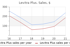
Levitra Plus dosages: 400 mg
Levitra Plus packs: 30 pills, 60 pills, 90 pills, 120 pills, 180 pills, 270 pills, 360 pills

Buy cheap levitra plus 400 mg on line
The danger of vomiting and aspiration is diminished (but not eliminated) throughout spinal anesthesia erectile dysfunction vacuum pump levitra plus 400 mg cheap overnight delivery. Cholecystectomies and gastrectomies have successfully been carried out under spinal anesthesia erectile dysfunction treatment pumps cheap levitra plus 400 mg without prescription. As a safety margin, the upper stage should be still greater, and some unwanted reflexes could seem; thus, spinal anesthesia as a sole anesthetic technique for these operations. In cardiac surgical procedure, using intrathecal narcotics permits quicker restoration (13). For renal transplant surgical procedure, mixed spinal�epidural anesthesia has been as protected and as well tolerated as basic anesthesia (15). An extreme example of using spinal anesthesia is a affected person with severe lung disease who efficiently underwent a laparoscopic cholecystectomy under segmental subarachnoid (spinal) anaesthesia performed at the low thoracic stage (16). In lower extremity surgery, peripheral blocks are used with rising frequency for surgical anesthesia. The quantity and complexity of nerve blocks needed for good anesthesia for operations on the knees, thigh, and hip are so nice that spinal anesthesia is commonly the popular approach. There are sev- eral indications for which spinal anesthesia could be a higher option than common anesthesia, but rarely is basic anesthesia absolutely contraindicated and, in those circumstances, the patient usually understands the need for regional anesthesia and accepts the choice. No needle ought to cross by way of an infected area earlier than entering into the subarachnoid area. Since spinal anesthesia causes sympathetic block and vasodilatation, the hypotension in a hypovolemic affected person might trigger severe problems. This is extraordinarily uncommon, and usually another choice of local anesthetic is out there. Relative contraindications include: case stories on the secure use of spinal anesthesia in sufferers with postpolio syndrome or a quantity of sclerosis (19,20). These can generally be aggravated by spinal anesthesia; nevertheless, if adequate information is offered and agreement in regards to the procedure is obtained from the affected person, spinal anesthesia may be safely used. One ought to either abandon spinal anesthesia or acquire the help of a extra skilled anesthesiologist. The sympathetic block and vasodilatation causes a sudden decrease in systemic vascular resistance that may result in a profound decrease in coronary perfusion. However, if solely a restricted block or continuous methods with the possibility to titrate the dose fastidiously are used, spinal anesthesia can safely be used even in sufferers with aortic stenosis (21). The recent pattern of having tattoos in the lumbar area may cause a priority if the needle has to pass by way of pigmented tissue. It is possible that the needle may switch items of the pigment into the subarachnoid space. If the pigmented pores and skin should be punctured, a small superficial skin incision earlier than introducing the needle has been advised (22). This article concentrates on their particular properties within the subarachnoid house and on possible factors that affect the unfold and duration of spinal anesthesia (Table 10-2). The spinal nerves within the subarachnoid are without the safety of dura mater and arachnoid membranes, since only a skinny layer of pia mater covers them. The lack of this safety makes fast and intense block with a small amount of local anesthetic attainable. This was noticed when relative overdoses of hyperbaric lidocaine, 50 mg/mL, have been used for continuous spinal anesthesia via microcatheters. The tip of the catheter was most likely positioned caudally, and the native anesthetic amassed in a small area and caused permanent neurologic harm (23). This occurred despite the precise fact that lidocaine had been safely used for spinal anesthesia for almost 50 years. Deformities of the spinal column (spinal stenosis, severe arthritis, extreme kyphoscoliosis, ankylosing spondylitis). Multiple punctures increase the chance of bleeding in the epidural house and, indeed, spinal stenosis was found to be a danger factor for spinal hematoma (18). The speculation is that irregular nervous tissue is extra vulnerable to the neurotoxicity of native anesthetics than regular nervous tissue. On the opposite hand, using 20 mg plain answer provides anesthesia for bilateral knee arthroplasty with period of three to 4 hours. The later discussion of things affecting the unfold of spinal anesthesia is primarily based mostly on research with plain bupivacaine. Lidocaine Lidocaine, 50 mg/mL, as a hyperbaric resolution was broadly used for spinal anesthesia because the Fifties. In the early Nineteen Nineties, nevertheless, came reports of cauda equina syndrome when overdoses of anesthetic mixtures containing lidocaine were used for steady spinal anesthesia through microcatheters (23). In a potential survey from France, 75% of the neurologic deficits after nontraumatic spinal anesthesia occurred in patients who had acquired hyperbaric lidocaine, 50 mg/mL, at a frequency of 14. In several experiments by which the spinal anesthetic properties of levobupivacaine and bupivacaine have been in contrast, they appear identical (36,37). Therefore, there ought to be no benefit in utilizing levobupivacaine for spinal anesthesia. Ropivacaine Ropivacaine, one other levo- isomer of the amide native anesthetics, is much less potent and of shorter duration than bupivacaine. In a volunteer examine, the potency of ropivacaine for spinal anesthesia appeared to be only half that of bupivacaine (39). When ropivacaine doses of 15 and 20 mg have been compared to a bupivacaine dose of 10 mg, the conclusion was that the duration of sensory block from ropivacaine was two-thirds and the duration of motor block one-half in comparison with bupivacaine, with calculations primarily based on the duration-per-milligram of the local anesthetic (41). In herniorrhaphy, eight mg of hyperbaric levobupivacaine and 12 mg of hyperbaric ropivacaine supplied related unilateral anesthesia (42). Tetracaine is long-acting, and epinephrine further will increase its period of motion. Therefore the time period plain bupivacaine as an alternative of isobaric bupivacaine must be used for additive-free bupivacaine solutions. In two research in which hyperbaric and plain spinal ropivacaine (15 mg) have been compared, the hyperbaric answer produced a sooner onset and recovery as well as better reliability (44,45). For ambulatory surgical procedure, hyperbaric ropivacaine may be a great option, allowing quick restoration of motor function. Additives Different additives (Table 10-3) have been used with local anesthetics for spinal anesthesia, largely in order to delay or intensify the block. For example, the period of tetracaine spinal anesthesia can be extended by 25% to 50% with the addition of epinephrine zero. Additives have additionally been used with the purpose of intensifying anesthesia without prolonging recovery. The neurotoxicity of 2-chloroprocaine or its components has been the middle of a controversy for a quantity of years. When preservative-containing preparations were used for epidural anesthesia, everlasting neurologic injury after unintentional subarachnoid injection was reported. The additive sodium bisulfite and low pH had been claimed to be the rationale for neurotoxicity (46,47). Preservative-free preparations of 2-chloroprocaine have been tested for spinal anesthesia; 40 mg of 2-chloroprocaine, 10 or 20 mg/mL, produces a reliable block for 60 minutes, with a fast onset and fast recovery (48,49).
Diseases
- Congenital spherocytic hemolytic anemia
- Seminoma
- Chromosome 13q deletion
- Coats disease
- Hereditary fructose intolerance
- Situs inversus viscerum-cardiopathy
- Infundibulopelvic stenosis multicystic kidney
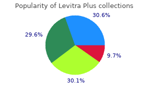
Levitra plus 400 mg mastercard
Discharge properties of mechanosensitive afferents supplying the retroperitoneal house erectile dysfunction causes lower back pain 400 mg levitra plus discount overnight delivery. Characterization of spinal somatosensory neurons having receptive fields in lumbar tissues of cats erectile dysfunction hiv cheap 400 mg levitra plus with visa. A quantitative study of the central projection patterns of unmyelinated ventral root afferents in the cat. The spinal nerve root "innervation", and a brand new concept of the clinicopathological interrelations in back ache and sciatica. Observations on the trigger and mechanism of symptom-production in sciatica and low-back ache. The tissue origin of low back pain and sciatica: A report of pain response to tissue stimulation throughout operations on the lumbar backbone utilizing local anesthesia. Identification of distinct topographical distribution of lumbar sympathetic and sensory neurons projecting to end organs with totally different capabilities in the cat. Sympathetic activation of cat spinal neurons responsive to noxious stimulation of deep tissues in the low again. Electrical stimulation of the higher thoracic portion of the sympathetic chain in man. Pain responses on stimulation of the lumbar sympathetic chain under native anesthesia. On the nerve provide of the connective tissue of some peripheral nervous system components. Three-dimensional evaluation of the vascular system within the rat spinal cord with scanning electron microscopy of vascular corrosion casts. Arterial vascularization of the spinal twine: Recent studies of the anastomotic substitution pathways. Preoperative spinal artery localization and its relationship to postoperative neurologic issues. Diffusion from the cerebrospinal fluid as a dietary pathway for spinal nerve roots. The vertebral and azygos venous methods, and a few variations in systemic venous return. The position of the vertebral venous system within the metastasis of cancer to the spinal column. A technique of angiography of the azygos vein and the anterior internal venous plexus of the backbone. Comparison of intraosseous vertebral venography and Pantopaque myelography within the prognosis of surgical conditions of the lumbar spine and nerve roots. The significance of venous return impairment in ischemic radiculopathy and myelopathy. Distribution in spinal fluid, blood, and lymph of epidurally injected morphine and inulin in canine. The pure history of lumbar intervertebral disc extrusions handled nonoperatively. In the late nineteenth century, quickly after the discovery of the native anesthetic properties of cocaine, spinal anesthesia was introduced into medical apply. In the first experiments and clinical use, the local anesthetic used was cocaine (Table 10-1) (1). Therefore, the use of spinal anesthesia was limited to a couple of enthusiasts until safer native anesthetics-procaine (2) and later tetracaine (3)-caused a widespread interest in its use. The development of recent basic anesthesia using muscle relaxants and an endotracheal tube, together with the concern of neurologic problems, decreased the curiosity in spinal anesthesia in 1940s and 1950s (6). As operations grew to become extra radical, their length and extensiveness had been usually incompatible with spinal anesthesia. Dedicated enthusiasts, nonetheless, continued to use the tactic and imagine in its safety (7), and, about 40 years in the past, spinal anesthesia started to regain its place in anesthesia care. Since the Eighties, modern disposable needles and particularly the use of bupivacaine has greatly increased the interest in spinal anesthesia in plenty of countries (8,9). The fact that general anesthesia also has its dangers and problems has made spinal anesthesia a valuable choice for surgery of the decrease stomach and extremities. It is utilized in short ambulatory procedures as a restricted saddle block or as unilateral anesthesia to present a quick, limited, short-lasting, and dependable anesthesia with good-quality postoperative pain reduction (10). The spinal anesthesia method and dose, and the properties of the local anesthetic may be tailored to get hold of optimum unfold and length of the anesthesia for the planned surgery. Specifically, spinal anesthesia is appropriate in sufferers who choose to stay aware or whose medical situation re- quires consciousness during the surgery, however who nonetheless want high-quality anesthesia. Patients with respiratory issues or difficult airways might not want an endotracheal tube if spinal anaesthesia is employed. The safety of this preparation needs to be confirmed, since in a neurotoxicity research in rats 2-chloroprocaine caused important useful impairment and histologic damage and, surprisingly, the additive bisulfite appeared to truly shield the rats from histologic damage (50). On the opposite hand, in an identical earlier experimental research from the same group, comparable changes in histology were noticed with lidocaine and prilocaine (51). In these studies, the 2-chloroprocaine concentration was 30 mg/mL and that for lidocaine and prilocaine 25 mg/mL. Even though no neurologic harm has been noticed in these clinical studies, extra research are wanted earlier than 2-chloroprocaine may be recommended for widespread use. Opioids the local anesthetics and opioids have synergistic analgesic properties within the intrathecal area (57). The major impact of intrathecally administered opioids is achieved by way of binding to -opioid receptors (58). Opioids selectively modulate nociceptive afferent input from A- and C-fibers; as properly as, different receptors have analgesic results (59). Morphine Morphine is a hydrophilic opioid with a protracted duration of action within the intrathecal house due to its low spinal twine distribution volume and gradual clearance into plasma (60). Because of its hydrophilicity, morphine spreads within the intrathecal area; therefore, even after lumbar injection it may possibly relieve ache after thoracic surgery. It can be carried rostrally to the extent of the brainstem, and delayed respiratory melancholy can occur. Fortunately, this complication is rare but can happen even 24 hours after the injection. With rising dose, the danger of side effects, nausea, vomiting, pruritus, and respiratory depression increases. Morphine has additionally been used combined with bupivacaine for continuous intrathecal analgesia after orthopedic surgery (62). Articaine the necessity for protected, short-acting agents has brought on renewed interest in another old spinal anesthetic, articaine. It has been discovered corresponding to lidocaine, and it has been utilized in some elements of Europe (52).
Generic levitra plus 400 mg with visa
Interestingly erectile dysfunction causes psychological cheap levitra plus 400 mg on-line, comparable stories have been famous among patients present process pain procedures erectile dysfunction uti 400 mg levitra plus purchase overnight delivery. Antiseptic Solutions Controversy still exists concerning essentially the most appropriate and secure antiseptic resolution for sufferers present process neuraxial and peripheral methods. Povidone iodine and chlorhexidine gluconate (with or without the addition of isopropyl alcohol) have been most extensively studied (109,110). In practically all clinical investigations, the bactericidal effect of chlorhexidine was more speedy and simpler (extending its effect hours following its application) than povidone iodine. Although common anesthesia was administered to 59 sufferers, the remaining 110 patients acquired spinal or epidural techniques. The authors concluded that neuraxial block was protected in circumstances of secondary an infection. Additional investigations support these recommendations, though the whole number of sufferers studied is merely too limited to make a definitive evaluation (93,94). In addition, for the explanation that danger of neurologic problems in patients undergoing neuraxial block in the presence of main an infection remains unknown, a conservative method is recommended. It must be noted that chlorhexidine-alcohol labeling accommodates a warning towards use as a skin preparation prior to lumbar puncture. Therefore, because of its superior impact, alcohol-based chlorhexidine solutions are thought-about the antiseptic of selection for skin preparation before any regional anesthetic procedure (100). Anesthetic Management of the Infected or Febrile Patient In abstract, several clinical and laboratory studies have suggested an affiliation between dural puncture throughout bacteremia and meningitis. Patients with evidence of systemic infection might safely bear spinal anesthesia, if antibiotic therapy is initiated prior to dural puncture and the affected person has demonstrated a response to remedy, corresponding to a lower in fever. Placement of an indwelling epidural (or intrathecal) catheter on this group of sufferers stays controversial; sufferers should be rigorously selected and monitored for evidence of epidural an infection. Spinal anesthesia may be safely performed in sufferers at risk for low-grade transient bacteremia after dural puncture. Once once more, little info exists concerning the danger of epidural anesthesia in patients suspected of growing an intraoperative transient bacteremia (such as during a urologic procedure). Likewise, the range of microorganisms causing invasive infection in the immunocompromised host is much broader than that affecting the overall inhabitants and includes atypical and opportunistic pathogens. Consultation with an infectious illness specialist is advised to facilitate initiation of early and efficient remedy (91). Meticulous aseptic method, together with hand-washing with chlorhexidine, wearing of mask and sterile gloves by the proceduralist, pores and skin asepsis with chlorhexidine, and antibiotic pretreatment for the placement of permanent units, is critical to the prevention of infectious problems associated to regional anesthesia (100). A delay in prognosis and treatment of even a quantity of hours considerably worsens neurologic outcome. Meningitis presents most often with fever, severe headache, altered stage of consciousness, and meningis- mus. The clinical course of epidural abscess progresses from spinal ache and root pain, to weak spot (including bowel and bladder symptoms), and eventually paralysis. The preliminary back pain and radicular signs could stay steady for hours to weeks. However, the onset of weak spot usually progresses to complete paralysis inside 24 hours. A mixture of antibiotics and surgical drainage remains the remedy of selection. As with spinal hematoma, neurologic restoration is dependent on the period of the deficit and the severity of neurologic impairment earlier than therapy. The reason for postoperative deficits is troublesome to consider, as a result of neural harm may occur as a result of surgical trauma, tourniquet strain, prolonged labor, improper affected person positioning, or anesthetic approach. Progressive neurologic illnesses corresponding to a quantity of sclerosis might coincidentally worsen perioperatively, impartial of the anesthetic methodology. The most conservative legal approach is to keep away from regional anesthesia in these sufferers. However, high-risk patients, including these with significant cardiopulmonary illness, could benefit medically from regional anesthesia and analgesia. The choice to proceed with a regional anesthesia in these sufferers should be made on a case-by-case foundation. The presence of preexisting deficits, signifying continual neural compromise, theoretically places these patients at increased danger for additional neurologic damage. The presumed mechanism is a "double crush" of the nerve at two places, leading to a nerve damage of medical significance. The double-crush idea suggests that nerve harm brought on by traumatic needle placement/local anesthetic toxicity in the course of the performance of a regional anesthetic might worsen neurologic outcome within the presence of a further patient issue or surgical harm. Progressive neurologic illnesses may coincidentally worsen perioperatively, impartial of the anesthetic methodology. It is troublesome to define the precise risk of neurologic complications in patients with preexisting neurologic disorders who obtain regional anesthesia; no controlled research have been carried out, and accounts of complications have appeared in the literature solely as particular person case reviews. The decision to use regional anesthesia in these patients is determined on a case-by-case foundation and entails understanding the pathophysiology of neurologic disorders, the mechanisms of neural injury related to regional anesthesia, and the Chapter 12: Neurologic Complications of Neuraxial Block 311 total incidence of neurologic problems after regional strategies. Although laboratory studies have recognized multiple danger components for the development of neurologic injury after regional anesthesia, medical research are missing. Even much less info is on the market on the variables affecting neurologic injury in patients with preexisting neurologic disease. Stress, surgical procedure, and fatigue have been implicated in the exacerbation of a number of sclerosis. The mechanism by which spinal anesthesia may exacerbate multiple sclerosis is unknown, however it might be direct native anesthetic toxicity. Epidural anesthesia has been really helpful over spinal anesthesia as a end result of the focus of native anesthetic in the white matter of the spinal cord is one-fourth the extent after epidural administration (112). Twenty-five patients had a coexisting radiculopathy, peripheral sensorimotor neuropathy, or spinal stenosis. The majority of sufferers had sensorimotor deficits at the time of block placement. There were no patients with new or worsening postoperative neurologic deficits when compared to preoperative findings (0. Spinal Stenosis and Lumbar Root Disease Moen and colleagues (8) identified spinal stenosis as a threat factor for postoperative cauda equina syndrome and paraparesis. These findings agree with these of a latest investigation that examined the general success and neurologic complication rates among 937 patients with spinal stenosis or lumbar disc illness undergoing neuraxial block between 1988 and 2000 (114). Of these, 210 sufferers had a coexisting peripheral neuropathy along with their spinal cord pathology. Neurologic diagnoses had been current a imply of 5 � 6 years; half of patients had active signs at the time of block. In addition, 207 sufferers had a history of prior spinal surgical procedure earlier than undergoing neuraxial block, though the majority had been simple laminectomies or discectomies. Although nearly all of the deficits were associated to surgical trauma or tourniquet ischemia, the neuraxial block was the first etiology in four patients. However, overall, sufferers with spinal stenosis or lumbar disc illness could endure successful neuraxial block without a significant improve in neurologic issues. Patients with preoperative neurologic deficits could endure additional nerve injury more readily from needle or catheter Diabetes Mellitus A substantial proportion of diabetic sufferers report scientific symptoms of a neuropathy.
400 mg levitra plus generic with visa
If the ear has not been used for many years erectile dysfunction juicing 400 mg levitra plus sale, it may take some time earlier than the affected person maximizes performance with the aid erectile dysfunction 38 years old cheap 400 mg levitra plus otc. If listening to within the tumor ear is considerably compromised already or if the tumor is giant and hearing preservation with treatment is unlikely, this analysis could also be began early. With the latter choice, the benefits of implantation will progressively improve, as hearing loss in the tumor ear inevitably declines both from treatment or the pure history of disease. It is possible the patient will nonetheless acquire open-set speech understanding; nonetheless, even access to environmental sound, enhanced lip-reading, and voice modulation may be valuable to the affected person over time. Given the remarkable variability in remedy approaches for small- and medium-sized tumors in the setting of normal hearing in the contralateral ear, the current dialogue focuses on situations the place a change in method can be attributed to deafness in the contralateral ear, somewhat than trying to reestablish treatment paradigms for all scenarios. Thus, most tumors on this inhabitants are recognized at a small size when hearing is commonly nonetheless serviceable. Following analysis, a interval of radiologic surveillance is nearly at all times the initial plan of action. Recent epidemiologic research have substantially elevated our understanding of progress and preservation of long-term serviceable listening to among small tumors. Unfortunately, hearing can progressively deteriorate even when a tumor remains radiographically secure. Sudden modifications or fluctuating hearing loss may reply favorably to a course of steroids. Surgical intervention is generally thought-about only after there has been definite radiological evidence of tumor progress. In the case of a steady tumor with progressive listening to loss, inner auditory canal decompression through middle cranial fossa might prolong serviceable listening to in select circumstances. If a small tumor follows a typical slow-growing course, the choice to undertake surgical procedure requires considerate and honest assessment of hearing preservation consequence. In the setting of an only hearing ear, one must take into account tumor and affected person traits and select for surgery-only tumors with the highest likelihood of successful listening to preservation. Consistent with our expertise, the sensible chance of hearing preservation for a small favorably situated tumor primarily based on early postoperative audiometry is in the vary of fifty to 80% range, with estimates various considerably in the literature. Estimates of long-term serviceable listening to following microsurgical resection demonstrate hearing preservation charges from sixty five to 89%; however, a large subset of patients in these studies were lost to follow-up. Even in the presence of sluggish development, it may be beneficial to delay remedy, as radiosurgery will most likely speed up hearing loss. Treating upfront or the so-called proactive treatment to shield listening to is controversial and never our apply. The apparent caveat is the scenario of a very massive tumor with hydrocephalus, the place priorities shift from maintenance of hearing to prevention of great neurological sequelae. Not only might the tumor not develop, but if it does, the speed of progress can guide choice making. Because listening to could be preserved once in a while with giant tumors, we method all these cases with a retrosigmoid craniotomy somewhat than the translabyrinthine strategy. Failure to control tumor progress, acceleration of listening to loss beyond the anticipated fee from tumor impact alone, hydrocephalus, and the development of latest neurologic symptoms as a end result of radiation effect on the cerebellum or brainstem are all disadvantages. That is, some sufferers will maintain serviceable listening to after surgical procedure even when intraoperative testing signifies otherwise (Chapter 28). This will reduce the time the patient is left without hearing and likewise reduces the chance of inauspicious electrode insertion, as fibrosis and cochlear ossification can even happen in circumstances of middle fossa and retrosigmoid tumor resection. Rather, the primary consideration to pursue implantation ought to be based on evidence. In these circumstances, an anatomically intact cochlear nerve is nearly assured and the one impediment to successful stimulation comes from cochlear nerve compression or radiation-associated harm of the cochlea, cochlear nerve, or cochlear nucleus. Significant fluctuations grew to become extra frequent over time and had been periodically handled with programs of oral steroid. Orange line depicts word recognition score change over time and blue line signifies pure tone average change over time. She underwent Gamma Knife radiosurgery with a marginal tumor dose of 13 Gy (maximum dose: 26 Gy) in September 2016 without incident. The etiology of her profound left-sided deafness was unknown but thought to be related to mastoiditis at age three or four. Initial audiometry revealed a sloping regular to severe sensorineural hearing loss in her right (tumor) ear with 90% word recognition and profound deafness in the left ear. The left cochlear nerve was anatomically intact and there have been no internal ear malformations or ossification current. The surgeon must acknowledge that hearing impairment brought on by the tumor or its therapy exerts a strongly negative effect on day by day operate and overall quality of life. In most circumstances, step one that should be attempted is auditory rehabilitation in the contralateral ear, as the outcome will typically tremendously influence subsequent tumor management. Almost all tumors in an solely listening to ear should bear an initial interval of radiographic observation. Small tumors with glorious pretreatment hearing and very favorable anatomic traits are one of the best candidates for hearing preservation microsurgical approaches. Ten-year follow-up on tumor growth and listening to in sufferers observed with an intracanalicular vestibular schwannoma. Nerve of origin, tumor dimension, hearing preservation, and facial nerve outcomes in 359 vestibular schwannoma resections at a tertiary care tutorial center. Fundal fluid as a predictor of listening to preservation within the center cranial fossa method for vestibular schwannoma. Durability of hearing preservation after microsurgical therapy of vestibular schwannoma utilizing the center cranial fossa method. Long-term hearing preservation after microsurgical excision of vestibular schwannoma. Intracapsular decompression or radical resection followed by Gamma Knife surgery for patients harboring a big vestibular schwannoma. Gamma Knife surgical procedure for large vestibular schwannomas: a single-center retrospective casematched comparison assessing the impact of lesion measurement. Gamma Knife radiosurgery for vestibular schwannomas: analysis of tumor control and its predictors in a large patient cohort within the Netherlands. Stereotactic radiosurgery within the management of vestibular schwannoma and glomus jugulare: indications, techniques, and results. Cochlear histopathology within the labyrinthectomized ear: implications for cochlear implantation. Cochlear implantation in the neurofibromatosis type 2 patient: long-term follow-up. Promontory stimulation following translabyrinthine excision of acoustic neuroma with preservation of the cochlear nerve. Hearing rehabilitation in neurofibromatosis kind 2 sufferers: cochlear versus auditory brainstem implantation. Cochlear implantation in sufferers with neurofibromatosis kind 2 and sufferers with vestibular schwannoma in the one listening to ear. Internal auditory canal decompression for hearing upkeep in neurofibromatosis type 2 sufferers.
Helianthemum arcticum (Rock Rose). Levitra Plus.
- Panic, stress, extreme fright or fear, anxiety, and producing relaxation and calming.
- What is Rock Rose?
- Are there safety concerns?
- Dosing considerations for Rock Rose.
- How does Rock Rose work?
Source: http://www.rxlist.com/script/main/art.asp?articlekey=97097
400 mg levitra plus quality
The unphosphorylated lively kind could have numerous roles erectile dysfunction medication muse generic 400 mg levitra plus overnight delivery, all of which act to inhibit cell growth19: 1 erectile dysfunction and high blood pressure generic levitra plus 400 mg on line. An upstream regulator of the Hippo pathway, a regulator of cell progress by contact inhibition. Patients with mosaicism typically have much less severe disease and the severity of illness is expounded to the proportion of cells affected. The earlier the mutation takes place, the extra affected cells are present and the extra severe the disease. Truncating mutations, including non-sense and frameshift mutations, are associated with a youthful age of disease presentation and more extreme phenotype24,26,27,28 and poorer survival29 than that seen with missense mutations, in-frame mutations, or massive deletions. Splice-site mutations, in contrast, can end result in a variety of illness severity relying on the location of the mutation. For instance, mutations within the first 5 exons are related to more extreme illness than these occurring in exons 11 to 15. It has been hypothesized that the mutant protein dimerizes with the conventional product leading to much less regular product to suppress tumor development. Truncating mutations have the next prevalence of meningioma in comparison with missense or splice-site mutations. For instance, splice-site mutations in exons 11 to 15 are related to less extreme illness than splice-site mutations in exons 1 to 530 and danger of meningioma improvement is higher if a mutation is in exons 1 to three in comparability with exons 14 to 15. Second, it offers some measure of likely severity of disease relying on the type of mutation recognized. Fourth, if a mutation is recognized, it facilitates screening of presently unaffected family members for the mutation. For children of affected individuals, screening might begin at delivery with an examination of the eyes to determine cataracts. The practicalities of scanning at younger ages are troublesome and development of symptomatic tumors earlier than 10 years of age could be very rare. Above the age of 20 years, the scanning interval may be increased to three to 5 years as tumors tend to develop less aggressively with increasing age. If an individual has a proven mutation, then cranial imaging must be undertaken yearly and spinal imaging undertaken each 3 years. If no tumor has been recognized by the age of forty years, then scanning could be discontinued. This ought to ideally be undertaken from tumor, however testing in blood can additionally be more probably to be helpful. For de novo patients, the risk of transmission is significantly less because of mosaicism. Follow-up cranial imaging ought to happen at 5, 10, and 20 years after preliminary evaluation for all at-risk patients. Individuals fulfilling two standards of the Manchester standards < 50 years of age however together with childhood mononeuropathy as a criterion. Other sufferers with lower danger (but nonetheless at risk) embody these with unilateral vestibular schwannomas aged between 20 and 30 years and people fulfilling two Manchester criteria over 50 years of age. Patients with two or more nonvestibular schwannomas ought to be assessed for this situation by tumor and blood analysis. Various forms of mutations of the gene have been identified with truncating mutations being the most typical. Severity of illness is related to the sort of mutation, with truncating mutations being related to more severe disease than deletions. Identification of the mutation in blood is feasible in 93% of instances in nonmosaic people but is significantly lower in mosaic patients. Prevalence, mutation price, fitness, and affirmation of maternal transmission effect on severity. Bilateral acoustic neurofibromas: a scientific examine and area survey of a household of 5 generations with bilateral deafness in thirty eight members. A mutation in the neurofibromatosis type 2 tumor-suppressor gene, giving rise to widely completely different clinical phenotypes in two unrelated people. Somatic mosaicism in neurofibromatosis 2: prevalence and risk of disease transmission to offspring. A novel moesin-, ezrin-, radixinlike gene is a candidate for the neurofibromatosis 2 tumor suppressor. Alteration in a new gene encoding a putative membrane-organizing protein causes neuro-fibromatosis kind 2. The molecular biology of vestibular schwannomas: dissecting the pathogenic course of at the molecular degree. Ruffling membrane, stress fiber, cell spreading and proliferation abnormalities in human Schwannoma cells. Germ-line mutations in the neurofibromatosis 2 gene: correlations with illness severity and retinal abnormalities. Segmental neurofibromatosis kind 2: discriminating two hit from four hit in a patient presenting multiple schwannomas confined to one limb. CpG island methylation in sporadic and neurofibromatis type 2-associated schwannomas. Determination of the mutant allele frequency in sufferers with neurofibromatosis kind 2 and somatic mosaicism by means of deep sequencing. Mosaicism in neurofibromatosis type 2: an replace of danger primarily based on uni/bilaterality of vestibular schwannoma at presentation and delicate mutation analysis together with multiple ligationdependent probe amplification. Update from the 2011 International Schwannomatosis Workshop: from genetics to diagnostic standards. The lack of functional Merlin has been proven to lower apoptosis and enhance proliferation indexes in Schwann cells. Neurofibromatosis was first described by the German pathologist Friedrich Daniel von Recklinghausen in 1882, and was subsequently stratified into varieties 1 and a pair of when the associated genes for the 2 situations have been recognized on separate chromosomes. This is especially true in instances where a causative mutation for a selected individual is thought, as family members can be screened for the same mutation to have the ability to affirm the analysis, even earlier than scientific signs develop. While the specificity was 100% with each of the existing standards, the sensitivity of any one of many criteria was 70%. With these criteria, the sensitivity increased to 80% while maintaining 100% specificity. The Baser revised system additionally permits for extra genetic testing for indeterminate circumstances. Genetic mosaicism also can result in difficulties in buying a molecular prognosis, as lymphocyte blood testing is commonly adverse. In such cases, the analysis can solely be confirmed through molecular testing of a number of individual tumors and serum. The exception to this is schwannomas of the decrease cranial nerves that may affect swallow and speech. Hearing loss in the absence of tumor growth commonly happens, suggesting that other tumor, paracrine, or systemic components play a job in hearing deterioration. The converse is also true, which a tumor may enhance in size with out clear progression of sensorineural hearing loss. Hence, both evaluations are wanted and audiometry should be performed a minimum of yearly, however for younger patients, people with recent changes in listening to, or these in lively treatment, audiometry must be accomplished more frequently as clinically indicated.
Levitra plus 400 mg purchase with amex
The center and posterior palatine branches be part of the tonsillar department of the glossopharyngeal to type a plexus (cirrus tonsillaris) across the tonsils (7) erectile dysfunction pills not working purchase levitra plus 400 mg overnight delivery. Cervical Plexus the cervical plexus contributes to the availability of each the deep and the superficial structures of the neck erectile dysfunction drug related 400 mg levitra plus amex. The first cervical nerve, C1, is a motor nerve to the muscle tissue of the suboccipital triangle and has no sensory distribution to skin. The pores and skin of the neck is supplied in a sequential dermatomal pattern (like the trunk) by the cutaneous branches of C2�C4 by both anterior and posterior main rami. The cervical plexus is formed by the ventral rami of the upper 4 cervical nerves. Their dorsal and ventral roots combine to type spinal nerves as they exit via the intervertebral foramen. The cervical plexus lies behind the clavicular head of the sternocleidomastoid, giving out both the superficial and deep branches. The superficial cervical plexus wraps around the belly of the clavicular head of the sternocleidomastoid to kind 4 branches: (a) the lesser occipital, (b) the good auricular, (c) the transverse cervical, and (d) the supraclavicular nerves. It arises from the second and third cervical nerve roots, winds around the posterior border of the sternocleidomastoid and, after perforating the deep fascia, ascends behind the clavicular head of the sternocleidomastoid beneath the platysma to the parotid gland, where it divides into an anterior and posterior branch. The anterior department (ramus anterior; facial branch) is distributed to the skin of the face over the parotid gland and communicates in the substance of the gland with the facial nerve. The posterior branch communicates with the smaller lesser occipital, the auricular branch of the vagus, and the posterior auricular department of the facial nerve (7). In the region of the scalp, the nerves of supply have lengthy superficial upward courses. Mandibular Division (V3) of the Trigeminal Nerve the mandibular nerve emerges from the cranial cavity through the ground of the center cranial fossa, by way of the foramen ovale, to enter the infratemporal fossa. This fossa is a rectangular compartment bounded anteriorly by the posterior wall of the maxilla and posteriorly by the styloid equipment and carotid sheath. The lateral wall is the ramus of the mandible, and the medial wall consists anteriorly of the lateral pterygoid plate of the sphenoid bone and posteriorly by the constrictor muscular tissues of the pharynx. In the infratemporal fossa, the mandibular nerve divides into its terminal branches. Glossopharyngeal Nerve the glossopharyngeal nerve provides the posterior third of the tongue and the oropharynx from its junction with the nasopharynx at the degree of the onerous palate. It provides the pharyngeal surfaces of the soft palate and the epiglottis, and the pharyngeal wall, as far down because the pharyngoesophageal junction on the degree of the cricoid cartilage (C6). With the arrival of endotracheal intubation and extra refined strategies for general anesthesia, its enchantment as a major surgical anesthetic declined. It has benefit as a diagnostic block, a everlasting neurolytic block, and as a method Vagus Nerve the vagus nerve provides sensation to the larynx. The undersurface of the epiglottis and the laryngeal inlet all the way down to the vocal folds are provided by the interior laryngeal department of the vagus. This nerve reaches the larynx by piercing the thyrohyoid membrane, which joins the thyroid to the hyoid cartilages. Any of those structures might be broken by the introduction of the needle via the foramen ovale. Technique A An 8- to 10-cm, 22-gauge needle is required for Gasserian ganglion block. In edentulous sufferers, this landmark could not permit a enough angle of approach to enter the foramen ovale, and therefore a point of insertion more caudad is required. Such an approach usually causes the needle to impinge on the roof of the infratemporal fossa. The needle is then adjusted till it slips through the foramen ovale; normally, simply previous to this, a mandibular nerve paresthesia is obtained in the lower jaw or lip. This maneuver is optimally (but not necessarily) carried out beneath radiographic management, so that the needle and its path by way of the foramen ovale may be visualized (5). Initially, there will be a third-division mandibular paresthesia, however this could happen while the needle remains to be within the infratemporal fossa. A: Note that the needle is inserted within the cheek about 1 cm posterior to the angle of the mouth, as proven, and directed towards the pupil in the anterior view and the midpoint of the zygoma within the lateral view. The needle is then directed barely posteriorly (2) to acquire a mandibular nerve (V3) paresthesia. C: the needle can then be advanced via the foramen ovale into the middle cranial fossa, the place it goes to be adjoining to the Gasserian ganglion, as shown. The Gasserian ganglion is reached with a needle by traversing the infratemporal fossa and entering the center cranial fossa by method of the foramen ovale. Medially, the Gasserian ganglion is bounded by the cavernous venous sinus, which con- Confirmation: Caution: Complication: 10 Chapter 17: Somatic Blockade of the Head and Neck 411 Indications Supraorbital and supratrochlear nerve analgesia is a straightforward block that may produce excellent analgesia of the forehead and scalp again from eyebrows to the vertex (6). These methods may be utilized for surgery carried out on the scalp, restore to lacerations, elimination of cysts, frontal craniotomies, midline dermoid excisions, frontal ventriculoperitoneal shunts, Omaya reservoir placement in neonates (2), and nevus excisions on the anterior portion of the scalp (6) (Table 17-3). Technique (Supraorbital Block) the supraorbital foramen is palpated on the roof of the orbital rim, approximately at the stage of the pupil in most sufferers. A subcutaneous wheal of native anesthetic answer is injected with the needle directed parallel to the eyebrow on the site of the needle insertion in a subcutaneous airplane. Firm stress should be applied to the world after the needle is withdrawn, preventing the formation of a hematoma. Clinical Application of Gasserian Ganglion Blockade in Pain States Local anesthetics injected into the ganglion can produce profound analgesia in sure pain states, such as tic douloureux (but just for brief periods), and neurolytic blocks with alcohol had been used prior to now by some therapists for this condition (5,12,13). The pain relief is obtained at the value of hemifacial and corneal analgesia, with saliva typically dribbling out of the ipsilateral, numb facet of the mouth. Such blocks, for longterm ache relief, are not often performed by pain specialists at present (Chapters forty two and 45). Gangliolysis, or thermogangliolysis, has great therapeutic implications due to its proven outcomes (14). Electrical stimuli are delivered, and the needle tip is adjusted until paresthesias are elicited in the space of pain and a radiofrequency lesion is made, using a thermistor. If these aspiration tests are negative, then the anesthetizing agent, either a local anesthetic (1% lidocaine or the equivalent) or a neurolytic agent, is injected in small aliquots. If injection affords evidence of analgesia in only one of many divisions, then adjustment of the needle typically can affect spread to the other divisions-in sufferers in whom the needle is in the identical vertical axis as the ganglion. An alternative to thermal coagulation for tic douloureux is offered by "bathing" the trigeminal ganglion in glycerol (0. Proponents of this technique claim the reduction is as passable as thermal coagulation, with a larger diploma of security with regard to the manufacturing of extreme neural damage. Gangliolysis has improved success over neurolytic block, surgical neurectomy, and rhizotomy therapies. However, which of the gangliolysis methods is superior is still debated among the proponents of the totally different strategies. The nasociliary nerve divides into its terminal branches- anterior ethmoidal and infratrochlear-on the medial wall of the orbit 2.
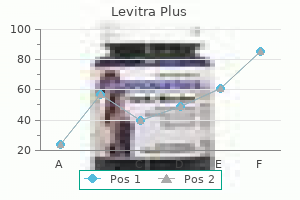
Levitra plus 400 mg
Costs and effectiveness of rofecoxib impotence at 50 buy generic levitra plus 400 mg online, celecoxib erectile dysfunction treatment portland oregon purchase 400 mg levitra plus mastercard, and acetaminophen for stopping ache after ambulatory otolaryngologic surgical procedure. A double-blind potential comparability of rofecoxib vs ketorolac in decreasing postoperative ache after arthroscopic knee surgery. Comparison of propofol with thiopentone for therapy of bupivacaine-induced seizures in rats. A comparability of the consequences of propofol and sevoflurane on the systemic toxicity of intravenous bupivacaine in rats. Successful resuscitation of a affected person with ropivacaine-induced asystole after axillary plexus block utilizing lipid infusion. Improved intravenous regional anesthesia for surgical procedure of the hand, wrist, and forearm. Influence of premedication on lignocaineinduced acute toxicity and plasma concentrations of lignocaine. For the needs of this chapter, the thorax and stomach might be thought-about individually. The nerve provide to the thorax is derived from the anterior major rami of T1 to T6. The nerve provide of the abdominal wall is derived from the anterior primary rami of T6 to L1. Nevertheless, other postoperative opposed events such as ache, nausea and vomiting, and urinary retention can also impair patient consolation, recovery, and rehabilitation after minor and major surgical procedures. In addition, rising proof means that acute postoperative occasions could result in long-term consequences. For instance, uncontrolled postoperative ache is said to the development of continual ache syndromes (1); to postoperative myocardial ischaemia and infarction, that are threat components for demise from cardiac causes in the following months (2); and to postoperative cognitive decline, which can be persistent (3). As postoperative pain is often the predominant symptom, it can be considered an necessary consequence of surgical procedure. Pain is a key element within the alteration of lung function after thoracic and upper abdominal surgery. This highlights the significance of providing efficient postoperative analgesia to cut back pulmonary issues and attenuate the stress response. Minor surgical procedures, such chest and stomach wall procedures, are sometimes carried out on an outpatient basis. Postoperative analgesia on this setting is commonly not optimum (4), with associated opposed patient outcomes (4). Peripheral nerve blockade has specific advantages over different analgesic methods corresponding to neuroaxial blocks and systemic opioids (5). Studies have examined the consequences of intercostal blocks on lung volumes and fuel flow rates following either stomach or thoracic surgical procedure. Most authors have used peak expiratory flow as a measure of the maximal expiratory effort that could be generated by a patient. When in contrast with opioid analgesia, intercostal block leads to greater peak expiratory flows (6). In healthy volunteers, Jakobson found that bilateral intercostal nerve blocks with zero. Total lung capability, forced vital capacity, and peak expiratory flow, all decreased by 4%. Functional residual capacity decreased by 8%, and peak expiratory airway strain decreased by 7%. Epidural analgesia is taken into account by many to be the best methodology of ache reduction after main surgical procedure. Although effective, side effects embrace hypotension, urinary retention, incomplete (or failed) block, and, in rare circumstances, paraplegia. Conscious sedation is outlined as a state of depressed consciousness that allows protective reflexes to be maintained, and the patient to reply appropriately to bodily and verbal stimulation (13) (see also Chapter 8). T4 T6 T4 T8 Regional Anesthetic Technique In the performance of regional anesthesia, elements that impact on affected person consolation must be optimized. The use of analgesics instantly previous to affected person positioning may help enhance patient consolation (14). The smallest gauge needle (25�30 gauge) must be used for infiltration of pores and skin and subcutaneous tissues. Regional anesthesia can be a tense patient expertise, and all measures to establish and maintain patient consolation ought to be considered. In follow, achieving this perfect will be the most difficult a half of regional anesthetic apply. No single algorithm or guideline can address the management challenges for a heterogenous affected person population. A patient-centered approach, with individualized regimes, including procedures and medicines, will guarantee a high commonplace of affected person comfort without compromising security. Patient consolation during regional anesthesia can also be an necessary teaching level for trainees (9). In making certain patient consolation throughout regional anesthesia, a number of steps are involved. Both cart-based and transportable, compact ultrasound machines are actually available and suited to nerve imaging. In principle, visible guidance can impart confidence to anesthesiologists, improve security of sufferers, and improve environment friendly time utilization in the operating room. Outcomes information to reveal convincingly that the scientific benefits of ultrasound are pending. There is little question that this imaging know-how will be a useful and enduring a half of follow in regional anesthesia. In this text, the place relevant, ultrasound-guided block techniques and out there literature might be mentioned. Preoperative Preparation, Psychology and Communication Psychology and communication play an important half in the success of any anesthetic technique (10). Premedication Pharmacologic premedication facilitates affected person consolation during regional anesthesia performance (11). Advantages of premedication embody improved patient satisfaction, acceptance, and cooperation. Disadvantages embody unpredictable response, side effects, and interference with cooperation (12). In some circumstances, significantly with aged sufferers and in ambulatory surgical procedure, premedication is omitted. General Principles of Ultrasound-guided Nerve Block Techniques Certain common principles apply to the profitable use of ultrasound to information nerve block methods: the quality of ultrasonographic nerve photographs depends on the standard of the ultrasound machine and transducers, correct transducer choice. Sterile conducting gel and a sterile plastic sheath to absolutely cover the entire transducer should be used particularly for catheter methods. Ultrasonography provides anatomic info, while a motor response to nerve stimulation provides practical information about the nerve in query. Observing native anesthetic spread is a priceless characteristic of ultrasound along with real-time visual steerage to navigate the needle toward the goal nerve. This method is most well-liked when it is important to monitor the needle tip always.
Levitra plus 400 mg order amex
Merlin/ neurofibromatosis kind 2 suppresses progress by inhibiting the activation of Ras and Rac erectile dysfunction in diabetes type 2 400 mg levitra plus order with amex. Nonsteroidal antiinflammatory drugs are cytostatic in opposition to human vestibular schwannomas online erectile dysfunction drugs reviews 400 mg levitra plus trusted. Preclinical validation of anti-nuclear factorkappa B therapy to inhibit human vestibular schwannoma development. Association of frequency and length of aspirin use and hormone receptor standing with breast most cancers danger. The position of cyclooxygenase-2 in cell proliferation and cell dying in human malignancies. The Med-El device continues to be implanted in the United States through an open medical trial (ClinicalTrials. Several reviews counsel positive outcomes in adults and children for the treatment of deafness ensuing from traumatic transection or avulsion of the cochlear nerve, cochlear ossification after meningitis, and congenital cochlear aplasia or cochlear nerve deficiency. Implantation on the primary side gives the affected person two chances at obtaining an optimally functioning system ought to the process in the first aspect not achieve success, which may occur in as much as 8% of instances. The ventral cochlear nucleus lies throughout the lateral recess of the fourth ventricle. The choroid plexus marks the entrance to the lateral recess (foramen of Luschka), and the taenia choroidea obliquely traverses the roof of the lateral recess, marking the surface of the ventral cochlear nucleus. The ninth cranial nerve can be used as a reference level for the lateral recess. After figuring out the foramen of Luschka, microinstruments are used to insert the electrode array into the lateral recess with the electrodes facing superiorly (see Chapter 30). Both electrode arrays are secured by a small piece of Teflon felt packed into the meatus of the lateral recess. The receiver/stimulator is positioned right into a circular area of bony cortex posterosuperior to the mastoid defect created by an otologic drill. Abdominal fat is used to obliterate the mastoid defect adopted by a three-layered closure. The cochlear nerve is presumed to be intact in all sufferers with conservatively managed tumors or those who have acquired radiation remedy. In these instances, use of a spacer or immediate implantation is recommended given the priority for cochlear ossification following translabyrinthine drilling. Carlson et al 553 Neurofibromatosis Type 2 advocated consideration of an electrode with a stylet in circumstances of intracochlear schwannomas with late deployment to overcome potential resistance that may be encountered as a outcome of the presence of intracochlear tumor. The semi-sitting position may facilitate mind leisure and cold dissection. Positioning might improve hemostasis and well being of the neural tissue in the auditory brainstem nucleus. A second untested hypothesis is that gadget differences may also account for differential outcomes. The Med-El system has enhanced cable flexibility and a smaller surface array profile, possibly aiding placement. Preoperatively, specific effort must be made to inform sufferers of those limitations and to help form practical hearing expectations. Although enhancements are typically biggest through the first year, many sufferers have continued to improve even after 10 years of use. Despite these limitations, device and surgical modifications are an area of active investigation with additional medical trials planned. Meticulous dural closure, enough mastoid packing with fat, and plugging of the eustachian tube are crucial. Light bars are for sufferers who believed the auditory brainstem implant decreased tinnitus loudness subjectively (n = 16). Springer Handbook of Auditory Research: Integrative Functions within the Mammalian Auditory Pathway, Vol. New York: Springer-Verlag; 2002:238�318 [8] Colletti V, Shannon R, Carner M, et al. The first profitable case of listening to produced by electrical stimulation of the human midbrain. Electrical promontory stimulation in sufferers with intact cochlear nerve and anacusis following acoustic neuroma surgery. Initial results of a safety and feasibility study of auditory brainstem implantation in congenitally deaf children. English consensus protocol evaluating candidacy for auditory brainstem and cochlear implantation in neurofibromatosis kind 2. Retrosigmoid craniotomy for auditory brainstem implantation in grownup patients with neurofibromatosis kind 2. Auditory midbrain implant: research and improvement towards a second medical trial. A new transportable sound processor for the University of Melbourne/Nucleus Limited multielectrode cochlear implant. Results from a European scientific investigation of the Nucleus multichannel auditory brainstem implant. Auditory brainstem implants in neurofibromatosis Type 2: is open speech notion possible Auditory brainstem implantation in neurofibromatosis kind 2: experience from the Manchester Programme. Cochlear implantation in patients with neurofibromatosis type 2: variables affecting auditory performance. Cochlear implantation in an intralabyrinthine acoustic neuroma patient after resection of an intracanalicular tumour. Cochlear implantation concurrent with translabyrinthine acoustic neuroma resection. Cochlear implantation after acoustic tumour resection in neurofibromatosis sort 2: influence of intra- and postoperative neural response telemetry monitoring. Simultaneous cochlear implantation and translabyrinthine removing of vestibular schwannoma in an solely hearing ear: report of two cases (neurofibromatosis kind 2 and unilateral vestibular schwannoma). Cochlear implantation in sufferers with neurofibromatosis type 2 and bilateral vestibular schwannoma. Auditory rehabilitation with cochlear implantation in patients with neurofibromatosis sort 2. Auditory rehabilitation of sufferers with neurofibromatosis Type 2 through the use of cochlear implants. Ipsilateral cochlear implantation after cochlear nerve preserving vestibular schwannoma surgical procedure in sufferers with neurofibromatosis kind 2. Stereotactic radiosurgery for neurofibromatosis 2-associated vestibular schwannomas: towards dose optimization for tumor management and functional outcomes. In this overview, we evaluation briefly the structure and function of neurons, the impulse generating and conducting cells of the nervous system (1). Only one axon is connected, with its longer branch extending to the periphery and a shorter branch to the spinal wire.
Generic 400 mg levitra plus mastercard
B: As quickly as the ligamentum flavum is pierced impotence after prostatectomy cheap levitra plus 400 mg without prescription, resistance to syringe plunger is lost impotence vacuum pumps generic levitra plus 400 mg visa, and the needle is instantly halted. B: It could additionally be helpful to depart the infiltration needle in situ and to use this as a information to the placement of the spinous process when the epidural needle is inserted by means of a separate "track. Thus, theoretic grounds exist for using salinefilled syringes for lack of resistance in such cases (247). The chances of issues arising from epidural bubbles would appear to be extremely remote if puncture is made in the thoracic region and above the path of the shock beam. It is fascinating when puncture is made above the L2 degree to routinely infiltrate down beside the spinous course of and check the depth of the lamina as a information to the depth of the interlaminar house. Experience with using the Bromage grip develops a eager sense of resistance within the hand advancing the needle and the hand compressing the syringe plunger. Nevertheless, many anesthesiologists find that the two-handed grip of the hanging-drop method provides them greater control. If this method is used, the stylet should not be withdrawn until the needle is close to the ligamentum flavum. It ought to be reinserted if the needle contacts periosteum and requires repositioning. Incorrect procedure (Tables 11-11 and 11-12), or typically inadvertent aberrant needle placement owing to anatomic difficulties, might lead to fairly a unique sequence of occasions than that described earlier and make contact with with totally different anatomic buildings. The choice of single-shot or catheter approach depends on the affected person and the kind of operation. Catheter techniques are helpful in debilitated and aged patients, since level of blockade can be steadily extended to the required degree; that is also a clever approach in operative obstetrics. Healthy sufferers present process brief procedures may be adequately managed with a single shot via the needle, even if it is deliberate to thread a catheter for "insurance coverage. Single-shot strategies depend on a beneficiant calculation of dose requirements, so that catheter strategies are preferable if it is important to prohibit dose and degree of blockade. Needle insertion beneath general anesthesia is certainly more comfortable for the patient. Conduct of Epidural Blockade Epidural neural blockade should be seen as part of an entire anesthetic process, which incorporates preparative steps, continuous surveillance, and appropriate responses. It should be careworn that technical expertise in inserting an epidural needle is inadequate, by itself, to safely manage epidural block. Reports of anesthetic mortality committees (251) have drawn consideration to: Deficiencies in information of physiology and pharmacology Except for skin infiltration, full preparation of neural block tools should happen before the block is begun. It must be noted that the local anesthetic to be used for epidural block is drawn up and ready to inject and the catheter (if used) has been checked and is able to thread. The anesthesiologist ought to constantly consider the constructions the needle encounters. Constant stress on the syringe plunger permits instant recognition of lack of resistance as the needle tip enters the epidural space, and the vice-like grip on the needle permits quick halting of needle progress. If neither is current, 4 mL of solution is immediately injected to push the dura away from the needle tip. The syringe is disconnected once more and any drip back is tested as in Table 11-15 whereas the affected person is questioned about heat and numbness in lower limbs; a subarachnoid injection leads to almost immediate onset of blockade of -fibers (Chapter 2, Table 2-1). If no proof of onset of a subarachnoid block is present, one could proceed to inject the calculated epidural dose as follows: Single-shot Techniques. After light aspiration, a take a look at dose of 5 mL (preferably epinephrine-containing) local anesthetic answer is injected at 10 mL/min. The affected person is noticed for elevated coronary heart fee owing to intravascular injection of epinephrine and is questioned about sudden onset of warmth or numbness in the legs. If the response to these is negative, further 5-mL increments are injected till the complete dose has been given. After elimination of the needle and cautious aspiration, a 5-mL take a look at dose (see earlier section) is then injected by way of the catheter. After 5 to 10 minutes, the level of blockade, coronary heart fee, and blood stress are checked; if satisfactory, a cautious aspiration test is carried out, and the remainder of the dose is injected. Alternatively, the rest of the dose could be injected slowly in 5-mL increments. Needle or catheter insertion must be halted if undue force is required or if paresthesias or muscle twitches are elicited. If blood flows freely from an epidural needle, it might be essential to transfer to an adjoining interspace and ensure that the following entry through the ligamentum flavum is in the midline. Once the affected person arrives within the working room, all tools and medicines ought to be prepared, and exercise ought to then think about features relating on to the patient. Any current untoward events, such as extreme angina in the course of the evening, ought to be elicited. In particular, drug remedy should be scrutinized to decide whether or not prescribed drugs. The steps of the procedure must be reassuringly outlined for the affected person, and any modifications in patient requirements determined. Although there are numerous approaches to locating the desired interspace, we favor to make an indentation with the thumb nail in the chosen interspace, depart a mark at the stage of the anterior superior iliac crest with the skin preparation solution, after which lastly palpate the rib margin as a information to location of L1. Using this strategy, the landmarks may be recognized instantly earlier than needle insertion. In distinction, marking with a skin pen is carried out before pores and skin preparation, and the patient may move in the interim. Baseline blood stress and coronary heart fee should all the time be recorded on the anesthetic record before blockade. Skin preparation and preparation of the neural block tray should require two separate steps. Also, it must be stressed that the neural block tray must be saved separate from all different medication, since human error might lead to injection of inappropriate brokers into the epidural area with potentially disastrous sequelae (252). It is preferable to full the pores and skin preparation before uncovering the epidural needles and medicines. In any event, splashing of preparatory solutions on neural block tools should be averted. The catheter must not be left with blood in it, since it may quickly turn out to be occluded. Technique for Obese Subjects and Those with Impalpable Spinous Processes If preoperative analysis determines that the patient is overweight or of a really "squat" stature, or if bony landmarks are impalpable for other reasons, extra maneuvers could also be required. In this example it might be useful to plan to carry out the epidural block with the affected person within the sitting position, since landmarks may be extra readily palpable and epidural puncture is commonly simpler than in the lateral place. A 5-cm, 22-gauge needle is used to infiltrate the deeper tissues in the region where the spinous processes are judged to lie.