

Hyzaar dosages: 50 mg, 12.5 mg
Hyzaar packs: 30 pills, 60 pills, 90 pills, 120 pills, 180 pills, 270 pills, 360 pills
Buy hyzaar 50 mg overnight delivery
A response that requires a free vitality input have to be coupled to one other reaction that releases no less than that much energy heart attack kid 50 mg hyzaar discount free shipping. Metabolic pathways consist of a collection of coupled reactions linked by widespread intermediates arrhythmia vs fibrillation 12.5 mg hyzaar discount with mastercard. In the absence of such processes, particular person reversible reactions eventually attain equilibrium, and the move of metabolites through a pathway ceases. For instance, a genetic defect or inhibitor that reduces manufacturing of B also decreases operation of the pathway from fuel! There are 5 common elements of metabolic pathways: response steps, regulated steps, unique characteristics, pathway interfaces, and clinical relevance. Acetyl CoA is utilized in fats synthesis, ldl cholesterol synthesis, ketone physique synthesis, and formation of acetylated molecules. Inner membrane: oxidative phosphorylation Acetyl CoA: product of fats and glucose oxidation Acetyl CoA: a focus in metabolism Glycolysis, glycogenesis, glycogenolysis, pentose phosphate shunt, fatty acid synthesis, steroid synthesis. Many intermediates within one pathway are substrates for other pathways, providing a way for various pathways to interact. Pyruvate carboxylase, which varieties oxaloacetate by carboxylation of pyruvate, is allosterically activated by acetyl CoA. A deficiency of any of those vitamins negatively impacts operation of the cycle and impairs vitality manufacturing. Cycle intermediates also take part in synthetic pathways leading to glucose, fatty acids, porphyrins, and amino acids (dashed arrows). Because all mitochondria within the zygote come from the ovum, these diseases exhibit maternal inheritance, by which affected mothers transmit the illness to all of their youngsters. Uncouplers short-circuit the proton gradient by transporting H� ions from the intermembrane house to the matrix, thereby abolishing the gradient. Pyruvate dehydrogenase is regulated by covalent modification with phosphorylation. Glycolysis interfaces with glycogen metabolism, the pentose phosphate pathway, the formation of amino sugars, triglyceride synthesis (by technique of glycerol 3-phosphate), the production of lactate (a dead-end reaction), and transamination with alanine. Pyruvate dehydrogenase interfaces with different pathways such because the citric acid cycle or fats synthesis by way of its product, acetyl CoA. Deficiencies in any of the pyruvate dehydrogenase enzymes produce lactic acidosis. Phosphorylation of glucose to glucose 6-phosphate, the primary regulated step in glycolysis, is irreversible and traps glucose contained in the cell. Glucokinase, current within the liver and pancreatic b cells, is very active solely at high glucose concentrations (high Km) and rapidly phosphorylates large amounts of glucose (high Vmax). Reversible conversion of fructose 1,6-bisphosphate to two 3-carbon intermediates by aldolase A b. The regulated steps in glycolysis are indicated by one-way arrows and boxed enzymes. Reversible conversion of 3-phosphoglycerate to 2-phosphoglycerate by phosphoglycerate mutase 8. This reaction happens in anaerobic glycolysis associated with shock and extreme train. All the kinase reactions are irreversible and serve a regulatory role in glycolysis. Glucokinase induction by insulin and lack of inhibition by glucose 6-phosphate promote clearance of blood glucose by the liver in the fed state. Acetyl CoA is a positive effector for pyruvate carboxylase, which favors technology of oxaloacetate as a substrate for gluconeogenesis. Glucose 6-phosphate, the first product formed in glycolysis, connects the glycolytic pathway to the pentose phosphate pathway and to glycogen synthesis, galactose metabolism, and the uronic acid pathway. In the fasting state, when glucose is in brief provide, pyruvate is carboxylated to oxaloacetate, providing carbon skeletons for gluconeogenesis. Enzymes which would possibly be required to bypass the three irreversible steps in glycolysis are discussed in Box 6-1. Simple reversal of the phosphoglucose isomerase response converts fructose 6-phosphate to glucose 6-phosphate. Lactate, alanine, and glycerol (boxes) are the first sources of carbon skeletons for gluconeogenesis. Reciprocal regulation ensures that gluconeogenesis or glycolysis predominates, stopping futile cycling of glucose to pyruvate and back once more to glucose. Acetyl CoA, a product of fatty acid oxidation, is a optimistic allosteric effector of pyruvate carboxylase, which diverts pyruvate into the gluconeogenic pathway somewhat than the citric acid cycle. High insulin and low glucagon ranges (fed state) (1) Leads to increased pyruvate kinase activity and elevated fructose 2,6-bisphosphate levels (2) Result: increased glycolysis (particularly in the liver) and decreased gluconeogenesis b. The liver is crucial site for gluconeogenesis, whereas the kidneys and the epithelium of the small intestine assume a less necessary function (mainly throughout starvation). Gluconeogenesis occurs partially within the mitochondria (pyruvate carboxylase reaction) and in part within the cytosol. Gluconeogenesis: preserve glucose in fasting state Gluconeogenesis sites: liver (major site), kidneys (starvation), epithelium of small gut Lactate provides approximately one third of the carbon skeletons utilized in gluconeogenesis. Glycerol is a crucial source of carbon atoms for gluconeogenesis in fasting or starvation conditions, when triacylglycerols in adipose tissue are mobilized. Glycogen is a extremely branched glucose polymer found primarily in liver and skeletal muscle. The branching enzyme, glucosyl (4:6) transferase, removes a block of glucose items from the nonreducing finish of a rising glycogen chain and reattaches glucose units in an a-1,6 linkage at a unique site, making a branch point. Phosphoglucomutase isomerizes glucose 6-phosphate to glucose 1-phosphate in a reversible reaction. Glycogen synthase, the rate-limiting enzyme, provides one glucose unit at a time, in a 1,4 linkage, to the nonreducing end of glycogenin. The ratio of glucose 1-phosphate to free glucose launched is determined by the number of department points and size of the branches. Phosphoglucomutase reversibly converts glucose 1-phosphate to glucose 6-phosphate, and is due to this fact useful in glycogenesis and glycogenolysis. The fate of glucose 6-phosphate derived from glycogenolysis differs in muscle and liver. Reciprocal regulation ensures that synthesis or degradation predominates, stopping the wasteful operation of each pathways simultaneously. High insulin (low glucagon), typical of the fed state, promotes glycogen synthase activity resulting in glycogen synthesis. Glycogen phosphorylase: rate-limiting enzyme of glycogen degradation Debranching enzymes: release free glucose Liver glycogenolysis: helps preserve blood glucose in fasting state Muscle glycogenolysis: uses glucose for its own power purposes Glycogen metabolism is reciprocally regulated, as is gluconeogenesis. Active protein kinase A phosphorylates glycogen synthase, which inactivates the enzyme and prevents glycogenesis.
Hyzaar 50 mg cheap without a prescription
Unfortunately studies concerning surgical remedy are missing as a result of most are case collection with a really low stage of evidence blood pressure normal value 50 mg hyzaar safe. Urethral stents have been proposed to keep away from restenosis blood pressure of 140 90 order hyzaar 12.5 mg without prescription, however their efficacy over time has not but been demonstrated. Additional various strategies are anecdotal, and their use must be reserved to highly chosen cases. Nine of 12 sufferers with lesions above T5 had signs of autonomic hyperreflexia throughout being pregnant and/or supply. The cesarean delivery fee for ladies with lesions above T5 was 47% and for ladies with lesions below that stage, 26%. Surgical procedures to keep away from bowel distension have an excellent level of evidence, however their use ought to be reserved for the uncommon instances of bowel program failure. It appears that the cesarean option might be preferred as a end result of it can be of shorter duration than vaginal supply. Incidence and clinical features of autonomic dysreflexia in patients with spinal twine damage. Autonomic dysreflexia in acute spinal wire damage: an under-recognized clinical entity. Regional sympathetic operate in high spinal wire harm throughout mental stress and autonomic dysreflexia. Cardiovascular penalties of loss of supraspinal management of the sympathetic nervous system after spinal twine harm. Autonomic control of the heart and renal vascular mattress throughout autonomic dysreflexia in excessive spinal wire damage. Atrial fibrillation related to autonomic dysreflexia in sufferers with tetraplegia. Spinal twine damage alters cardiac electrophysiology and will increase the susceptibility to ventricular arrhythmias. Autonomic dysreflexia and sudden dying in folks with traumatic spinal wire injury. Can epidural fentanyl control autonomic hyperreflexia in a quadriplegic parturient Prevalence and etiology of autonomic dysreflexia in children with spinal wire accidents. The impact of nifedipine on cystoscopy-induced autonomic hyperreflexia in sufferers with high spinal wire injuries. Comparison of sublingual captopril and nifedipine in immediate treatment of hypertensive emergencies: a randomized, single-blind scientific trial. Comparison of sublingual captopril and sublingual nifedipine in hypertensive emergencies. A comparability of safety and efficacy of sublingual captopril with sublingual nifedipine in hypertensive disaster. Comparison of sublingual captopril, nifedipine and prazosin in hypertensive emergencies throughout hemodialysis. Evaluation of captopril for the management of hypertension in autonomic dysreflexia: a pilot study. Management of life-threatening autonomic hyperreflexia utilizing magnesium sulphate in a patient with a high spinal wire damage in the intensive care unit. A study of the alpha-1 adrenoceptor blocker prazosin in the prophylactic administration of autonomic dysreflexia in high spinal cord injury patients. Autonomic dysreflexia throughout a bowel program in patients with cervical spinal twine damage. Lidocaine anal block limits autonomic dysreflexia during anorectal procedures in spinal wire damage: a randomized, doubleblind, placebo-controlled trial. Effect of controlled-release oxybutynin on neurogenic bladder perform in spinal twine damage. Combined intravesical and oral oxybutynin chloride in adult patients with spinal wire damage. Reiterated intravesical instillation ` ` of capsaicin in neurogenic detrusor hyperreflexia: a 5-years experience of 100 instillations [in French]. Intravesical capsaicin versus ` ` resiniferatoxin for the treatment of detrusor hyperreflexia in spinal twine injured patients: a double-blind, randomized, managed examine. Intravesical capsaicin versus resiniferatoxin in patients with detrusor hyperreflexia: a prospective randomized examine. The function of capsaicin-sensitive afferents in autonomic dysreflexia in sufferers with spinal wire harm. Electromotive drug administration of lidocaine to anesthetize the bladder earlier than intravesical capsaicin. Intravesical electromotive administration of oxybutynin in patients with detrusor hyperreflexia unresponsive to normal anticholinergic regimens. Denys P, Even-Schneider A, Thiry Escudie I, Ben Smail D, Ayoub N, ChartierKastler E. Efficacy of botulinum toxin A for the remedy of detrusor hyperreflexia [in French]. Botulinum toxin for remedy of urinary incontinence as a end result of detrusor overactivity: a systematic evaluation of effectiveness and antagonistic effects. Botulinum-A toxin for treating detrusor hyperreflexia in spinal wire injured patients: a model new alternative to anticholinergic drugs Botulinum toxin type A for refractory neurogenic detrusor overactivity in spinal cord injured sufferers in Singapore. Heart conduction problems in a tetraplegic patient attributable to a single therapeutic dosage of baclofen. Delayed issues of discontinuation of intrathecal baclofen therapy: resurgence of dyssynergic voiding, which triggered off autonomic dysreflexia and hydronephrosis. Endoscopic sphincterotomy in paraplegic sufferers: retrospective instances in Geneva [in French]. Transurethral sphincterotomy offers important relief in autonomic dysreflexia in spinal twine injured male sufferers: long-term followup outcomes. Sphincterotomy and the therapy of detrusor-sphincter dyssynergia: current status, future prospects. Clinical features of transurethral anterior sphincterotomy and urological management of sufferers with cervical spinal wire damage [in Japanese]. Transurethral electroresection of the external urethral sphincter within the urological management of male tetraplegics [in Japanese]. Management of female neurogenic bladders caused by cervical spinal twine injuries-cutaneous vesicostomy [in Japanese]. The mesh Wallstent in the treatment of detrusor external sphincter dyssynergia in males with spinal twine harm: a 12-year follow-up. Prospective comparison of exterior sphincter balloon dilatation and prosthesis placement with external sphincterotomy in spinal wire injured men. The use of the Memokath stent within the treatment of detrusor sphincter dyssynergia in spinal twine damage sufferers: a single-centre seven-year expertise. Surgical therapy of neurogenic detrusor overactivity (hyperreflexia) in paraplegic sufferers by sacral deafferentation and implant driven micturition by sacral anterior root stimulation: strategies, indications, outcomes, issues, and future prospects. The draw back of ileocystoplasty for individuals with cervical spinal twine harm and an indwelling urinary catheter. Augmentation enterocystoplasty for the management of voiding dysfunction in spinal cord harm sufferers.
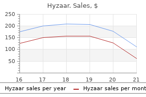
Order 12.5 mg hyzaar otc
In ladies blood pressure drop symptoms hyzaar 50 mg buy amex, this must be roughly 95 to one hundred ten levels blood pressure risks hyzaar 12.5 mg purchase with amex, whereas in males, this must be 90 to ninety five degrees. The columellar-lobular angle is outlined as the angle fashioned by the intersection of a line tangential to the columella and a line tangential to the infratip lobule. A nostril with poor support may require cartilaginous struts to counteract the inherently weakened tip from the rhinoplasty. The impact of facial animation supratip break, the columellar-lobular angle, and the two tipdefining factors (the most projected portion of the nasal tip). There are a quantity of strategies to decide whether or not the nasal tip projection is sufficient. Essentially the length of the upper lip (from subnasale to labrale superioris) ought to equal the nasal projection (measured from subnasale to pronasale). If the nasofrontal angle is between 36 degrees and forty levels, then the length of a perpendicular line by way of the nasal tip must be zero. A vertical line by way of essentially the most projected a half of the higher lip should divide the nostril into two equal components. Some sufferers have overactive depressor septi nasi muscle tissue, which end in a drooping nasal tip on smiling. The columella present on a lateral view should be 3 to 4 mm beneath the inferior alar rim. If the width of the nasal dorsum is considerably greater than 80%, then lateral nasal osteotomies should be thought-about. The eyebrows should gracefully move into the nasal dorsum analogous to a gull wing in flight. The alar rims and columella also needs to be a gently curving line that seems as a fowl in flight. Oblique View the oblique view is most pure and sometimes extra revealing than commonplace pictures. Nasal airflow via both the interior and the exterior nasal valves must be evaluated. Rhinoscopy with a nasal speculum could be performed each before and after the administration of a topical decongestant. The commonplace facial pictures ought to include frontal, right, and left lateral views; proper and left oblique views; and a high and low basal view. The pictures are useful from a medicolegal standpoint, they usually also permit the surgeon to research the nose in more detail and to develop a surgical plan. Before injecting the nostril, cottonoids or cottontipped applicators soaked in 4% cocaine or oxymetazoline are placed in every nostril to constrict the mucous membranes of the turbinates. If the rhinoplasty is to be performed under sedation, cocaine is preferred due to its anesthetic properties. If the procedure is performed beneath common anesthesia, oxymetazoline is sufficient. Three cottonoids are placed in every nostril: one alongside the center turbinate, one alongside the superior nasal vault, and one alongside the inferomedial septum. Multiple incision techniques are used to acquire entry to the cartilage and bone support of the nose. Complete Transfixion this incision supplies entry to the caudal septum, medial crura, and nasal backbone. It is a useful incision to acquire access to the nasal septum if only a septoplasty is to be carried out. The incision is made a number of millimeters cephalad to the caudal edge of the septum. Partial Transfixion this incision is much like the whole transfixion incision besides that it stops on the level of the medial footpads of the lower lateral cartilages. Intercartilaginous Incision this incision is made on the junction of the upper and decrease lateral cartilages. A, A complete transfixion incision is made caudal to each the medial crura and through the membranous septum. B, A partial transfixion incision is comparable besides the incision stops short of the medial footpads of the medial crura. The intercartilaginous incision is made on the junction of the upper and decrease lateral cartilages. The blade ought to cross below the decrease lateral and above the higher lateral cartilage. This incision may be mixed with bilateral intercartilaginous incisions for a cartilage delivery method in endonasal rhinoplasty or combined with a transcolumellar incision for an external rhinoplasty. Intracartilaginous Incision this incision is made through both the vestibular nasal mucosa and a portion of the decrease lateral cartilages. This incision in impact performs an entire cephalic strip of the lower lateral cartilages with out the need for delivering the cartilage. The incision could be made with a notched V within the middle of the columella or as a "stair step. Each of those techniques are described generally terms, in the order by which the authors perform them. Other surgeons could carry out the sequence in a special order (Tables 66-3 and 66-4). Closure, taping, and splinting Rim/Marginal Incision this incision parallels the caudal edges of the lower lateral cartilages. The incision is used in combination with an intercartilaginous incision in an endonasal rhinoplasty. The two incisions allow the lower lateral cartilage to be delivered and visualized. The Cottle elevator is specifically designed to elevate the nasal envelope without perforation. Access to the nasal septum in an endonasal strategy is through a partial-transfixion incision, which is connected to bilateral intercartilaginous incisions. The partial-transfixion incision could be extended to the nasal flooring on the side on which the septoplasty is to be carried out. After completing the incisions, the caudal aspect of the nasal septum is uncovered by dissecting the mucoperichondrium from one aspect. Two tunnels might be developed, one superior and the other inferior, that can finally be joined so that broad exposure of the septum is obtained. Once the septum is uncovered, it may be treated in certainly one of 4 ways: (1) resection, (2) morselization, (3) segmental transection, and (4) swinging door flaps. Fomon scissors may be used to make the superior and inferior cuts through the bony septum. If no cartilage is needed for the rhinoplasty, the resected cartilage may be morselized and replaced. Some 4-0 gut mattress sutures may be positioned through the septum to help in realignment.
Hyzaar 50 mg lowest price
The anatomic location of the mandibular canal: its relationship to the sagittal ramus osteotomy hypertension diagnosis jnc 7 cheap hyzaar 12.5 mg free shipping. A retrospective analysis of lingual nerve sensory changes after mandibular bilateral sagittal split hypertension 14080 discount hyzaar 12.5 mg visa. Accuracy of utilizing the antilingual as a sole determinant of vertical ramus osteotomy position. Blindness as a complication of Le Fort osteotomies: function of atypical fracture patterns and distortion of the optic canal. Postoperative computed tomography scan examine of the pterygomaxillary separation through the Le Fort I osteotomy using a micro-oscillating noticed. Intraoperative issues of sagittal osteotomy of the mandibular ramus: incidence and administration. Further refinement and analysis of the intraoral vertical sub-condylar osteotomy. A comparative examine of bicortical screws and suspension versus bicortical screws in giant mandibular developments. Soft tissue healing after parasagittal palatal incisions in segmental maxillary surgery: a evaluation of 311 sufferers. Maxillary perfusion during Le Fort I osteotomy after ligation of the descending palatine artery. A modified intraoral sagittal splitting technic for correction of mandibular prognathism. Alterations in nasal respiration and nasal airway dimension following superior repositioning of the maxilla. Nasal airway changes after Le Fort I impaction and development: anatomical and useful findings. Complications of, the mandibular sagittal ramus osteotomy related to the presence or absence of third molars. Presence of impacted enamel as a figuring out factor of unfavorable splits in 1256 sagittal split osteotomies. Factors influencing condylar place after the bilateral sagittal break up osteotomy fixed with bicortical screws. Mobility of the mandible following mandibular development and maxillomandibular or inflexible inner fixation: an experimental investigation in Macaca mulatto. Long-term effects of orthognathic surgery on the temporomandibular joint: comparability of rigid and non-rigid fixation strategies. Condylar torque as a potential cause of hypomobility after sagittal split osteotomy. Signs of temporomandibular issues in patients with horizontal mandibular deficiency. Condylar remodeling and resorption after Le Fort I and bimaxillary osteotomies in patients with anterior open bite. Stability after vertical subcondylar ramus osteotomy for correction of mandibular prognathism. Auriculotemporal syndrome secondary to vertical sliding osteotomy of the mandibular rami: report of a case. Modified external reference measurement technique for vertical positioning of the maxilla. Rigid versus wire fixation for mandibular advancement skeletal and dental changes after 5 years. Comparison of relapse in bilateral sagittal split osteotomies for mandibular advancement: inflexible internal fixation (screws) versus inferior border wires with anterior skeletal fixation. The coordination of care between restorative dentistry, surgery, orthodontics, and prosthodontics is critically essential during this part of reconstruction of the cleft lip and palate affected person. Many kids with cleft lip and palate have malformed teeth, dental crowding, lacking tooth, supernumerary tooth, and ectopic eruption that regularly require cautious analysis and treatment by pediatric or basic dentists through the phases of combined dentition and into the early permanent dentition. Many kids have underdeveloped, or lacking, maxillary lateral incisors and may be candidates for dental implants and/or different prosthetic reconstruction. Sometimes ignored in interdisciplinary care of these sufferers is psychological counseling. A important number of these children have self-esteem points associated with their facial deformities, especially during the formative preteen years, and households must keep an open mind and focus on these social issues related to their cleft lip and palate child. A thorough team discussion in regards to the consultations that may be obtained, and the providers that can be provided, must happen to ensure that every team member has access to all elements of care and to ensure that patient expectations are affordable and appropriate. Orthognathic care in the cleft lip and palate patient begins with growth of the maxilla in the transitional dentition, and during this time, the orthodontist should take each alternative to develop the transverse dimension of the maxilla. Occlusal view of an 18-year-old who had inappropriate administration of cleft dentofacial anomaly. The surgeon could have a tough enough time manipulating the maxilla with out having additional issues associated with poorly bonded orthodontic home equipment with small arch wires and without adequate surgical lugs for intraoperative intermaxillary fixation. These small issues can present an more and more irritating surgical procedure that may end in insufficient treatment in fixation. Discussions about early orthognathic surgical procedure must include an understanding by the household and patient that, in most situations, the patient would require secondary surgery after the cessation of facial progress. Early within the historical past of orthognathic surgical procedure, the applying to kids with cleft lip and palate dentofacial anomalies proved to be difficult. There is often adjunctive bone grafting and a few form of extra fixation utilized in these circumstances of huge maxillary developments within the cleft affected person to prevent relapse. Subsequently, the mandible was then repositioned posteriorly to obtain a category I canine and molar relationship with the maxilla. In many patient situations, the projection of the mandible could additionally be in a comparatively normal position, and essentially, the strategy of treating to the deformity was employed. The patient was adopted by a "cleft" surgeon, however with out interplay from other cleft group members. Statistically, the variety of youngsters who need to have orthognathic surgery and have had unilateral cleft lip and palate is roughly 25%. Lateral cephalogram of a cleft patient presenting for correction of a severe dentofacial anomaly. Lateral cephalogram of a 12-year-old bilateral cleft lip and palate affected person before orthognathic surgical procedure. Lateral cephalogram of the identical 12-year-old with early intervention maxillary advancement. The elevated help supplied by maxillary development for the nostril and lip changes the beauty appearance without direct surgery on these constructions. Other presurgical orthognathic concerns for patients with bilateral cleft lip and palate embrace using palatal splints, particularly in conditions in which the bone graft is less than enough in the alveolar cleft to assist prevent fracture of the bone graft in this space and after the down-fracture and manipulation of the maxilla. Occlusal splints must be fabricated earlier than surgical procedure with consideration of whether overcorrection of the jaw actions will be planned. Posnick and Ewing7 confirmed that 24 patients with out pharyngoplasty with imply maxillary developments of 6. Note recurrent maxillary hypoplasia, not secondary to relapse, however continued mandibular progress.
Cheap hyzaar 50 mg free shipping
The anterior circle represents the place of suture placement through the galea heart attack film cheap hyzaar 12.5 mg without a prescription, which elevates the lateral forehead towards the bone tunnel blood pressure chart english hyzaar 12.5 mg cheap free shipping. The patient before an endoscopic brow carry (left) and 3 years after the raise (right). Extensive launch of inside tissues was carried out alongside the entire orbital rims superiorly with transection of the depressor muscles to acquire a really stable carry over time. Redundant tissue (forehead skin) created by a mean of 1 cm of brow elevation is definitely distributed evenly over the posterior 15 to 20 cm of elevated scalp, which essentially absorbs or redistributes this excess tissue with few to no indicators of bunching. Because of this phenomenon, the endoscopic brow and forehead lift tends to elevate the hairline solely a really small quantity in contrast with the open pores and skin excising coronal approach. Interestingly, in a survey carried out in 1998 of American Society of Plastic Surgeons members, of the total 6951 brow lifts performed by 570 members who returned the questionnaire, 3534 involved a coronal technique and incision and 3417 have been carried out endoscopically essentially the most famous difference was the higher risk of hair loss with the coronal method; nevertheless, each strategies loved very low general complication charges. Realistically, the procedure has a quantity of potential problems that have kept the process from becoming extraordinarily popular. The most worriesome drawback is that the vector of dissection runs precisely perpendicular to the trail of the frontal nerve making the potential for motor nerve injury presumably larger than different techniques. Also, the try and obtain vital carry in this tenacious area of tissue adhesion could be difficult and end in early relapse or inadequate lifting. Albeit challenging, a well-performed temporal lift could be the ideal procedure for the right patient. The ideal patient for an isolate temporal forehead lift has lateral forehead hooding and gentle midface ptosis however very little issues within the forehead or glabellar regions. Deep dissection under the nerve is inherently safer however requires extra launch of retaining ligament to get hold of sufficient lateral brow and ckeek lift. A subcutaneous flap above the frontal nerve is extra prone to harm hair follicles or nerves, however acquiring adequate release for forehead elevation is much easier and may be carried out with the utilization of endoscopes or lighted retractors. Representative incisions for typical brow lifting procedures: (1) direct brow raise, (2) midforehead carry, (3) trichophytic brow lift, (4) coronal forehead raise, and (5) endoscopic forehead carry. Although that is most likely the least used of all the strategies described, it may be a sensible alternative for the elderly patient with skinny eyebrows and deep horizontal rhytids who requires a brief procedure beneath native anesthesia. New techniques for forehead and brow rejuvenation fill the literature and offer doubtlessly exciting methods to achieve aesthetic enchancment with less threat than with current procedures. A few such procedures include lateral brow lifting with temporal incisions solely, denervation techniques by way of small punctures around the brow, and direct approaches by way of an upper blepharoplasty incision. Many of the "minimally invasive" procedures reap the advantages of the proximity of the local depressor muscle. For occasion, the transpalpebral or transblepharoplasty strategy for forehead rejuvenation gains access to the local depressors through an upper eyelid incision. Likewise, the orbicularis can be incised and subperiosteal dissection performed above the orbital rim to elevate the lateral brow through this same local incision. Suture plication of the periosteum above the rim may additional elevate the lateral forehead. Another adjunctive method in the upper third of the face is that of fat grafting in areas of age-related fat atrophy. Fat can essentially be grafted wherever; nonetheless, caution is required within the glabellar region where occasional local necrosis can happen from fats infiltration. Regardless of dilution, the total dosage in items of botulinum toxin and its proper placement determine the outcome. However, the larger muscle tissue of the glabella (procerus and corrugators) require at least 15 items of the toxin and up to 50 items for optimum results. Treatment of horizontal forehead strains typically requires between 15 and 25 models. It should be noted that simultaneous treatment of horizontal forehead strains from the frontalis could decrease or remove brow elevation that in any other case could have been created by botulinum toxin treatment of the depressor muscles. Moreover, extreme toxin therapy of horizontal strains close to the eyebrows (within 1 cm) ought to typically be avoided owing to the risk of true ptosis of the brow, forehead, and higher eyelids. Botulinum toxin has also been recommended to help longterm stability of the surgical brow and forehead carry. The concept involved is that management of the downward pull of the depressors (by temporarily paralyzing them chemically) gives the periosteum time to connect securely in an elevated position. Such therapy of depressor muscular tissues within the glabellar area can help elevate the medial forehead. Of course, as with surgical forehead lifting, overcorrection in the medial brow could lead to an irregular facial expression. Prior to any resurfacing procedure such as laser pores and skin resurfacing, chemical peels, or dermabrasion, the affected person must be treated with topical pores and skin medicines to decrease the chance of scarring and pigment problems. Retinoic acid� type preparations used for ideally 6 weeks earlier than resurfacing and 4% hydroquinone for patients with darker skin tones (Fitzpatrick three or higher) are two prospects (see Chapter 70). Simultaneous resurfacing procedures may be completed with brow lifting, provided the surgical plane of dissection is subperiosteal or subgaleal and not subcutaneous. True complications embrace poor scar look, wound dehiscence, hematoma, pores and skin sloughs or perforations, asymmetries, sensory disturbances, facial paralysis, eyelid ptosis, corneal abrasions, dry eye syndrome, hair loss (alopecia), infection, relapse, irregular facial expressions, and contour irregularities. Of all these potential problems, everlasting facial paralysis and main tissue loss are the most devastating. Some problems such as corneal abrasions could be very concerning to the patient owing to the severe pain and may be practically eradicated by correct method and perioperative consideration to element. All severe pain requires instant evaluation, and suspected abrasion should be handled by appropriate ophthalmic drops for ache and patching of the affected eye for 12 to 24 hours. Appropriate ophthalmologic consultation is required for persistent or uncontrollable eye pain, persistent dry eye symptoms, or unusual adjustments in imaginative and prescient. The downside is the shortcoming to predict whether the numbness a affected person has will partially, absolutely, or not go away, and just how quickly it may be alleviated. With proper method, an endoscopic forehead and brow raise has a excessive fee of sensory nerve recovery, but full recovery might take a number of months and require affected person reassurance. Most patients have early sensation of the brow but numbness in the posterior scalp provided by the deep branch of the supraorbital nerve. This is typically not a significant concern to the patient and slowly improves over a period of eight months. The ink is relatively everlasting but usually requires touch-ups owing to some fading over the first three to 5 years. However, session with a surgeon earlier than micropigmentation is essential because placement of a everlasting forehead tattoo in a more elevated position might create issues if the patient needs a surgical brow carry later. The stress helps to limit edema and hematoma formation while presumably enhancing fixation. The patient ought to be instructed to restrict activity and to use chilly compresses over the eyes and brows. Avoidance of antiplatelet medication preoperatively, a careful surgical approach, and the instant postoperative use of cold compresses, elevation, and restricted strenuous activity significantly decrease postoperative therapeutic time. The comparatively snug postoperative dressing could additionally be removed on postoperative day 1 to visually inspect the surgical website for any issues.
12.5 mg hyzaar generic with visa
Chest radiograph exhibits a proper hilar mass (M) with upward bowing of the minor s sure (small arrows) blood pressure medication heart rate buy hyzaar 12.5 mg visa. Adenocarcinomas often appear ill defined on chest radio graphs because of their irregular margin hypertension and renal failure 50 mg hyzaar buy with amex. They fre quently have an irregular and spiculated margin because of related lung fibrosis. When occurring in a subpleural location, this may end in thin linear extensions to the pleural surface. A: On a chest radiograph, a big, thick-walled cavitary mass is visible in the proper upper lobe. A: Chest radiograph in a affected person with adenocarcinoma showsanill-de nednoduleintheleftlungapex(arrow). Adenocarcinomas typically seem unwell de ned on radio graphs due to their irregular and spiculated edge. This tumor could be classi ed as a Tl carcinoma in the lung most cancers staging system. The bubbly lucencies represent cystic air-filled areas within the tumor termed pseudocavitation. They might resemble both tumor radiographically, showing as a solid nodule, a nodule of ground-glass opacity, a nodule of blended attenuation, or as a diffuse or multifocal abnormality. Small Cell Carcinoma Small cell carcinoma is the third most common histologic variety of primary lung cancer have scanty cytoplasm (Table 3-8 and 3-9; see 3-22). It is unclear whether this sample results from multicentric origin of the tumor or endobronchial unfold. Although lepidic development is present in such sufferers, with tumor cells lining alveolar partitions, mucin produced by the tumor fills the alveoli, resulting within the radiographic appear ance of consolidation. Along with carcinoid tumor and atypical carci noid tumor, small cell carcinoma is taken into account to be a type of neuroendocrine carcinoma and is described additional below. These nodules are usually centrilobular; they characterize air-space or acinar nodules and are frequent with diffuse bronchioloaveolar carcinoma. Diffuse bronchioloalveolar carcinoma involving each higher lobes, with air-space consolidation and air bronchograms. Distinction from poorly differentiated squamous cell or adenocarcinoma can 15%-20% of lung cancers Strongly associated with smoking Neuroendocrine carcinoma Paraneoplastic syndromes commonly associated Most happen in major or lobar bronchi Extensive peribronchial invasion Large hilar or parahilar mass Bronchial narrowing Lymph node enlargement Metastases at diagnosis in >90% Prognosis very poor be difficult, and actually many cases classified as large cell carcinoma on the idea of light microscopy are reclassified as other cell sorts if electron microscopy is used. It tends to present as a large peripheral mass; than 60% are larger than four cm at presentation. It is just like adenocarcinoma in its radiologic char acteristics (except for its massive size), histologic ultrastructure, and survival statistics. Small cell carcinoma tends to occur in the main or lobar bronchi and is related to in depth peribronchial inva sion and a big hilar or parahilar mass. Endobron chial tumor masses are less frequent than with squamous cell carcinoma, but the large tumor mass incessantly com presses bronchi. Large cell neuroendocrine carcinoma is an important subtype of enormous cell carcinoma, differing histologically from different neuroendocrine tumors such as small cell carcinoma and atypical carcinoid tumor. This tumor is usually related to marked mediasti nal lymph node enlargement. Presentation as a lung nodule could be very unusual, accounting for less than Adenosquamous Carcinoma Adenosquamous carcinoma has blended histologic character istics of both adenocarcinoma and squamous cell carcinoma. If electron microscopy is used, as many as one third of all lung cancers have mixed traits. These tumors often present as lots within the lung periphery and are indis tinguishable from adenocarcinoma or giant cell carcinoma. Although the tumor is relatively radiosensitive, its prognosis may be very poor because of the frequent presence of distant metastases at the time of analysis. Reported circumstances of small cell carcinoma that current as lung nodules or plenty, and that have been cured at sur gery, could actually represent misclassified cases of atypical carcinoid. Interstitial thickening characterized by interlobular septal thickening within the middle lobe indicates native lymphangitic spread of tumor. Carcinoma With Pleomorphic, Sarcomatoid, or Sarcomatous Features this and disparate group tissues of tumors big consists of cell those characterized pathologically by a mixture of epithelial mesenchymal. These tumors are rare and should current as polypoid endobronchial lots or giant lung plenty. Carcinoid tumors are discussed intimately under because of their distinct medical and radiographic appearances. Carcinomas of Salivary Gland Type Salivary gland kind carcinomas, also referred to as bronchial gland carcinomas, embrace adenoid cystic carcinoma (cylin droma) and mucoepidermoid carcinoma. These are much like salivary gland tumors in their histologic characteristics and arise from glands within the tracheal or bronchial wall. Carcinoid Tumor Carcinoid tumor originates from neuroendocrine cells in the bronchial wall. Typical carcinoid tumor is a low-grade malignancy and accounts for a few p.c of all major lung malignancies. This tumor most frequently happens within the central bronchi, end result ing in an endobronchial mass, and is locally invasive. Typical carcinoid tumor, atypical carcinoid tumor, large cell neu roendocrine carcinoma, and small cell carcinoma are con sidered to be several types of neuroendocrine carcinoma. Although lung cancer can manifest in quite lots of methods, a short listing of radiographic abnormalities is usually seen. These abnormalities replicate the location and manner during which lung most cancers arises and the sites to which it most com monly spreads. Such abnormalities embrace the presence of a lung nodule, evidence of bronchial obstruction with collapse or consolidation of a lobe or lung, a hilar or mediastinal mass, and benign or malignant pleural effusion. Radiographic findings related to carcinoid tumor and the bron chial gland carcinomas are discussed later in this chapter because of their considerably different organic behavior and X-ray appearances. This measurement can also be used to distinguish a Tl carcinoma (3 cm or less in diameter) from a T2 carcinoma (larger than 3 cm). Lung cancers presenting as a solitary pulmonary nodule could have particular radiographic appearances that recommend the 1. The radiographic assessment of a solitary pulmonary nodule is a common and essential drawback; this subject is mentioned in higher detail in Chapter 9. The differential diagnosis of a solitary nodule is reviewed in Table 9-1 in Chapter 9. Approximately 5% of lung cancers occur in the superior sulcus; any cell type could additionally be responsible. Superior sulcus tumors are commonly related to symptoms because of their propensity to invade constructions in the thoracic inlet, together with the brachia! Diameter >2 cm Most common within the higher lobes Ill-defined, irregular, or spiculated margin Lobulated or irregular in shape Containing air bronchograms or bubbly lucencies (pseudocavitation) Cavitation with a thick 3-14), and vertebral column.
Buy 50 mg hyzaar otc
Transverse measurements are made with a Boley gauge at the intersection of the vertical and horizontal strains on the bottom of the model blood pressure in spanish 50 mg hyzaar cheap amex, on the mesiobuccal cusps of the first molar hypertension and headaches hyzaar 12.5 mg visa, and at the canine. Additional measurements are made at the cusps of the tooth on either side of the interdental osteotomy and on the roots (the intersection of the vertical and horizontal strains on the interdental cut). Even with separation at the cusps, the roots may converge with sure tipping actions. After all marking and measuring is complete, the maxillary cast is sectioned in the areas of the interdental osteotomy. This is done with the thinnest diamond disk out there and, within the area closest to the teeth, is best done by just scoring the solid with a #15 blade and breaking this last little bit of the forged alongside the rating (after sectioning the rest of the cast). This will stop any removal of stone in areas of the tooth and creating an unrealistic simulation of the surgery. Once the cast is sectioned, the subsequent step is establishment of the maxillary arch form the proper maxillary arch kind is the set on the mandible. Feasibility of the planned actions is set by evaluating the postmovement measurements to the pre sectioning measurements described previously. Once the maxillary arch type is set, the rest of maxillary strikes could be accomplished is similar fashion as for a nonsegmental surgical procedure. Alternatively, a palatal fashion splint can be fabricated, without occlusal coverage, and a separate interocclusal splint could be fabricated to position the remainder of the maxillary actions. In a two-jaw orthognathic case, which involves segmental maxillary surgery, the ultimate splint should be made first on this manner. Because this splint shall be wired into place on the maxilla and left, postsurgically, for a period of time, the intermediate splint is made as a "piggy-back" splint or a "splint within a splint. The highly polished ultimate maxillary splint must be adequately lubricated to stop curing to the intermediate splint. If the maxilla is the primary jaw to be mobilized in mixed surgical procedure, the following steps are carried out in the mannequin surgical procedure. Final interocclusal splint with a transpalatal strap, fabricated over a layer of wax to present aid and never impinge on the palatal mucosa and blood provide as quickly as inserted and secured. Holes are made in the flange to enable for the splint to be secured to the orthodontic brackets. The beforehand described vertical and horizontal measurements are made on the casts and recorded on the models and plaster as nicely as on the laboratory knowledge sheet (orthognathic roadmap). The base of the solid is trimmed sufficiently to accommodate impaction, development, cant correction, or rotation. The motion is correlated to the medical database and pictures to place the maxillary midline in concordance with the facial midline. Any cant correction is achieved by correcting vertical measurements which could be unequal at first. For instance, if on medical examination, the maxillary left canine is 2 mm lower than the right canine, this is leveled within the last place of the forged. The vertical position of the maxilla can be prescribed by the scientific orthognathic database. A number of strategies can be utilized to maintain the maxillary cast in this new position whereas making the movements and verifying via measurements. Three small balls of white dental wax can be used during the "move and measure" part followed by inserting yellow sticky wax in between the model and the base to secure the maxilla to the bottom more securely in that place once the proper position is obtained. Some clinicians use dental plaster whereas others utilize a glue gun for the same function. Following the location of maxilla in its new prescribed place; the mandibular solid is separated from its base, positioned into the deliberate occlusion, and a model new base is created for it, ideally in a stone of a special shade as opposed to the original mounting. Ideally, occlusal adjustments that are needed are performed before impressions are obtained; nevertheless, if this was unanticipated, these changes at the second are achieved on the casts and marked with purple pencil to find a way to facilitate replication at the beginning of surgical procedure. Once that splint is full, the mandibular mannequin can be repositioned onto the preliminary base and the intermediate splint is constructed. Postsurgical measurements are then obtained and recorded on the roadmap for surgical procedure. Sequence of bimaxillary orthognathic surgery the model surgery for bimaxillary surgery will depend on which sequence the surgeon will perform the osteotomies within the operation. The mannequin surgical procedure shall be carried out in the identical sequence to allow for proper intermediate splints to be fabricated. The majority of surgeons prefer to function on the maxilla first, fixate it in opposition to the unoperated mandible using an intermediate splint, and then transfer the mandible to occlude with this new maxillary position for the final maxillamandible relation. Specific conditions by which this sequence may be altered and the mandible operated on first embrace cases during which there might be counterclockwise rotation of the maxillomandibular advanced and a big (temporary intraoperative) anterior open chunk created if the maxilla is moved first. Splint Fabrication Most surgical splints are made with cold cure acrylic material. Most clinicians prefer protection of the splint to include at least the first molar tooth. However, this will allow extrusion of the second molars earlier than splint elimination; therefore, protection of all teeth in the arch is beneficial. The steps concerned in splint fabrication begin with software of separating media on the casts. The regions that will accommodate the incisal tips of the enamel are marked with pencil to facilitate trimming. Holes are positioned between the teeth within the flange of the splint for wires to be handed. Marks could additionally be made with a pencil at the areas of the cusp tip indentations to prevent extreme trimming. While performing bimaxillary procedures that contain a segmental maxilla, the intermediate splint is a composite splint. In segmental maxillary surgery, the ultimate splint is often created with an extra palatal strap to stop collapse of segments in the quick postoperative section. This splint is commonly left in place for several weeks to permit help for the segments. Three-Dimensional Virtual Model Surgical Simulation Introduction Traditional mannequin surgical planning has supplied oral and maxillofacial surgeons with the ability to meticulously plan and perform orthognathic surgical procedures. Limitations and alternatives for the introduction of error through conventional model planning remain, however. B, Intermediate splint (red) and ultimate splint (blue) interdigitation on mounted casts. Error may be launched via an inaccurate facebow switch, chunk registration, mounting, or in acquiring model measurements. It also involves assimilating a quantity of unrelated knowledge sets (such as the cephalometric film and clinical photos), that are two-dimensional representations.
Hyzaar 50 mg effective
These have been first derived from murine embryos123 arrhythmia recognition quiz proven 12.5 mg hyzaar,124 and have subsequently been produced from human embryos hypertension foods to avoid discount hyzaar 50 mg otc. Furthermore, the timing and precise methodology of such interventions stay to be optimized. Increasingly, bioengineering approaches are being employed in experimental approaches to restore the injured spinal twine. Many approaches have shown promise; nevertheless, none as but appears prepared for human translation. In general, bioengineered materials can be classed as absorbable or nonabsorbable. They may be designed with specific eluting properties for drug or trophic issue supply, as a permissive substrate for neural regeneration, a way of increasing floor area for delivery of a cell-replacement therapy, or a means of altering the neural setting to promote restore. Biocompatibility, or the flexibility to induce minimal immune response, is crucial, as resulting astrogliosis might remove the benefits of this strategy. Their molecular constructions could be readily altered for dynamic and optimal physical properties throughout storage, administration, and in vivo use. Also of great significance is the ability to control their eluting properties for managed launch of a substance over a desired timeframe (an strategy that, of course, assumes that we perceive the specified timeframe for an intervention to be applied). An additional necessary benefit of such an area delivery strategy is the avoidance of systemic unwanted aspect effects. As properly, this technology would have necessary advantages over local infusion pumps at present employed for such focused administration, which risk neural injury and dislodgment and may require refilling or removal. Synthetic steering channels have been created in an effort to help the regeneration of spinal wire axons throughout the location of an injury, in parallel with efforts in peripheral nerve harm. Efforts to induce regeneration and repair of the injured twine have led to translatable therapies directed at inhibiting myelin inhibitors, concentrating on intracellular messenger systems that mediate development cone dynamics, and degrading the glial scar. Additionally, cell transplantation methods have monumental momentum as a outcome of unparalleled scientific and public curiosity, though arguably many scientific and scientific features of a therapeutic transplantation paradigm still require decision. Changes in axonal physiology and morphology after continual compressive damage of the rat thoracic spinal wire. Many groups have taken the extra step of impregnating their gadgets with therapeutic cells. It additionally appears that the regeneration induced by these conduits may be tract-specific. Beneficial results of modest systemic hypothermia on locomotor operate and histopathological damage following contusion-induced spinal wire injury in rats. Effects of epidural hypothermic saline infusion on locomotor consequence and tissue preservation after average thoracic spinal twine contusion in rats. Asialoerythropoietin is a nonerythropoietic cytokine with broad neuroprotective activity in vivo. Increased cerebral infarct volumes in polyglobulic mice overexpressing erythropoietin. Inflammation and its function in neuroprotection, axonal regeneration and useful restoration after spinal cord injury. Cellular inflammatory response after spinal wire injury in Sprague-Dawley and Lewis rats. Cellular localization of tumor necrosis factor-alpha following acute spinal twine damage in adult rats. Systemically administered interleukin-10 reduces tumor necrosis factor-alpha manufacturing and significantly improves functional recovery following traumatic spinal cord harm in rats. Tumor necrosis factors protect neurons in opposition to metabolic-excitotoxic insults and promote upkeep of calcium homeostasis. Tumor necrosis factor receptor deletion reduces nuclear factor-kappaB activation, cellular inhibitor of apoptosis protein 2 expression, and practical recovery after traumatic spinal cord damage. Pharmacological remedy of acute spinal wire injury: current status and future prospects. Methylprednisolone inhibits early inflammatory processes but not ischemic cell demise after experimental spinal cord lesion in the rat. Methylprednisolone inhibits production of interleukin1beta and interleukin-6 in the spinal twine following compression harm in rats. Glucocorticoid receptor-mediated suppression of activator protein-1 activation and matrix metalloproteinase expression after spinal cord injury. Inhibition of monocyte/macrophage migration to a spinal twine harm website by an antibody to the integrin alphaD: a possible new anti-inflammatory treatment. Activated microglia contribute to the upkeep of persistent ache after spinal cord damage. Protective autoimmunity: regulation and prospects for vaccination after brain and spinal twine accidents. Harnessing the immune system for neuroprotection: therapeutic vaccines for acute and chronic neurodegenerative issues. Passive or energetic immunization with myelin fundamental protein promotes restoration from spinal twine contusion. Autoimmune T cells protect neurons from secondary degeneration after central nervous system axotomy. Clinical experience utilizing incubated autologous macrophages as a therapy for full spinal wire harm: phase I examine results. Peripheral nerve-stimulated macrophages simulate a peripheral nerve-like regenerative response in rat transected optic nerve. Transplantation of activated macrophages overcomes central nervous system regrowth failure. Spinal axon regeneration evoked by changing two growth cone proteins in grownup neurons. Corticospinal neurons upregulate a range of growth-associated genes following intracortical, but not spinal, axotomy. Axonal elongation into peripheral nervous system "bridges" after central nervous system harm in adult rats. Axonal regeneration after crush harm of rat central nervous system fibres innervating peripheral nerve grafts. Spinal cord repair in adult paraplegic rats: partial restoration of hind limb function. Differential expression of immediate early genes in rubrospinal neurons following axotomy in rat. Dissociated neurons regenerate into sciatic however not optic nerve explants in tradition no matter neurotrophic factors. Identification of myelin-associated glycoprotein as a major myelin-derived inhibitor of neurite progress. A novel role for myelin-associated glycoprotein as an inhibitor of axonal regeneration. Recovery from spinal wire damage mediated by antibodies to neurite development inhibitors. Nogo-A-specific antibody therapy enhances sprouting and practical recovery after cervical lesion in grownup primates. Anti-Nogo-A antibody therapy enhances sprouting of corticospinal axons rostral to a unilateral cervical spinal twine lesion in grownup macaque monkey.