

Decortin dosages: 40 mg, 20 mg, 10 mg, 5 mg
Decortin packs: 30 pills, 60 pills, 90 pills, 120 pills, 180 pills, 270 pills, 360 pills

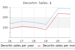
20 mg decortin generic overnight delivery
Typical luminal cells must be adverse beneath the circumstances used for traditional assays allergy medicine 72 cheap 10 mg decortin free shipping. The tumor cells have large allergy medicine hydroxyzine hcl generic decortin 10 mg without a prescription, round nuclei with outstanding nucleoli and abundant eosinophilic granular cytoplasm. The presence of nuclear pleomorphism and mitotic figures should raise the potential for malignancy. For problematic circumstances, immunostudies can affirm the presence of myoepithelial cells and exclude invasion. Neuroendocrine Carcinoma: Poorly Differentiated Mucinous Carcinoma (Left) Mucinous carcinomas, significantly the hypercellular variant, commonly express chromogranin and synaptophysin. Asioli S et al: Working formulation of neuroendocrine tumors of the skin and breast. Crona J et al: Metastases from neuroendocrine tumors to the breast are extra widespread than previously thought. Kawasaki T et al: Neuroendocrine cells associated with neuroendocrine carcinoma of the breast: nature and significance. Righi L et al: Neuroendocrine differentiation in breast cancer: established information and unresolved problems. The absence of hormone receptors in a well-differentiated tumor ought to at all times elevate the potential for a special prognosis. Thus, these tumors appear to have an aberrant expression pattern not present in normal breast cells. Neuroendocrine Carcinoma: Well Differentiated Neuroendocrine Carcinoma: Poorly Differentiated (Left) the scant cytoplasm related to carcinomas with small cell morphology ends in nuclear molding. This is an unusual appearance for breast carcinomas and the potential for a metastasis must be considered and excluded, if potential. This poorly differentiated carcinoma has brief spindle cells reminiscent of strong papillary carcinoma. Invasive Breast Carcinoma With Neuroendocrine Differentiation Invasive Carcinoma With Neuroendocrine Differentiation: Synaptophysin (Left) this poorly differentiated carcinoma of no particular kind has cells with massive markedly pleomorphic nuclei with prominent nucleoli. There is at present no cause to carry out these markers unless a metastasis to the breast is under consideration. Immunohistochemical studies are useful to affirm the absence of myoepithelial cells. However, the patient had a history of breast carcinoma, and this was a metastasis. Additional research confirmed expression of hormone receptors and different breast markers. Metastatic breast cancer should be thought of in this setting, as these cancers could be effectively palliated with hormonal therapy. Metastatic Breast Cancer to Bone Metastatic Carcinoma to Bone: Chromogranin (Left) Women with breast most cancers often current with bone metastases. Subsequent imaging and biopsy confirmed the affected person had a major breast carcinoma. The extravasated blood and hemosiderin give the tumor a characteristic red-brown shade. This hemorrhage leads to the gross shade and mammographic appearance as a radiodense mass. The related carcinoma is mostly of no special kind (ductal) but can be a selection of particular types. Alrahbi S et al: Extent of margin involvement, lymphovascular invasion, and extensive intraductal component predict for residual illness after wide local excision for breast cancer. Although all carcinomas start as carcinoma in situ, some cancers may rapidly overgrow these areas. The best measurement used for staging usually requires medical, radiologic, gross, and microscopic data. If the carcinoma only invades into the pectoralis, T classification is predicated on tumor measurement. T4b: Invasive Carcinoma, Skin Ulceration T4b: Invasive Carcinoma, Satellite Skin Nodules (Left) Skin ulceration as a outcome of direct invasion is typically associated with very large domestically advanced carcinomas and is classified as T4b. They are usually associated with dermal lymph-vascular invasion and are a form of intramammary metastasis. The clinical look is as a result of of extensive obstructive dermal lymphvascular invasion. This well-differentiated carcinoma has good tubule formation, low nuclear grade, and a low mitotic index. This sort of carcinoma usually has a poor prognosis though a subset will respond well to chemotherapy. Invasive Carcinoma, Grade 3 Lymph-Vascular Invasion (Left) Lymph-vascular invasion is related to elevated likelihood of lymph node involvement as well as disease recurrence. Dermal Lymph-Vascular Invasion Invasive Tubular Carcinoma (Left) Special histologic forms of invasive breast most cancers have prognostic significance. Invasive Carcinoma, Mitotic Count Invasive Carcinoma, Ki-67 (Left) Proliferation is a vital prognostic factor and can be determined using a quantity of different methods. The enumeration of mitoses in cancers is used for histologic grading and correlates well with different measures of proliferation. This welldifferentiated carcinoma exhibits < 5% constructive cells, which would predict for little if any profit from adjuvant chemotherapy added to hormonal therapy. Desmedt C et al: Uncovering the genomic heterogeneity of multifocal breast most cancers. Karanlik H et al: Preoperative chemotherapy for T2 breast most cancers is associated with improved surgical end result. Shaikh T et al: Multifocal and multicentric breast most cancers is associated with elevated native recurrence no matter surgical procedure kind. Tot T et al: Molecular phenotypes of unifocal, multifocal, and diffuse invasive breast carcinomas. Cabioglu N et al: Increased lymph node positivity in multifocal and multicentric breast most cancers. Tot T et al: the distribution of lesions in 1-14-mm invasive breast carcinomas and its relation to metastatic potential. Tot T: the metastatic capability of multifocal breast carcinomas: in depth tumors versus tumors of limited extent. In some instances, the prebiopsy imaging size may provide the most effective estimate of actual size. This complicates measurement willpower, as adding collectively the scale in every specimen can overestimate measurement.
Diseases
- Brachydactyly elbow wrist dysplasia
- Pfeiffer Singer Zschiesche syndrome
- Hepatic ductular hypoplasia
- Bare lymphocyte syndrome
- Marburg fever
- Keratoderma hypotrichosis leukonychia
- Chromomycosis
- Neurocutaneous melanosis
- Iridocyclitis
- Beemer Langer syndrome
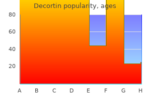
Decortin 40 mg buy generic
The superior mesenteric vessels move behind the neck of the pancreas and in entrance of the third portion of the duodenum allergy forecast jackson ms decortin 10 mg purchase otc. The root of the transverse mesocolon and small bowel mesentery come up from the floor of the pancreas and transmit the blood vessels to the small bowel & transverse colon allergy treatment vivite vibrance therapy by allergan purchase decortin 5 mg visa. Anatomically, the pancreatic axis from head to tail is directed superiorly and to the left. The transducer is tilted slightly cranially and laterally to the left to observe the pancreatic axis, thus imaging the pancreas in its entirety. The splenic vein courses alongside the posterior pancreas and provides a wonderful landmark in locating the pancreas. This picture was taken with a small quantity of cranial tilt so that blue signifies flow towards the transducer and red away from the transducer. The splenic vein is pink in its proximal portion but exhibits blue color distally, owing to its course. Power Doppler is extra delicate for detecting vascular flow but fails to present information on circulate path. Note the connection of the pancreatic head with the posteriorly positioned inferior vena cava. Note the superior mesenteric vein coming into view; it is a good landmark for locating the neck of the pancreas on the sagittal ultrasound. The origin of the superior mesenteric artery arising from the abdominal aorta is brought into view. The abdomen lies superiorly and may be crammed with fluid for use as an acoustic window. The oblique course of the psoas muscles leads to the lower pole of the kidney mendacity lateral to the upper pole. The proper kidney normally lies 1-2 cm decrease than the left, as a result of inferior displacement by the liver. The adrenal glands lie above and medial to the kidneys, separated by a layer of fats and connective tissue. The right kidney abuts the liver and the hepatic flexure of the colon and duodenum, whereas the left kidney is in close contact with the pancreas (tail), spleen, and splenic flexure. It then divides into 5 segmental arteries, only 1 of which (the posterior segmental artery) passes dorsal to the renal pelvis. The segmental arteries divide into the interlobar arteries that lie within the renal sinus fat. Each interlobar artery branches into four to 6 arcuate arteries that observe the convex outer margin of every renal pyramid. The arcuate arteries give rise to the interlobular arteries that lie throughout the renal cortex, including the cortical columns (of Bertin) that invaginate between the renal pyramids. The window levels and work station controls have been set to optimally show the renal accumulating system. The color scale is bigoted; on this case, opacified urine is displayed as white. Less dense urine throughout the renal tubules in the pyramids and the diluted urine throughout the bladder are displayed as red. In the supine place at quiet breathing, the higher poles of the kidneys normally lie in entrance of the twelfth ribs. Medial to the kidneys, the course of the renal fascia is variable (and controversial). However, the renal and lateroconal fasciae are laminated buildings that may be distended with fluid collections to form interfascial planes that do talk throughout the midline and in addition inferiorly to the extraperitoneal pelvis. Inferiorly, the anterior and posterior renal fasciae come shut collectively at about the stage of the iliac crest. This strategy normally supplies wonderful visualization of the right kidney and is beneficial for measuring bipolar renal length. The plane of this picture is angulated extra medially in comparison with the previous image. If the renal parenchyma is brighter than regular liver, renal parenchymal illness must be suspected. This analysis of the place of major vessels is helpful to avoid major vessels during renal interventional procedures, such as renal biopsy or nephrostomy. Scanning via the posterior method is beneficial while performing interventional procedures, corresponding to nephrostomy or renal biopsy. However, visualization/image high quality may be impaired by thick paraspinal muscular tissues and rib shadowing. Note that the proper renal artery lies posterior to the renal vein and inferior vena cava. Note that the image high quality is restricted by interference from gas within the stomach and bowel. The presence of bowel loops anteriorly additionally limits using this approach for interventional procedures on the left kidney. Note the relationship of the spleen to the higher pole of the left kidney, allowing it to be used as an acoustic window, particularly in patients with splenomegaly. Note the proximity of the kidney to the skin floor and the absence of intervening bowel loops. This view is helpful for performing renal interventional procedures, similar to renal biopsy or nephrostomy. This evaluation of the place of main vessels is beneficial for avoiding major vessels when performing renal interventional procedures, such as renal biopsy or nephrostomy. Note the proximity of the kidney to the skin surface and absence of intervening bowel/other major structures, making this a suitable strategy for renal interventional procedures. There is variability in venous velocity consequent upon cardiac and respiratory exercise. The sagittal plane can be steered electronically to obtain the lengthy axis of the kidney. Note the comparatively central location of the small gut compared with the peripherally positioned massive intestine. Most of the bowel segments are intraperitoneal, apart from the 2nd to 4th parts of the duodenum, the ascending and descending colon, and middle 1/3 of the rectum, that are retroperitoneal. Note that the lesser curvature and anterior wall of the abdomen contact the underside of the liver and the gallbladder abuts the duodenal bulb. The higher curvature is hooked up to the transverse colon by the gastrocolic ligament, which continues inferiorly because the greater omentum, overlaying many of the colon and small bowel. The lesser omentum carries the portal vein, hepatic artery, widespread bile duct, and lymph nodes.

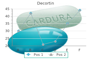
Decortin 10 mg order on-line
The sticky mucus admixed with blood can differentiate bloody present from antepartum bleeding allergy testing locations discount 20 mg decortin with mastercard. Placenta previa allergy testing tempe az decortin 10 mg, placental abruption, and vasa previa are all related to antepartum bleeding. This is a uterine contraction pattern of extreme number of contractions or tachysystole. There are seven contractions within the 9-minute window illustrated and late decelerations. The use of a betamimetic agent such as terbulatine will bring about uterine leisure and hopefully resolve the late decelerations. A fetal scalp stimulation inducing an acceleration would be reassuring and allow continued remark of this tracing. This 18-year-old nulliparous affected person is progressing into the energetic section of labor. She is having repetitive deep variable decelerations and an amnioinfusion would help to alleviate the wire compression and hopefully, enable for a vaginal delivery. Studies have shown that amnioinfusion for variable decelerations reduces the chance for cesarean. This affected person is having late decelerations likely because of the hypotension from the epidural analgesia. The corrective actions often result in resolution of the late decelerations fairly rapidly. The mechanism of the motion of epiduralinduced hypotension is sympathetic blockade resulting in vasodilation. In basic, latent labor happens when the cervix is less than 6 cm dilated and energetic labor when the cervix is >6 cm dilated. Early decelerations are mirror images of uterine contractions, caused by fetal head compressions. Late decelerations are gradual in shape and are offset from the uterine contractions, attributable to uteroplacental insufficiency (hypoxia). N ormal labor, supply, and postpartum care: anatomic concerns, obstetric and analgesia, and resuscitation of the new child. Her antenatal history is unremarkable except for a urinary tract infection handled with an antibiotic 2 weeks in the past. The affected person was famous to be anemic on her prenatal screen with a hemoglobin level of 9. The analysis of the anemia contains: ferritin stage: 90 mcg/L (normal 30-100); serum iron: one hundred forty mcg/dL (normal 50-150); hemoglobin electrophoresis: Hb A1 of 95% and Hb A2 of 5. Understand that deficiency of folate and vitamin B12 are causes of macrocytic anemia. Considerations this pregnant affected person has a mild anemia, for the reason that hemoglobin level is less than 10. Typically, with a mild microcytic anemia within the absence of danger components for thalassemia (such as Southeast Asian ethnicity), a trial of iron supplementation and recheck of the hemoglobin in 3 weeks would be the subsequent step. In this case, iron studies have been performed which had been normal/ high normal, thus eliminating iron deficiency as a trigger. The hemoglobin electrophoresis research strongly counsel -thalassemia trait (heterozygous for -thalassemia) with the elevated A2 hemoglobin. If the patient had -thalassemia homozygous illness, there would have been problems and scientific manifestations since childhood. The patient ought to now be endorsed about her laboratory findings, and referred for genetic counseling, and instructed that her baby has a one in four threat for -thalassemia illness if the father of the infant additionally has -thalassemia trait. This process might lead to ineffective erythropoiesis, hemolysis, and ranging degrees of anemia. It is most frequently as a outcome of iron deficiency, partially because of decreased iron shops prior to pregnancy and elevated demands for iron (due to fetus want and expanded maternal blood volume). Iron Deficiency A gravid woman who presents with gentle anemia and no risk factors for hemoglobinopathies (African-American, Southeast Asian, or Mediterranean descent) could also be treated with supplemental iron and the hemoglobin level reassessed in 3 to 4 weeks. Persistent anemia necessitates an evaluation for iron shops, similar to ferritin stage (low with iron deficiency) and hemoglobin electrophoresis. Hemoglobinopathies the dimensions of the pink blood cell could give a clue in regards to the etiology. A microcytic anemia is mostly as a result of iron deficiency, although thalassemia may also be causative. Results from a hemoglobin electrophoresis can differentiate between the two, and may also point out the presence of sickle cell trait or sickle cell anemia. The different sorts of thalassemias are categorised based on the deficient peptide chain. A neonate born with -thalassemia major might seem healthy at delivery, however as the hemoglobin F degree falls (and no -chains are capable of substitute the diminishing -chains of the fetal hemoglobin), the toddler could turn into severely anemic and fail to thrive if not adequately transfused. W hereas the thalassemias are quantitative defects in a hemoglobin chain production, sickle cell illness involves a qualitative defect that results in a sickle-shaped and rigid hemoglobin molecule. Sickle cell anemia is a recessive dysfunction attributable to a degree mutation within the -globin chain during which the amino acid glutamic acid is changed with valine. Patients with sickle cell illness usually deal with signs related to anemia (ie, fatigue and shortness of breath) and pain. In being pregnant, girls with sickle cell disease typically have a extra intense anemia, extra frequent bouts of sickle cell crisis (painful vaso-occlusive episodes), and more frequent infections and pulmonary issues. Careful consideration must be taken when a pregnant sickle cell patient presents in crisis as a outcome of a few of the symptoms may mimic other widespread occurrences throughout being pregnant (ectopic pregnancy, placental abruption, pyelonephritis, appendicitis, or cholecystitis), and they could additionally be missed. Also, these patients have a higher incidence of fetal progress retardation and perinatal mortality; subsequently, serial ultrasonography is beneficial. Macrocytic Anemia Macrocytic anemias may be as a result of vitamin B12 and folate deficiency. Because vitamin B12 shops final for a couple of years, megaloblastic anemias in being pregnant are more likely to be attributable to folate deficiency. N itrofurantoin is a standard medication utilized for uncomplicated urinary tract infections. Iron deficiency Folate deficiency Vitamin B12 deficiency Physiologic anemia of pregnancy 2. She famous dark-colored urine after taking an antibiotic for a urinary tract infection. Which of the next best describes the chance that their unborn child will have sickle cell illness On hospital day 2, she develops acute dyspnea, and has an oxygen saturation degree of 85% on room air. Macrocytic anemias embody folate deficiency and vitamin B12 deficiency; nevertheless, folate deficiency is more generally seen in pregnancy than vitamin B12 deficiency. Physiologic anemia of pregnancy is a results of the physiologic hemodilution that happens within the vasculature.
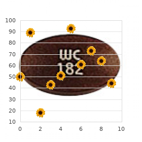
Purchase decortin 5 mg without a prescription
At low power allergy x for dogs best decortin 20 mg, the differential prognosis definitely contains angiosarcoma and other vascular tumors allergy zyrtec doesn't work purchase decortin 10 mg on line. Additional retraction might artificially give the impression of dilated vascular spaces. Small, well-formed balls of neoplastic cells are separated by a really delicate fibrovascular plexus. Tumors with delicate zellballen structure typically profit from an immunohistochemistry analysis. Hypocellular and hypercellular areas with ectatic pseudovascular spaces are present in giant cell angiofibroma. Note the increased variety of mast cells and wavy collagen deposited within the stroma. The uniform spindle and epithelioid tumor cell inhabitants has ovoid-to-round nuclei that may be slightly atypical; tumor cells regularly encircle thin-walled blood vessels. Solitary Fibrous Tumor "Patternless sample" of spindle tumor cells with investing stromal collagen 5. Both within the rib and within the vault of the cranium, such lesions may have a sunburst appearance radiographically. Conventional intraosseous hemangioma shows large ectatic and anastomosing vascular spaces with attenuated walls of flat endothelial cells. The central coarsened trabeculae type a "polka dot" pattern, characteristic of these lesions once they happen within the vertebral body. These lesions are usually well demarcated, clearly distinct from normal cancellous bone, and provides off a pink, hemorrhagic look with coarse trabeculation. Faint linear ossifications seen inside the lesion, especially at its periphery, symbolize retained inner cancellous bone. The branching lucency within the lesion may symbolize a dilated vascular channel. The gross look displaying a well, demarcated, pink, hemorrhagic, and cystic lesion is basic. The lesion includes the neck of the femur, which has been dissolved by the distinguished osteoclastic activity induced by the Gorham illness. The "polka dot" sample is illustrated right here, as properly as the proliferation of huge ectatic vascular spaces. The course trabeculae are notable for extensive remodeling and osteoclastic exercise toward the perimeters. The lumina are lined by a single layer of flat, uniform, cytologically bland endothelial cells. Solid areas of epithelioid endothelial cells with intensely eosinophilic cytoplasm admixed amongst a milieu of inflammatory cells make vascular differentiation tough to acknowledge on biopsy. The characteristic thickened sclerotic bony trabeculae is characteristic of benign vascular tumors of bone. Benign vascular tumors of bone will all show important overlap in radiographic options. Heterogeneity is a result of vascular enhancement combined with hypointense bony trabeculae. The posterior cortex is expanded and thinned; residual rebuttressed trabeculae are vertical in orientation. Expansion of the cortices by a large multiloculated, lytic lesion and separated by septa of reactive sclerotic bone is visible. The 2nd metatarsal is expanded within the distal diaphysis and metaphysis by a circumscribed, trabeculated, lytic lesion with sclerotic. Multiple lytic are seen in the lesions distal tibia, fibula, talus, and calcaneus. More well-defined and circumscribed vascular lesions are typically much less aggressive. These lesions could contain a number of sites inside the identical bone before they coalesce into a solitary multiloculated lesion. The tumor has eroded the outer and internal tables and produces fullness of the overlying delicate tissues. Other separate lesions within the adjacent head of the calcaneus and tibia are also seen. The areas of strong pink tumor have a multinodular architecture separated by thinned bony septa. Tumor lobules are separated by white fibrous septa and are predominantly nicely demarcated. The strong purple, hemorrhagic tumor appears well circumscribed, with enlargement of the cortical bone. The woven bone may be rimmed with osteoblasts, which may trigger confusion with osteoblastoma. Consideration of more aggressive lesions must also be thought-about in these situations. When present, the vessel is lined by epithelioid endothelial cells and is surrounded by small-caliber neoplastic vessels. Their presence can cause confusion with giant cell tumor of bone and aneurysmal bone cyst. This diploma of hypercellularity is a diagnostic pitfall for extra aggressive tumors, such as epithelioid hemangioendothelioma and angiosarcoma. The tumor cell nuclei could also be hyperlobated and include grooves and prominent nucleoli. The tumor entails the medullary cavity and cortex and is surrounded by reactive bone. The lesions involve many bones and seem as well-circumscribed radiolucencies of varying sizes. The tumor has well-defined margins, and reactive periosteal new bone is seen proximally and distally. A tumor typically mistaken for low-grade angiosarcoma or malignant hemangioendothelioma. Chordoma Rarely multifocal Arise in axial skeleton Neoplastic cells are cohesive and are embedded in extracellular myxoid matrix Cells stain for epithelial markers and brachyury Negative for endothelial markers 17. The mass erodes the posterior cortex and has induced a thin shell of reactive bone. The tumor has obliterated the diploic space, eroded the outer desk, and destroyed the inside desk and bulges into the delicate tissues. In this case, the hyalinized tumor has infiltrated an arterial wall and fills and occludes the lumen of a dilated vein.
Moringa Oleifera (Drumstick Tree). Decortin.
- What is Moringa?
- Are there safety concerns?
- Dosing considerations for Moringa.
- How does Moringa work?
Source: http://www.rxlist.com/script/main/art.asp?articlekey=97185
Discount decortin 10 mg visa
Prophylactic antibiotics given during surgery decrease the chance of changing into infected allergy medicine overdose buy 10 mg decortin with mastercard. In addition to opening the wound allergy-x for dogs reviews buy discount decortin 10 mg, the patient ought to bear dressing modifications and be began on antimicrobial brokers. In cesarean wound infections, there are two distinct populations of organisms that might be concerned: skin organisms versus vaginal organisms. A Gram stain of the wound might direct towards the proper antibiotic routine that may be efficient for the potential bacteria. Cesarean delivery significantly will increase the danger of endometritis because of the reality that the patient most probably had prolonged rupture of membranes, numerous vaginal examinations, and an intrauterine pressure monitor due, for instance, to an arrest of labor. Peptostreptococcus and peptococcus are the more than likely pathogens 45% of the time and bacteriodes 9%. The main organisms responsible for postcesarean endomyometritis are anaerobic bacteria with essentially the most generally isolated organisms embrace peptostreptococcus, peptococcus, and Bacteroides species. Antibiotic remedy and heparin are an accepted remedy for septic pelvic thrombophlebitis. She states that 3 weeks beforehand, she underwent a normal spontaneous vaginal supply. She had been breastfeeding without problem until 2 days in the past, when she famous progressive pain, induration, and redness to the best breast. Her proper breast has induration on the higher outer area with redness and tenderness. Next step in therapy: Incision and drainage of the abscess and antibiotic remedy. Understand that the presence of fluctuance within the breast in all probability represents an abscess that needs incision and drainage. This is a typical presentation of a breast an infection, since mastitis often presents within the third or fourth postpartum week. Induration and redness of the breast accompanied by fever and chills are additionally consistent. The treatment for this condition is an antistaphylococcal agent corresponding to dicloxacillin. Affected ladies are instructed to continue to breast feed or drain the breast by pump. They include cracked nipples, breast engorgement, mastitis, breast abscesses, and galactoceles. Cracked nipples often come up from dryness, and may be exacerbated by harsh soap or watersoluble lotions. Treatment contains air drying the nipples, washing with gentle soap and water, the use of a nipple shield, and the appliance of a lanolin-based lotion. Breast engorgement is usually noted in the course of the first-week postpartum and is as a outcome of of vascular congestion and milk accumulation. The patient will typically complain of breast pain and induration, and will have a low-grade fever. Postpartum mastitis is an an infection of the breast parenchyma, affecting about 2% of lactating women. Other signs and signs embody malaise, fever, chills, tachycardia, and a pink, tender, swollen breast. Importantly, there must be no fluctuance of the breast, which might indicate abscess formation. If the patient has a penicillin allergy, then clarithromycin orally for 10 to 14 days has been efficient. Breastfeeding or pumping ought to be continued to stop the event of abscess. A tradition of the breast milk despatched previous to initiating treatment is helpful for figuring out bacterial sensitivities and nosocomial surveillance. About 1 in 10 cases of mastitis is sophisticated by abscess, which should be suspected with persistent fever after 48 hours of antibiotic therapy or the presence of a fluctuant mass. The purulent assortment is finest treated by surgical drainage, or alternatively by ultrasound-guided aspiration; antistaphylococcal antibiotics should also be used. The milk accumulates in a quantity of breast lobes, resulting in a nonerythematous fluctuant mass. Breast milk incorporates almost all of the vitamins required aside from several nutritional vitamins (K and D), and is extra easily tolerated than formulation. She is breastfeeding and notes that the child prefers to breastfeed from the right breast. On the left breast, she notes a 3-day historical past of a tender mass on the higher outer quadrant. The left breast has a fluctuant mass of 4 � eight cm of the upper outer quadrant with out redness. This girl has had persistent tenderness and redness of the breast regardless of not lactating and not having trauma to the breast; these signs have worsened despite antibiotic remedy. There is a concern about inflammatory breast carcinoma (see Case 47), and she ought to undergo biopsy. Inflammatory breast most cancers presents with redness, tenderness, and heat and might mimic mastitis. It is an aggressive kind of malignancy with cancer cells located within the pores and skin lymphatics. A galactocele forms when a milk duct is blocked and the milk accumulates in a number of breast lobes, resulting in a nonerythematous fluctuant mass. Bromocriptine is an ergot alkaloid that blocks the discharge of prolactin from the pituitary (typically within the setting of a prolactinoma), largely as an attempt to enable a lady to be ready to have normal menstrual cycles. The American Academy of Pediatrics recommends that except contraindicated, every infant be breastfed solely for the first 6 months of life due to the well being benefits to the baby. Breast-fed infants have much less infections including meningitis, urinary tract infections, and sepsis thought to be because of immunoglobulin and leukocytes within the breast milk. Breast milk consists of two proteins, whey and casein, and has lower casein proportion than method milk, allowing for easier digestion. The presence of fluctuance in a red, tender, indurated breast suggests abscess, which wants surgical drainage. The best treatment of cracked nipples is air drying and the avoidance of utilizing a harsh cleaning soap. Her obstetric historical past is significant for two first trimester being pregnant losses occurring 1 and three years beforehand. Fetal heart fee tracing: Baseline of 140 bpm with decreased variability and recurrent late decelerations, indicative of severe hypoxia or acidosis. Describe the maternal and fetal issues associated with pregestational diabetes.
40 mg decortin trusted
Representative photomicrographs of the immunofluorescence and electron microscopy from this case are pictured beneath allergy treatment toddler 20 mg decortin generic fast delivery. There is marked mesangial matrix expansion however no appreciable mesangial hypercellularity jewelry allergy treatment buy generic decortin 20 mg on-line. The glomerular capillary loops and mesangial matrix include quite a few subendothelial and intramembranous lipid of differing density, dimension, and form. This produces the thickened foamy look of the capillary loops on silver stains by light microscopy. Ultrastructural elements of a model new syndrome with specific reference to lesions within the kidneys and the spleen. There is delicate mesangial matrix enlargement and a mild improve in mesangial hypercellularity without apparent vacuolization. There is delicate mesangial matrix enlargement with out obvious vacuolization or hypercellularity. This is as a outcome of of extraction of the lipid deposits during processing for paraffin embedding. Severe Glomerular Involvement With Vacuolated Mesangial Matrix Severe Mesangial Lipid Deposition (Left) There is distinguished mesangial matrix enlargement and vacuolization. This ends in a vacuolated appearance to the mesangial matrix and capillary loop basement membranes. The matrix contains electron dense lamellar material and lucencies resulting from incomplete lipid extraction. The mesangial matrix has a vacuolated appearance with lucent and lamellar material. The rounded deposits are stable or have a lamellar substructure and are unusually electron dense. Fat Stain Shows Lipid in Glomeruli Lipid "Thrombi" (Left) In lipoprotein glomerulopathy, glomerular capillaries are full of vacuolated lipoprotein thrombi. Goto S et al: Marked elevation of serum hyaluronan levels in collagenofibrotic glomerulopathy. Alchi B et al: Collagenofibrotic glomerulopathy: clinicopathologic overview of a uncommon glomerular disease. The tannic acid highlights the randomly organized, curved, and banded collagen fibers throughout the mesangium. The interstitium also stains, however this is a regular function of interstitial fibrosis. The capillary loop basement membrane reveals marked thickening and irregularity with basement membrane lucencies that contain tiny cell remnants. There is a small segmental sclerosing lesion with hyaline accumulation and tiny lipid droplets. There is diffuse podocyte foot process effacement with microvillus transformation. The glomerulus on the left exhibits delicate mesangial matrix enhance with delicate mesangial hypercellularity. However, it does show gentle mesangial hypercellularity with out capillary loop alterations. The basement membrane is diffusely thickened and "split" leading to a double lamina densa layer. There is impressive irregularity and thickening of the lamina rara interna and lamina rara externa. One glomerulus reveals pronounced mesangial matrix increase and capillary loop collapse. A 2nd glomerulus reveals mild mesangial sclerosis, and the 3rd seems basically regular. Marked Variability in Mesangial Matrix Severe Mesangial Sclerosis (Left) this three yr old with Galloway-Mowat syndrome and diffuse mesangial sclerosis reveals a severely affected glomerulus and a minimally affected glomerulus. There is a markedly thickened arteriole current with a few adjacent atrophic tubules. Epithelial hyperplasia is absent, presumably due to the terminal stage of the method. There is generalized epithelial cell hyperplasia that includes both the parietal and visceral epithelial cells. There could be very distinguished epithelial cell hyperplasia involving both parietal and visceral epithelial cells. One glomerulus reveals extreme mesangial matrix enhance and diffuse capillary loop collapse. The different glomerulus shows mesangial sclerosis with residual, however compromised, capillary loops. Most of the capillary loops are utterly obliterated with extensive capsular adhesions. There can be gentle variability within the thickness of the capillary loop basement membrane with distinguished scalloping along the inside floor of the capillary loop. Diffuse Podocyte Foot Process Effacement Subepithelial Basement Membrane Abnormalities (Left) Electron microscopy exhibits the interface with the mesangium that features portions of 2 mesangial cells. The capillary loop basements present irregular thickening with a scalloped contour on each the inner and outer features and lucencies with granularities. The segmental sclerosing lesion incorporates hyaline and lipid with overlying epithelial hyperplasia. Segmental Sclerosis With Capsular Adhesion in Galloway-Mowat Syndrome Sclerotic Glomerulus in Galloway-Mowat Syndrome (Left) this is one other segmental sclerosing lesion. There is residual mild epithelial cell hyperplasia, a feature that can disappear over time. The capillary loop basement membrane is fairly uniform in thickness, but mild irregularity is current. There is an adhesion composed of strands of matrix materials that connects the collapsed capillary loop to the Bowman capsule. The podocyte has an enlarged lacy clear cytoplasm, full of lipid that has been dissolved in processing. This is often the most conspicuous change in routine stains that raises the query of Fabry disease. I-Cell Disease � Podocyte deposits constructive for colloidal iron Arteriosclerosis � Vascular disease in Fabry illness similar to usual arteriosclerosis in hypertension � If lipid in podocytes (or other sites) not appreciated, analysis could additionally be missed 17. Subclinical Fabry Disease (in Patients With Other Renal Diseases) � Lipid deposits could additionally be "incidental" finding in biopsies for other ailments. T�ndel C et al: Foot process effacement is an early marker of nephropathy in younger basic Fabry patients without albuminuria. Demonstration of the lipid accumulation is finest proven by electron microscopy or 1 m sections. Diffuse fine interstitial fibrosis is current and commonly accompanied by patchy tubular atrophy.
Decortin 10 mg buy with mastercard
Sensitive allergy symptoms penicillin order 40 mg decortin visa, confidential allergy symptoms milk cheap decortin 40 mg without a prescription, however direct questioning is one of the best strategy: " Are you being harm or threatened by anyone A safety plan may be mentioned including packing a bag upfront, having personal documents prepared, having an extra set of car/ house keys, establishing a code with friends/ household, and having a plan of where to go. Prior to prescribing emergency contraception, which of the following is most important to order A pink rash is noted on the introital region, and also some bruising on the vulvar and perineal space. Usually intimate companion violence lessens during pregnancy due to concern about hurting the fetus. She has just lately immigrated to the nation and has not been vaccinated in opposition to hepatitis B. This patient has indicators of neglect such as bedsores and unkempt look, and likely extended soiling without diaper changes. Intimate associate violence will increase in being pregnant and can lead to preterm supply, low start weight, and placental abruption. H omicide, normally in the first trimester, is the second main cause of injury-related deaths to pregnant women after motorized vehicle accidents. Sexual assault is a traumatic experience and allowing the patient to dictate the order of occasions at analysis is necessary. Emergency contraception is best if given throughout the first 72 hours from the assault. Repetitive visits, falls, and poor control of medical situations may be indicators of elder abuse. Mandatory reporting is required in conditions the place impending murder or baby abuse is suspected. Ulipristal acetate: evaluation of the efficacy and security of a newly permitted agent for emergency contraception. Know the widespread presentations of ureteral and bladder accidents after gynecologic surgical procedure. Considerations this affected person has a medical picture equivalent to pyelonephritis; nevertheless, as a outcome of she has recently undergone a hysterectomy, damage to or obstruction of the ureter is of paramount concern. Endometriosis tends to obliterate tissue planes, making ureteral damage extra likely. If the same medical picture were current without the latest surgery, then the most likely prognosis could be pyelonephritis and the next step can be intravenous antibiotics and urine tradition. Finally, the wound incisions are normal, which argues in opposition to a wound infection causing the postoperative fever. Laparoscopic hysterectomies could cause harm to the ureter by mechanical ligation, as an example, if a stapling gadget were used. Thermal damage also can cause ureteral injury both directly to the ureter, or thermal unfold. Various procedures, such as placement of stents into the ureters, can be carried out. Cancer, extensive adhesions, endometriosis, tubo-ovarian abscess, residual ovaries, and interligamentous leiomyomata are threat elements. Any gynecologic process, including laparoscopy or vaginal hysterectomy, could result in ureteral damage; nevertheless, the overwhelming majority of the injuries are related to stomach hysterectomy. The most typical location for ureteral harm is at the cardinal ligament, where the ureter is only 2- to 3-cm lateral to the cervix. The ureters are within 2- to 3-cm lateral to the internal cervical os and can be injured upon clamping of the uterine arteries. Ureteral accidents include suture ligation, trans-section, crushing with clamps, ischemia-induced harm from stripping the blood supply, and laparoscopic harm. This procedure is carried out in the hope that the ureter is kinked however not occluded. The choice for quick ureteral restore versus initial percutaneous nephrostomy with later ureteral restore ought to be individualized. Right hydronephrosis is reflected by dilation of the renal amassing system and hydroureter, whereas the left accumulating system is regular (A). Delayed movie of the same patient exhibits the right hydroureter more prominently (B). Prevention of Complications an important intervention in stopping surgical web site infections after hysterectomy is the usage of preoperative antibiotics, sometimes a first era cephalosporin agent 15 to 60 minutes previous to incision time (see Case 33). During the procedure, the best ureter is meticulously and cleanly dissected free and a Penrose drain is positioned round it to guarantee its safety. She is asymptomatic until postoperative day 9, when she develops profuse nausea and vomiting, and is noted to have ascites on ultrasound. There are many risk components associated with ureteral damage; nonetheless, the majority are associated with laparoscopic hysterectomies. Other danger elements include: cancer, in depth adhesions, endometriosis, tubo-ovarian abscess, residual ovaries, interligamentous leiomyomata, and most gynecological procedures. Also, the presentation of fever and flank tenderness after surgical procedure makes the prognosis of ureteral ligation most likely compared to the opposite options. When the ureter is ligated, the affected person is at an elevated danger of hydronephrosis and/ or hydroureter. Antibiotic remedy and aid of the obstruction must be administered promptly to avoid the situation on this scenario of pyelonephritis. Patients with a bladder perforation injury typically current with gross hematuria, pain, or tenderness within the suprapubic region and issue in voiding. Over dissection of the ureter might result in devascularization injury as a result of the ureter receives its blood provide from numerous arteries along its course and flows along its adventitial sheath. Urine is leaked into the belly cavity and causes irritation to the intestines and induces nausea and emesis. With a vesicovaginal fistula, urine is repeatedly leaking out the vagina, however not into the belly cavity. In bladder perforation accidents, patients current with pain within the suprapubic area. Constant urinary leakage after pelvic surgical procedure is a typical history for vesicovaginal fistula (see Case 34-Urinary Incontinence). As with the patient identified with a ureteral ligation, this affected person presents with fever and flank tenderness. The incontrovertible reality that the process on this situation was performed using bipolar cautery, the probability that the symptoms deal with thermal damage versus ligation is far larger. Meticulous ureteral dissection can result in devascularization injury to the ureter for the reason that vascular channels run alongside the adventitia of the ureter.
Purchase decortin 5 mg overnight delivery
Acyclovir and analogous agents given in pregnancy during main episodes can lower the length of viral shedding and period of lesions allergy testing st cloud mn cheap decortin 20 mg without prescription. The patient states that four weeks previously allergy treatment 5 shaving 5 mg decortin generic otc, after she had engaged in sexual intercourse, she experienced some vaginal recognizing. Understand that the ultrasound examination is an effective technique for assessing placental location. Because of the painless nature of the bleeding and lack of risk factors for placental abruption, this case is extra more probably to be placenta previa, defined because the placenta overlying the interior os of the cervix. Placental abruption (premature separation of the placenta) often is associated with painful uterine contractions or extra uterine tone. The historical past of postcoital spotting earlier during the pregnancy is according to previa because vaginal intercourse might induce bleeding. The ultrasound examination is carried out before a vaginal examination as a result of vaginal manipulation (even a speculum examination) might induce bleeding. Complete placenta previa (A), marginal placenta previa (B), and low-lying placentation (C) are depicted. The two most typical causes of serious antepartum bleeding are placental abruption and placenta previa Table 10� 1). When the affected person complains of antepartum hemorrhage, the doctor should first rule out placenta previa by ultrasound even earlier than a speculum or digital examination, since these maneuvers might induce bleeding. At occasions, transabdominal sonography might not be capable of visualize the placenta, and transvaginal ultrasound is necessary and is more dependable for visualizing the interior cervical os. H ence, a woman with a preterm gestation and placenta previa is often observed on bed rest and full pelvic rest in an effort to delay gestation and avoid morbidity of fetal prematurity. The bleeding from previa rarely leads to coagulopathy, versus that of placental abruption. Because the decrease uterine section is poorly contractile, postpartum bleeding might ensue. Several danger components have been cited including parity, elevated maternal age, smoking, multiple gestations, prior curettage, and prior cesarean delivery. Of note, placenta accreta (invasion of the placenta into the uterus) is extra frequent with placenta previa, notably within the presence of a uterine scar corresponding to after a cesarean supply. Timing of supply is dependent upon clinical circumstances for placenta previa and placenta accreta. The N ational Institutes of H ealth concluded that elective supply is right at 36-37 accomplished weeks for these sufferers, but practices still vary. An ultrasound is carried out revealing that the placenta is masking the internal os of the cervix. Schedule an amniocentesis at 34 weeks and deliver by cesarean if the fetal lungs are mature C. Multiple gestation, with the increased surface space of placentation, is a threat factor for placenta previa. Polyhydramnios, as a outcome of the excess amount of amniotic fluid in the amniotic sac, can also be a risk factor for placenta abruption. Salpingitis entails inflammation and an infection of the fallopian tubes and over time could lead to everlasting scarring of the tubes. The main distinguishing issue between a previa and abruption is the presence or absence of pain. With abruption, painful uterine contractions are usually the chief criticism, whereas previa is painless. Even in the setting of an Rh� mother with an Rh+ fetus, an intrauterine transfusion earlier than supply would pose a considerably higher threat to the mom and baby than waiting to consider the state of affairs after start. Ultrasound must be performed first to rule out previa, speculum examination second to assess the cervix and search for lacerations, and eventually digital examination. Performing either a speculum examination or digital examination earlier than evaluating the patient with ultrasound places the patient at risk for hemorrhage. In the setting of a previa, the decrease uterine segment and cervix are highly vascularized, and varices of the cervix could also be visualized on speculum examination in some conditions; however, the speculum itself may trigger trauma to these varices and induce bleeding. A blind digital examination may end in additional separation of the placenta from the uterus, which may also trigger significant bleeding. Very usually, a marginal or low-lying placenta previa at the early second trimester will resolve by transmigration of the placenta. It is just too early to talk about scheduling a cesarean delivery because the placenta previa may resolve and permit for vaginal delivery. An ultrasound must be repeated within the third trimester to see whether or not the placenta has migrated. During the cesarean, the doctor will be succesful of assess the extent of the placental implantation and base management on how far the placenta has penetrated by way of the uterine wall. Placenta percreta and increta are usually recognized during a cesarean supply and never radiographically. Even if the patient has a placenta previa on the time of supply, both the mother and baby have a superb prognosis if a cesarean supply is performed. Ultrasound is the diagnostic test of selection in assessing placenta previa and must be carried out before digital or speculum examination. Placenta previa, within the face of prior cesarean deliveries, increases the risk of placenta accreta. When placenta previa is identified at an early gestation, similar to second trimester, repeat sonography is warranted since many instances the placenta will transfer away from the cervix (transmigration). A late-preterm, early-term stratified evaluation of neonatal outcomes by gestational age in placenta previa: defining the optimum timing for delivery. Magnetic resonance imaging of clinically steady late being pregnant bleeding: beyond ultrasound. Effectiveness of timing strategies for delivery of individuals with placenta previa and accreta. She states that she has been experiencing average vaginal bleeding, no leakage of fluid per vagina, and has no historical past of trauma. The fundus reveals tenderness, and a average quantity of darkish vaginal blood is noted in the vaginal vault. Complications that may happen: H emorrhage, fetal to maternal bleeding, coagulopathy, and preterm supply. Best management for this condition: Delivery (at 35 weeks, the dangers of abruption significantly outweigh the risks of prematurity). Understand that placental abruption and placenta previa are major causes of antepartum hemorrhage. Considerations the affected person complains of painful antepartum bleeding, which is consistent with placental abruption.

Generic 10 mg decortin visa
No myoepithelial cells are identified utilizing p63 (brown nuclei) allergy shots chronic sinusitis decortin 40 mg buy overnight delivery, displaying that the circumscribed nests of cells are invasive carcinoma allergy forecast eau claire wi generic 10 mg decortin with amex. Note the thick bands of dense fibrosis separating the nests of tumor cells, a characteristic characteristic of secretory carcinoma. Secretory Carcinoma Secretory Carcinoma: Ki-67 (Left) this secretory carcinoma shows strong nests of cells with a microcystic growth sample. The cells have uniform round nuclei with minimal pleomorphism, small nucleoli, and abundant granular cytoplasm. However, not like other triple-negative breast cancers, these tumors have a great prognosis. Mitoses are usually absent, and the Ki-67 proliferation index is quite low, as seen here. Secretory Carcinoma Secretory Carcinoma (Left) Secretory carcinomas are strongly positive for S100 protein, displaying each nuclear and cytoplasm staining. This eosinophilic secretory is optimistic for periodic acid-Schiff (diastase resistant) and is also constructive by Alcian blue. Cells with basophilic granular cytoplasm, if current, favor acinic cell carcinoma. Acinic Cell Carcinoma Acinic Cell Carcinoma (Left) At least a few of the cells in acinic cell carcinoma have ample cytoplasmic with basophilic granules. Features that differentiate cystic hypersecretory carcinoma from secretory carcinoma usually embrace higher grade nuclei and scant cytoplasm. Invasive Cribriform Carcinoma Invasive Apocrine Carcinoma (Left) Invasive cribriform carcinoma, like secretory carcinoma, invades as islands of cells with rounded areas. Tubule formation is typically absent, and the presence of eosinophilic secretions can be very uncommon. In this case, > 10% of the cells present full membrane positivity, but the depth is weak to moderate (a 2+ equivocal result). This outcome basically at all times correlates with excessive levels of protein expression in all the tumor cells. However, due to the dyscohesive nature of the cells, edge enhancement can be seen. In this case the immunoreactivity appears to be in the cracks between the cells, rather than true membrane immunoreactivity. Without microdissection, misguided results could presumably be obtained from assays such as the 21-gene recurrence rating. Heterogeneity could additionally be a supply of resistance to targeted remedy as negative clones might preferentially survive remedy. Benign nonapocrine cells must be negative under the circumstances used for medical assays. Bonev V et al: Long-term follow-up of breast-conserving remedy in patients with inflammatory breast most cancers treated with neoadjuvant chemotherapy. This causes obstruction, leading to edema fluid distending lymphatics, diffuse thickening of the dermis, and swelling of the breast. Inflammatory Carcinoma, Dilated Lymphatics Inflammatory Carcinoma, D2-40 (Podoplanin) (Left) In most circumstances, the presence of tumor cells within lymphatic areas is readily obvious on H&E. For problematic cases, staining for D2-40 (podoplanin) can help to spotlight lymphatic endothelial cells. Large B-Cell Lymphoma, Lymph Node Bacterial Abscess (Left) Tumors, similar to this huge B-cell lymphoma, can fill axillary nodes. The blockage of lymphatic drainage can lead to breast edema and erythema closely mimicking inflammatory breast carcinoma. Metaplasia makes evaluation of in situ lesions troublesome as a end result of the cells seem monomorphic because of the metaplasia. Lobular Neoplasia, Apocrine Features Invasive Apocrine Carcinoma (Left) Strict histologic criteria for invasive apocrine carcinoma include giant cells with ample eosinophilic granular cytoplasm, a nuclear:cytoplasmic ratio of 1:2 or greater, and huge vesicular nuclei with outstanding nucleoli in > 90% of the most cancers. This could account for the frequent observation of apocrine metaplasia in breast tissue and the expression of comparable proteins. The invasive carcinomas are usually similar in histologic sort, grade, and tumor markers. These foci may give rise to smaller foci of invasion (intramammary metastases) which are equivalent in appearance to the primary carcinoma. Multiple Foci of Invasive Carcinoma: Satellite Foci Multiple Foci of Invasive Carcinoma: 2 Biologically Separate Carcinomas (Left) Smaller satellite tv for pc foci are generally seen around the border of an invasive carcinoma. The satellite tv for pc foci are likely connected to the principle carcinoma in one other airplane of section by way of invasion down fibrovascular septa. However, a most cancers must penetrate beyond these muscular tissues into the chest wall in order to be classified as T4a. Tryfonidis K et al: Management of regionally superior breast cancerperspectives and future instructions. Yalcin B: Overview on locally advanced breast cancer: defining, epidemiology, and overview on neoadjuvant remedy. Ly M et al: High incidence of triple-negative tumors in sub-saharan Africa: a prospective research of breast cancer traits and risk factors in Malian women seen in a Bamako college hospital. G�th U et al: Breast most cancers with non-inflammatory pores and skin involvement: present information on an underreported entity and its problematic classification. Massidda B et al: Molecular alterations in key-regulator genes among sufferers with T4 breast carcinoma. Tada K et al: Skin invasion and prognosis in node adverse breast most cancers: a retrospective research. These small cancers doubtless have a better prognosis than the extra typical very massive T4b cancers. Breast Cancer With Ulceration and Satellite Nodules: T4b Breast Cancer With Satellite Skin Nodule: T4b (Left) this central ulcerating carcinoma is related to numerous satellite tv for pc skin nodules. Breast Cancer With Skin Edema, Mammography: T4b Male Breast Cancer (Left) this subareolar carcinoma is associated with skin edema. Dermal lymphvascular invasion in the absence of pores and skin adjustments is inadequate for this prognosis. However, within the absence of the medical indicators of inflammatory carcinoma, this finding alone is insufficient for classification as T4d. These cancers sometimes have a diffusely infiltrative pattern and are tough to diagnose. In order to be categorised as chest wall invasion (T4a), the carcinoma must go through the pectoralis and invade into the muscles of the ribs. However, if a most cancers responds well to treatment, there may be benefit from surgical resection. However, cancers with pores and skin invasion are related to the next frequency of lymph node metastases. It is usually related to a scale crust, and skin erosion can occur in in depth circumstances. However, the biopsy confirmed an abscess as a result of squamous metaplasia of lactiferous ducts. In order to find all macrometastases (> 2 mm), each node have to be thinly sectioned (at 2-3 mm) and all slices examined.