

Cozaar dosages: 50 mg, 25 mg
Cozaar packs: 28 pills, 56 pills, 112 pills, 224 pills, 30 pills, 60 pills, 90 pills, 120 pills, 180 pills, 270 pills, 360 pills
50 mg cozaar purchase
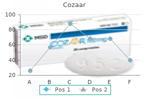
Chromosome inversions occur when two breaks occur on a chromosome adopted by a 180-degree flip of the phase and reinsertion at its original breakpoints diabetes symptoms videos buy discount cozaar 50 mg on line. Inversions diabetes pills vs insulin 50 mg cozaar otc, however, do intrude with pairing at meiosis and can end result in gametes with chromosome abnormalities. Isochromosomes happen as a end result of the chromosome dividing alongside the horizontal axis quite than the longitudinal axis on the centromere. Isochromosomes involving autosomes are generally deadly as a outcome of the resultant conception might be each trisomic and monosomic for genetic info. However, an isochromosome involving the lengthy arm of the X chromosome (iso Xq) is appropriate with life. Finally, deletions and duplications of chromosome segments arise from unequal crossing over at meiosis or from crossing over throughout pairing of inversions or reciprocal translocations. Breaks leading to loss of chromosome material on the tip are called terminal deletions, and lack of chromosome material between two breaks within a chromosome is identified as an interstitial deletion. There are a quantity of well-documented terminal deletion syndromes, including cri du chat (5p-) syndrome, characterized by microcephaly, profound psychological retardation, growth retardation, a singular facial look, and a particular catlike cry. Notice that there are solely 45 chromosome constructions, however this male is genetically diploid. Phenotype correlations, significantly with balanced translocations and inversions, could also be highly variable and dependent on whether the chromosomal break points disrupt critical genes. Experience from pregnancies achieved by assisted reproductive technology signifies that 15% of fertilized ova fail to divide. Another 15% fail to implant, and 25% to 30% are aborted spontaneously prior to formation of villi. Of the roughly 40% of fertilized ova that survive the primary missed menstrual interval, as many as one fourth are aborted spontaneously, so only about 30% to 35% of fertilized ova really end in live-born infants. Chromosome and lethal genetic abnormalities play a significant function in early losses (Zinaman, 1996). In persevering with pregnancies, chromosome abnormalities occur in 1 out of each 154 stay births (0. The breakdown by sort of abnormality is as follows: 22% autosomal trisomy (most commonly trisomy 21, followed by a a lot decrease incidence of trisomy 18 and trisomy 13), 37% intercourse chromosome aneuploidy, and 41% structural abnormalities (including translocations, insertions, inversions, isochromosomes, duplications, and deletions). Trisomy 21 is the most commonly occurring singular genetic abnormality in stay births, occurring at a rate of 1 in 830 reside births (0. The dialogue within the earlier part described only phenotypes that have been associated with chromosome abnormalities visible with traditional cytogenetic methods and lightweight microscopy that imply involvement of huge segments of chromosomes containing a big quantity (hundreds) of genes. High-resolution chromosome banding and advances in molecular cytogenetic technology have revealed a new class of chromosome syndromes often identified as microdeletions. The phenotypes of those circumstances are because of the absence (or duplication) of multiple contiguous genes throughout the concerned area. The discovery of microdeletion/duplication or contiguous gene syndromes has been essential in clinical genetics and genetic counseling in that it lastly supplies a analysis, recurrence threat, and prenatal diagnosis for a big group of syndromes that beforehand had no cytogenetic affirmation. Moreover, these "naturally occurring" sequestered small areas of contiguous genes have provided a strong tool for developmental geneticists to decipher the critical genes for normal human growth. For instance, the 22q11 region seems to be rich in genes liable for particular congenital heart defects and craniofacial anomalies. In the case of aneuploidy screening, the assayed markers only pertain to a subset Location Chromosome Abnormality Deletion Deletion and duplication Deletion Duplication and deletion Deletion Duplication Duplication Deletion Size (Mb) 2. Measuring the value of screening exams is historically based mostly on sensitivity (detection rate), false-positive rate, and optimistic predictive value inside a inhabitants. Testing for a specific disease could additionally be warranted by family historical past or ethnic background, and prenatal diagnosis might doubtlessly allow interventions before the disease would have been detected in the youngster clinically (Table 2. In some uncommon cases, diagnosis in utero might enable the opportunity to preclude irreversible adjustments in early improvement. However, the supplier must relay the scope and accuracy of available testing modalities. For occasion, a check that detects a phenotypic manifestation of disease (such as biochemical screening of hexosaminidase exercise to assess for Tay-Sachs) is extra sensitive for detecting an affected fetus than a genetic sequence panel that may embody solely a subset of the disease-causing mutations. The growing availability of these "expanded carrier screening panels" has prompted the American College of Obstetricians and Gynecologists to issue pointers for patient consent prior to offering expanded screening (Edwards, 2015): 1. Results of genetic testing are confidential and guarded in health insurance and employment by the Genetic Information Non-Discrimination Act of 2008. Many are related to vital antagonistic outcomes such as cognitive impairment, decreased life expectancy, and wish for medical or surgical intervention. In most instances, being a service of an autosomal recessive situation has no clinical penalties for the person provider. Referral to an applicable specialist for medical administration and genetic counseling is indicated in such circumstances to evaluate the inheritance patterns, recurrence dangers, and medical options. Genetic Counseling and Risk Assessment Ordering and sending a genetic check is easy, but the counseling time to ensure appropriate interpretation of the outcomes (positive or negative) is essential and often time consuming. The position of the care suppliers (genetic counselors and physicians) is to conduct nondirectional counseling of screening and diagnostic choices to provide prospective mother and father with information to optimize pregnancy outcomes based on their private values and preferences. All patients considering genetic screening or testing should have the chance to meet with providers who can carry out formal pedigrees, conduct patient education and counseling on benefits and downsides of testing, and talk about the provision of screening and invasive diagnostic testing when needed. Obstetrics & Gynecology Books Full 2 Reproductive Genetics Before screening or testing, the affected person needs to understand the choices that will ensue from a positive test result. Biochemical and Sonographic Screening Multiple screening options can be found for detecting fetuses in danger for the most common aneuploidy syndromes, namely trisomies 21, 18, and 13. The choice of test will depend upon the gestational age at presentation, number of fetuses, earlier obstetric history, household historical past, availability of sonologists or sonographers licensed to detect or measure check parameters, and options or preferences for pregnancy termination. Nuchal translucency measurements are validated for the precise gestational age window from 104/7weeks to 136/7 weeks as determined by crown-rump length, and pointers for measurement are standardized, which should be followed for the test to keep revealed detection charges. A nuchal translucency measurement lower than 3 mm is considered normal (Malone, 2005). The primary benefit of first trimester screening is earlier outcomes, which allow for greater privacy and broader choices of diagnostic testing and reproductive decisions. There are numerous iterations of screening, which combine parts of both first and second trimester screening measurements, and efficiency statistics of those methods depend upon the precise analytes included, whether the measurements are impartial of each other, and the timing of danger calculation. Abnormal outcomes of a few of the particular person parts assayed in aneuploidy screening are also predictive of antagonistic pregnancy outcomes. For example, abnormal biochemical markers on each first and second trimester screens have been related to fetal 37 growth restriction, intrauterine fetal demise, preterm supply, Smith-Lemli-Opitz syndrome (low estriol), and oligohydramnios-albeit with low predictive worth (Smith, 2006). To date, this expertise has been unsuitable for scientific utility due to a quantity of technologic obstacles such as limited numbers of fetal cells, unreliable recovery of fetal cells, and proof that the cells persist lengthy after pregnancy, thus complicating specificity within the setting of subsequent pregnancies (Bianchi, 2002). The performance statistics for Down syndrome detection in a high-risk inhabitants are much better than any other out there strategies (Table 2.
Cozaar 50 mg with amex
The shade of the fluid obtained via aspiration varies from clear to grossly bloody diabetes type 1 eating plan cozaar 50 mg cheap on line. If the cyst recurs diabetes symptoms yahoo best cozaar 25 mg, imaging ought to be performed to confirm their benign nature and reaspiration carried out underneath ultrasound guidance. Less than 20% of cysts recur after a single aspiration, and fewer than 10% recur after two aspirations. A biopsy should be performed on cysts that recur within 2 weeks or that necessitate more than one repeat aspiration. In cases of a stable mass, a set specimen is obtained and submitted for cytopathologic analysis. Several passes are made through the mass with continuous suction from the syringe. Moving the needle inside a single tract will give a passable cellular yield within the majority of circumstances. Complications of needle aspiration are rare and include hematoma formation and an infection. The theoretic danger of spreading most cancers alongside the needle track has not been substantiated. Amore definitive histologic evaluation including tumor grade angiolymphatic invasion and hormone receptor status could be made. In addition, core needle biopsy usually provides adequate tissue for genomic evaluation or cancer profiling. Vacuum-assisted directional biopsy can be utilized to acquire a larger volume of tissue. Following administration of native anesthetic, a small pores and skin incision is made and the core biopsy needle is inserted. A biopsy clip have to be positioned on the time of the biopsy for future localization of the lesion. Mammographic or stereotactic steerage is primarily used for biopsy of calcifications. The breast is imaged at 30-degree angles with 2D mammography, and the lesion is localized utilizing computer-assisted positioning and focusing on devices. Nonpalpable breast lesions found by breast imaging techniques, including screening mammography, require preoperative localization by the radiologist. Image-guided methods play an important position in preoperative staging of breast cancer sufferers and in the planning of definitive surgery. Tumor grade is based on tubule formation, nuclear pleomorphism, and mitotic counts using the Nottingham score to determine low, intermediate, or excessive grade. Initially, breast carcinoma could also be divided into invasive and in situ lesions (Table 15. Invasive ductal carcinoma accounts for the majority, roughly 80%, of invasive carcinomas. Other subtypes embody mucinous, tubular, medullary, micropapillary, and papillary. Both in situ and invasive carcinomas are sometimes found in the identical quadrant of the breast. Cytologic features vary from bland to highly malignant, and tumors are graded primarily based on architectural and cytologic traits. The degree of fibrous response as a outcome of the invading malignant cells is liable for the firm palpable mass, radiologic density, and texture during biopsy. Diagnosis is confirmed with a core needle biopsy, normally utilizing stereotactic guidance. The histologic diagnosis of ductal carcinoma in situ includes a heterogeneous group of tumors with varying malignant potential. Classification is based on architectural pattern (comedo, micropapillary, cribriform, or solid), tumor grade (high, intermediate, or low), and evidence of necrosis. Identification of microinvasion, a minute focus of stromal invasion, is essential as remedy recommendations could change. Treatment approaches embrace surgical procedure, radiation remedy, and adjuvant endocrine remedy. Breast-conserving surgery (lumpectomy, partial mastectomy) followed by radiation therapy has proven equivalent survival compared with mastectomy. This neoplasia tends to have a multicentric origin in the same breast and tends to involve each breasts more typically than infiltrating ductal carcinoma. Histologic subdivisions of infiltrating lobular carcinoma embody small cell, round cell, and signet cell carcinomas. Neoplastic cells infiltrating the stroma and adipose tissue in a single-file trend. This kind is acknowledged clinically as a quickly rising malignant carcinoma with extremely angiogenic and angioinvasive characteristics. The scientific picture of a scaly, raw, or ulcerated lesion of the nipple and areola is a results of an infiltrating ductal carcinoma that invades the epidermis. In approximately 85% of the patients, an underlying breast cancer is current with Pagetdiseaseofthebreast. Intraepithelial adenocarcinoma cells (Paget cells) are famous on histology, presenting both singly or in small groups throughout the epidermis of the nipple. Thesetumorsareusually estrogen and progesterone negative, excessive grade, have a excessive rate of p53 mutations, and have a poor prognosis. It is just like basal-type duct cells by way of expression of extra myoepithelial gene profiling. A fifth type of breast cancer, claudin-low tumors, constitutes approximately 10% of breast cancers. Systemic remedy consists of chemotherapy, endocrine remedy, biologic therapy, or a mixture of those regimens. In cases of invasive disease, remedy is based on the stage-appropriate guideline for invasive carcinoma. The main goals of treating breast carcinoma are management of native illness, treatment or prevention of distant metastasis, and improved high quality of life for ladies treated for the disease. With a number of therapeutic choices in each local and systemic remedy for breast carcinoma, ladies have an active function in deciding their very own treatment routine. Breast conservation with lumpectomy or quadrantectomy is a frequent alternative for the control of native illness. Sentinel node biopsy has turn out to be normal practice in the therapy of early stage breast cancer. Emphasisonconservative surgery plus radiation remedy to control multifocal most cancers in the same breast and on reconstructive surgery after mastectomy has improved the quality of life for girls with breast carcinoma. Identification of tumor receptor standing is critical as endocrine remedy is utilized both for adjuvant therapy and within the management of advanced disease. The genomic evaluation of tumors has led to the molecular subtyping of breast cancers. Subsequently, the luminal group was further differentiatedintoluminal-Aandluminal-Bsubgroups.
Cozaar 25 mg on line
The M�llerian ducts and nearby mesenchymal tissue form the majority of the female reproductive tract treatment of diabetes insipidus 50 mg cozaar discount visa. The M�llerian duct is derived from the coelomic epithelium during fetal improvement diabetes insipidus and lithium cozaar 50 mg buy amex. The metaplasia speculation postulates that the coelomic epithelium retains the power for multipotential growth. The decidual response of isolated areas of peritoneum during pregnancy is an example of this course of. It is well known that the floor epithelium of the ovary can differentiate into several different histologic cell types. Endometriosis has been found in prepubertal girls, ladies with congenital absence of the uterus, and barely in males. Metaplasia occurs after an "induction phenomenon" has stimulated the multipotential cell. The induction substance could additionally be a combination of menstrual debris and the influence of estrogen and progesterone. It has been hypothesized that the histogenesis of endometriosis in peritoneal pockets of the posterior pelvis results from a congenital anomaly involving rudimentary duplication of the M�llerian system (Batt, 1989). Molecular profiling of experimental endometriosis recognized gene expression patterns in widespread with human disease. The mesothelium is labeled with monoclonal antibody to cytokeratin and stained with diaminobenzidine (arrows). An endometrial stromal cell (arrowhead) passing via the mesothelium is believed to symbolize the preliminary step of invasion into the stroma of the peritoneum. Whole explants of peritoneum and endometrium: a novel model of the early endometriosis lesion. Similarly, it has been postulated that metaplasia of the coelomic epithelium that invaginates into the ovarian cortex is the pathogenesis for the event of ovarian endometriosis (Nisolle, 1997). Lymphatic and Vascular Metastasis the speculation of endometrium being transplanted by way of lymphatic channels and the vascular system (Halban, 1925) helps to explain rare and distant websites of endometriosis, such because the spinal column and nostril. Endometriosis has been noticed in the pelvic lymph nodes of roughly 30% of girls with the disease. Iatrogenic Dissemination Endometriosis of the anterior belly wall is usually discovered in girls after a cesarean delivery. The hypothesis is that endometrial glands and stroma are implanted through the procedure. Multiple investigations have instructed that modifications within the immune system, particularly altered function of immune-related cells, are directly associated to the pathogenesis of endometriosis. Whether endometriosis is an autoimmune disease has been intensely debated for many years. Studies have demonstrated abnormalities in cell-mediated and humoral components of the immune system in both peripheral blood and peritoneal fluid. Most likely the primary immunologic change entails an alteration within the function of the peritoneal macrophages so prevalent in the peritoneal fluid of patients with endometriosis. Conversely, girls who develop endometriosis have more peritoneal From McLaren J, Deatry G, Prentice A, et al. Decreased ranges of the potent regulator of monocyte/macrophage activation, interleukin-13, in the peritoneal fluid of sufferers with endometriosis. These hyperactive cells secrete multiple growth factors and cytokines that improve the event of endometriosis. Following the theory of various macrophage populations in endometriosis is the discovering that the destroying of normally extruded endometrial cells in endometriosis could also be deficient. Another attractive principle is the discovering of a protein just like haptoglobin in endometriosis epithelial cells called Endo 1. Further compounding the proliferative activity of endometriosis lesions are angiogenic components that are elevated in lesions. Altered maturation and function of peritoneal macrophages: attainable function in pathogenesis of endometriosis. An investigation by Simpson and coworkers demonstrated a sevenfold enhance in the incidence of endometriosis in relations of girls with the disease in contrast with controls (Simpson, 1980). One of 10 women with extreme endometriosis may have a sister or mom with clinical manifestations of the disease. The incidence of endometriosis in first-degree relations, girls with severe endometriosis, has been thought to be 7%. Studies have recognized deletions of genes, most particularly increased heterogenicity of chromosome 17 and aneuploidy, in ladies with endometriosis compared with controls (Kosugi, 1999). Loci on 7p and 10q have additionally been found to enhance the susceptibility for endometriosis (Painter, 2011; Treloar, 2005). The expression of this genetic legal responsibility most likely depends on an interaction with environmental and epigenetic components, with many elements being involved. Preliminary knowledge recommend some bilateral ovarian endometrial cysts could arise independently from completely different clones. Although no consistent abnormality has been found in girls with endometriosis, there are several candidate genes. This is particularly striking in Asian women, in whom a ninefold increase has been suggested (Jacoby, 2010). The ovaries are the most common site, being concerned in two of three ladies with endometriosis. The pelvic peritoneum over the uterus; the anterior and posterior cul-de-sac; and the uterosacral, round, and broad ligaments are additionally frequent sites where endometriosis develops. Brosens has emphasised the significance of distinguishing between superficial and deep lesions of endometriosis (Brosens, 2000). Deep lesions, penetrations of greater than 5 mm, symbolize a more progressive form of the illness. Distinguishing superficial implant lesions on peritoneal surfaces, together with the ovary, from deep endometriotic ovarian cysts and cul-de-sac nodules is important for therapy (discussed later finish parens here The native manufacturing of estrogen through aromatase exercise explains why progression of lesions might occur even with ovarian suppression. This is occasioned by a dysregulation of the isoform B of the progesterone receptor in most endometriotic lesions the place levels could also be undetectable. Endometriosis Association has offered suggestive evidence of the upper prevalence of other autoimmune ailments. Depending on the quantity of associated scarring, endometriosis of the bowel could additionally be troublesome to differentiate grossly from a main neoplasm of the large gut. Gross pathologic changes of endometriosis exhibit extensive variability in shade, shape, measurement, and related inflammatory and fibrotic adjustments. The visible manifestations of endometriosis in the female pelvis are protean and have many appearances. Increased awareness and anticipation have focused on the delicate lesions of endometriosis.
50 mg cozaar proven
This structure is the evolutionary remnant of the tail-wagging muscles in decrease animals blood glucose spike order 25 mg cozaar fast delivery. The endopelvic fascia is one other term typically used interchangeably with the pelvic diaphragm diabetic diet while traveling 50 mg cozaar cheap mastercard. The pelvic diaphragm and endopelvic fascia are terms used to characterize the connective tissue, the assist for the pelvis, and the pelvic ground. The muscle tissue of the pelvic diaphragm are interwoven for power, and a continuous muscle layer encircles the terminal parts of the urethra, vagina, and rectum. The levator ani muscles constitute the greatest bulk of the pelvic diaphragm and are divided into three elements, that are named after their origin and insertion: pubococcygeus, puborectalis, and iliococcygeus. This grouping of the intermediate part of the levator ani muscle lies posterior to the pubic bone and could also be visualized as pubovaginalis, puboanalis, and puboperinealis muscle bundles. The main muscle tissue that compose this funnel-shaped sling are the coccygeus and the levator ani. The coccygeus is a triangular muscle that occupies the world between the ischial backbone and the coccyx. The paired levator ani muscle tissue act as a single muscle and functionally are essential within the control of urination, in parturition, and in sustaining fecal continence. Anteriorly, the urethra is suspended from the pubic bone by continuations of the fascial layers of the urogenital diaphragm. The free fringe of the diaphragm is strengthened by the superficial transverse perineal muscle. Posteriorly, the urogenital diaphragm inserts into the central point of the perineum. The urogenital diaphragm has two layers that enfold and cover the striated, deep transverse perineal muscle. This muscle surrounds each the vagina and the urethra, which pierce the diaphragm. The pudendal vessels and nerves, the external sphincter of the membranous urethra, and the dorsal nerve to the clitoris are also discovered inside the urogenital diaphragm. The deep transverse perineal muscle is innervated by branches of the pudendal nerve. Anatomists call the retroperitoneal fascia subserous fascia, whereas surgeons check with this fascial layer as endopelvic fascia. The connective tissue is denser immediately adjacent to the lateral partitions of the cervix and the vagina. They turn out to be contiguous with the uterine serosa, and thus the uterus is contained inside two folds of peritoneum. These peritoneal folds enclose the loose, fatty connective tissue termed the parametrium. The broad ligaments afford minor support to the uterus however are conduits for necessary anatomic constructions. Within the broad ligaments are found the following constructions: oviducts; ovarian and spherical ligaments; ureters; ovarian and uterine arteries and veins; parametrial tissue; embryonic remnants of the mesonephric duct, Wolffian physique, and secondary two ligaments; the mesovarium; and the mesosalpinx. It attaches to the superoanterior facet of the uterus, anterior and caudal to the oviduct, and runs via the broad ligament to the lateral pelvic wall. The spherical ligament crosses the exterior iliac vessels and enters the inguinal canal, ending by inserting into the labia majora in a fanlike fashion. In the fetus a small, finger-like projection of the peritoneum, generally identified as Nuck canal, accompanies the spherical ligament into the inguinal canal. Cardinal Ligaments the cardinal, or Mackenrodt, ligaments prolong from the lateral features of the higher part of the cervix and the vagina to the pelvic wall. They are a thickened condensation of the subserosal fascia and parametria between the interior portion of the 2 folds of peritoneum. Note the 2 layers of the urogenital diaphragm enfolding the deep transverse perineal muscle. B, Schematic views of the perineum demonstrating superficial buildings and deeper structures. The cardinal ligaments help to maintain the anatomic place of the cervix and the higher a part of the vagina and provide the most important assist of the uterus and cervix. Uterosacral Ligaments the uterosacral ligaments lengthen from the upper portion of the cervix posteriorly to the third sacral vertebra. They are thickened near the cervix after which run a curved course around each side of the rectum and subsequently thin out posteriorly. The external floor of the uterosacral ligaments is fashioned by an inferoposterior fold of peritoneum at the base of the broad ligaments. Surgical restore of a displacement or tear of the rectovaginal fascia and levator ani muscle tissue ensuing from childbirth is important throughout posterior colporrhaphy. Normal position of the female pelvic organs in the pelvis is dependent upon mechanical support from each fascia and muscle tissue. Note the posterior aspect of the rectouterine fold, known as the cul-de-sac, or pouch, of Douglas. The round ligament is a vital surgical landmark in making the preliminary incision into the parietal peritoneum to gain entry to the retroperitoneal space. Direct visualization of the retroperitoneal course of the ureter is an important step in many pelvic operations, including dissections in girls with endometriosis, pelvic inflammatory disease, giant adnexal lots, broad ligament plenty, and pelvic malignancies. When a great amount of fluid is placed within the stomach cavity, postoperative bilateral labial edema might develop in some ladies because of patency of the canal of Nuck. During pelvic surgery, traction on the uterus makes the uterosacral and cardinal ligaments more prominent. There is a free area roughly 2 to 4 cm beneath the superior fringe of the broad ligament. Often gynecologic surgeons utilize this space to facilitate clamping of the anastomosis between the uterine and ovarian arteries. The course of the left ureter is much like its counterpart on the proper aspect in that it runs downward and medially alongside the anterior surface of the psoas major muscle. The proper ureter tends to cross on the bifurcation of the widespread iliac artery, whereas usually the left ureter crosses 1 to 2 cm above the bifurcation. The ureters follow the descending, convex curvature of the posterolateral pelvic wall towards the perineum. At approximately the extent of the ischial backbone, the ureter changes its course and runs forward and medially from the uterosacral ligaments to the bottom of the broad ligament. In this location the ureter is approximately 1 to 2 cm lateral to the uterine cervix and is surrounded by a plexus of veins. A cross-sectional examine by Hurd and colleagues, using computed tomography of girls with regular anatomy, evaluated the space from the ureter to the lateral aspect of the cervix.
25 mg cozaar discount otc
A systematic review and meta-analysis (King diabetes insipidus fluid restriction cozaar 50 mg with visa, 2008) discovered an elevated price of melancholy diabetes type 1 lada buy cozaar 25 mg without prescription, nervousness problems, and substance abuse and dependence in lesbian, gay, and bisexual individuals. Lesbian and bisexual women had a very excessive fee of alcohol and drug dependence and, again, this was more pronounced in adolescents. A number of studies have now documented that lesbian women use psychological health providers at excessive charges, with 70% to 80% having been in therapy, primarily for depression and relationship problems. Yes No Persistent lack, delay, or reduced intensity of orgasm despite high arousal Yes No Recurrent pain from tried or completed intercourse Yes Difficulty with vaginal entry, variable worry, avoidance, muscle tightening Lesbians have a better price of cardiovascular danger factors, corresponding to obesity and smoking. Since the 1990s, lesbian girls have turn into increasingly willing to disclose their sexual orientation to well being care providers and to seek routine physical examinations. Nonetheless, their rates of routine physicals and Pap smears are lower than nationwide pointers and decrease than these of their heterosexual peers, and adolescents in particular have issue disclosing their sexual orientation to physicians. Lesbians favor feminine and ideally lesbian well being care suppliers and regularly use different well being care providers similar to nonphysicians, acupuncturists, and therapeutic massage therapists. Lesbian girls are 10 times much less prone to be screened for cervical cancer than heterosexual women, even though their risk of creating the illness is comparable. This has been thought to be because of lack of concern and a perception that lesbian women were at low danger for cervical most cancers. Sexually transmitted diseases also happen in lesbian girls, notably those that have had male partners. Lesbians are more and more having children through artificial insemination and adoption. Couples wishing to have youngsters through donor insemination have concerns about coming out to their obstetrician, involving the nonpregnant companion, authorized issues, household help, and parenting issues. The proportion will increase the youthful the adolescent is for involuntary first intercourse. Twenty to 25% of girls in faculty have been victims of actual or tried sexual assault during faculty. Of the rape victims who got here to the emergency room, two thirds had basic body trauma. This sort of crime, nonetheless, is often underreported, and the actual incidence may be a lot greater. Victims are often reluctant to report sexual assault to the authorities due to embarrassment, concern of retribution, feelings of guilt, assumptions that little might be carried out, or just lack of expertise of their rights. Homeless ladies and women with mental sickness are particularly susceptible to sexual assaults in contrast with the final inhabitants. In the previous, society has held many misconceptions about the rape sufferer, significantly female victims. Furthermore, the feeling that rape was a sign of basic promiscuity was extensively held. Sexual assault happens to individuals of all ages and races in all socioeconomic groups. The very young, the mentally and physically handicapped, and the very old are notably prone. Sexual violence may be undesirable touching and rape, however it additionally includes nonphysical distressing acts of sexual harassment, threats, peeping, and taking nude photos with out consent. Although the perpetrator could additionally be a stranger, she or he is commonly an individual well-known to the sufferer. These include marital rape, which involves pressured coitus or related acts with out consent however within the marital relationship, and "date rape. Date rape is often not reported as a end result of the sufferer could believe she contributed by partially participating. Almost all states have statutes that criminalize coitus with females underneath sure specified ages. Consent is irrelevant because the feminine is defined by statute as being incapable of consenting. During a rape, the victim loses management over his or her life for that interval and regularly experiences nervousness and concern. When the assault is life threatening, shock with associated bodily and psychological signs could occur. Sexual assault encompasses many acts, together with rape and unwanted genital touching. Rape is a authorized time period and refers to any penetration of a physique orifice with threat of pressure or precise force and nonconsent. The charges were 8% to 13% in North America with higher rates in sub-Saharan Africa and lower in Asia. In separating out by female age teams, 11% of highschool adolescents reported having been forced to have Obstetrics & Gynecology Books Full 9 Emotional Aspects of Gynecology complete lack of emotional management to a well-controlled habits pattern. The actual reaction may rely upon a number of factors, together with the relationship of the sufferer to the attacker, whether drive was used, and the size of time the sufferer was held in opposition to his or her will. Generally, the sufferer appears disorganized and should complain of each physical and emotional symptoms. Physical complaints embody particular injuries or common complaints of soreness, eating issues, complications, and sleep disturbances. Behavior patterns may embrace fear, mood swings, irritability, guilt, anger, despair, and difficulties in concentrating. The second section of the rape-trauma syndrome entails long-term adjustment and is designated the reorganization phase. During this time, flashbacks and nightmares could proceed, but phobias may also develop. These could additionally be directed towards members of the offending sex, the sex act itself, or nonrelated circumstances, corresponding to a newly developed fear of crowds or heights. During this period the sufferer could institute numerous necessary way of life modifications, together with job, residence, friends, and significant others. The reorganization period might final from months to years and generally involves an try on the a part of the sufferer to regain management over his or her life. During this time, medical care and counseling should be nonjudgmental, sensitive, and anticipatory. An estimated 40% to 50% of these girls have a historical past of physical or sexual abuse. It is necessary to obtain knowledgeable consent before inspecting the patient and collecting specimens. In addition to addressing legal requirements, it helps the sufferer 179 to regain control over her physique and her life. After acute accidents have been decided and stabilized, a cautious history and physical examination ought to be performed. It is necessary to have a chaperone current while taking the historical past, performing the examination, and accumulating the specimen, to reassure the sufferer and to present support.
25 mg cozaar for sale
Judicious use of a water-based lubricant can facilitate a more comfy exam for the affected person diabetes symptoms in toddlers cozaar 25 mg discount without a prescription. The speculum is then inserted by putting the transverse diameter of the blades within the anteroposterior position and guiding the blades through the introitus in a downward movement with the tips pointing towards the rectum diabetes type 2 reversal diet generic 25 mg cozaar amex. Because the anterior wall of the vagina is backed by the pubic symphysis, which is rigid, pressure upward causes the patient discomfort. Also, within the resting state the vagina lies on the rectum and actually extends posteriorly from the introitus. Placing two fingers into the introitus and urgent down might facilitate the process. Once the blades are inserted, the speculum should be turned in order that the transverse axis of the blades is in the transverse axis of the vagina. The blades ought to be inserted to their full size after which opened so that the doctor could inspect for the place of the cervix. Once the blades are inserted and the cervix is visualized, the speculum should be opened and the introitus widened so that the cervix can be adequately inspected and any indicated specimens obtained. The vaginal canal is inspected in the course of the insertion of the speculum and upon its elimination. Obstetrics & Gynecology Books Full 7 History, Physical Examination, and Preventive Health Care proof of erythema or lesions. Vaginal lesions, similar to areas of adenosis (see Chapter 11), clear cystic constructions (Gartner cysts), or inclusion cysts on the strains of scars or episiotomy incisions, must be famous. Saline moist mount permits for visualization of normal vaginal epithelial cells as nicely as any irregular findings such as motile trichomonads, clue cells (vaginal epithelial cells studded with adherent coccobacilli, indicative of bacterial vaginosis), or polymorphonuclear leukocytes (indicative of inflammation). A potassium hydroxide wet mount includes the whiff-amine take a look at, which, if optimistic for a distinct fishy odor, may indicate bacterial vaginosis. In addition, inspection of the slide might reveal hyphae and budding yeast, indicative of Candida vaginitis. Occasionally, glandular epithelium could also be present on the portio vaginalis, shifting the transformation zone onto the portio. Generally, that is cleared by a means of metaplasia, in which squamous epithelium covers the columnar epithelium. This course of, nevertheless, could go away small areas of irregularities and inclusion cysts, known as nabothian cysts, which can be seen in numerous dimensions and shapes. They are typically translucent and range from a couple of millimeters to as much as 3 cm in size. These cysts are widespread benign findings and require no further analysis or remedy. Cervical ectropion occurs when the endocervical epithelium is uncovered to the vaginal environment and takes on a reddish appearance, just like granulation tissue. Any lesions of the cervix ought to be famous and, the place acceptable, a biopsy ought to be performed. In a patient contaminated with human papillomavirus, warts (condylomata acuminata) on the cervix can also be noticed. In 1943, Papanicolaou and Trout printed their now basic monograph demonstrating the value of vaginal and cervical cytology as a screening tool for cervical neoplasm. With the use of the Pap smear in screening programs, the incidence of invasive cervical most cancers has been reduced by 50%. Repeat co-testing happens each 5 years, whereas Pap testing alone continues on an every 3-year foundation. Pap smear screening is now not really helpful in girls after age 65, if she has had regular enough testing over the previous 10 years and he or she has not been treated for high-grade dysplasia within the past 20 years. It is essential to educate ladies that this prolonged interval between Pap smears is predicated on long-term, excellent evaluation of the prevailing data. No Pap smear screening is important after a whole hysterectomy carried out for benign situations. However, if a supracervical hysterectomy was performed, the same screening pointers pertain as if there had been no hysterectomy, because the cervix remains in situ. The goal of the Pap smear is to gather cells from the transformation zone of the cervix. The presence of adequate endoand ectocervical cells ensures that this space is captured within the specimen. After excess mucus is gently removed (routine swabbing might trigger inadequate cells to be sampled), the endocervical canal is sampled with a Cytobrush, which is positioned into the canal and rotated. A single broomlike sampling gadget can be used to gather both populations of cells in a single step. Chapter 28 discusses cervical dysplasia, classifications of abnormalities, surveillance, and therapy options. The lubricated index and middle fingers of the dominant hand are placed within the vagina, and the thumb is folded underneath in order not to trigger the patient misery within the space of the mons pubis, clitoris, and pubic symphysis. The fingers are inserted deeply into the vagina so that they relaxation beneath the cervix in the posterior fornix. The doctor ought to be in a snug place at this point, usually with the leg on the aspect of the vaginal analyzing hand on a table carry and the elbow of that arm resting on the knee. The doctor then elevates the uterus by pressing up on the cervix and delivering the uterus to the belly hand so that the uterus may be placed between the 2 palms, thereby figuring out its place, dimension, shape, consistency, and mobility. In the conventional and nonpregnant state, the uterus is approximately 6 cm � four cm and weighs roughly 60 g. Size may be estimated in centimeters or by evaluating with weeks of regular gestational age. The uterus could additionally be retroverted, by which the complete uterus ideas posteriorly, and may also be retroflexed, in which the fundus factors posteriorly as properly. The general form of the uterus is that of a pear, with the broadest portion on the upper pole of the fundus. Generally, the uterus is mobile, and if it fails to transfer, it might be mounted by adhesions. The floor ought to be easy; irregularities may indicate the presence of uterine leiomyomas (fibroids). The consistency of the uterus is mostly agency however not rock onerous, and this ought to be famous within the examination. If the proper hand is the pelvic hand, the primary two fingers of the best hand are then moved into the best vaginal fornix as deeply as they can be inserted. The stomach hand is positioned just medial to the anterior superior iliac backbone on the right, the 2 arms are brought as close together as possible, and with a sliding movement from the world of the anterior superior iliac spine to the introitus, the fingers are swept downward, permitting for the adnexa to be palpated between them. When the adnexa are palpated, its size, mobility, and consistency should be described. Adnexa are often not palpable in postmenopausal girls due to involution and retraction of the ovary to a place higher within the pelvis. Obstetrics & Gynecology Books Full Pel vic sid ewa ll 7 History, Physical Examination, and Preventive Health Care the perianal space can reveal abnormalities corresponding to genital warts, hemorrhoids, or pores and skin lesions. It should be potential to palpate as many as 70% of distal bowel lesions with the rectal finger.
Diseases
- Herpes zoster oticus
- Jeune asphyxiating thoracic dystrophy
- Renal calculi
- Pseudohypoaldosteronism type 1
- Aniridia ataxia renal agenesis psychomotor retardation
- Poikiloderma hereditary acrokeratotic Weary type
Safe 50 mg cozaar
Thus the mesonephros features as a fetal kidney diabetes medications and hair loss cozaar 50 mg purchase without a prescription, producing urine for about 2 or 3 weeks diabetes mellitus symptoms in cats buy cozaar 50 mg. Usually about forty mesonephric tubules operate on both side of the embryo at any given time. The metanephros, or everlasting kidney, begins its development early within the fifth week of gestation and begins to function late in the seventh or early in the eighth week. The metanephros develops both from the metanephrogenic mass of mesoderm, which is the most caudal portion of the nephrogenic cord, and from its duct system, which is derived from the metanephric diverticulum (ureteric bud). It is a cranially growing outpouching of the mesonephric duct near where it enters the cloaca. The metanephric duct system offers rise to the ureter, the renal pelvis, the calyces, and the accumulating tubules of the adult kidney. A important course of in the improvement of the kidney requires that the cranially growing metanephric diverticulum meets and fuses with the metanephrogenic mass of mesoderm in order that formation of the kidney can take place. Originally the metanephric kidney is a pelvic organ, however by differential progress it turns into situated in the lumbar area (Moritz, 1999). Starting within the second trimester, fetal urine is a significant contributor to amniotic fluid quantity. The fetus could swallow the amniotic fluid and recirculate it by way of the digestive system. Congenital abnormalities that impair normal improvement or function of the fetal kidneys typically result in little or no amniotic fluid (oligohydramnios or anhydramnios), whereas structural abnormalities of the gastrointestinal tract or neuromuscular situations that prevent the fetus from swallowing can lead to extra amniotic fluid (polyhydramnios). The urogenital sinus, in flip, is split into three components: the cranial portion (the vesicourethral canal), which is steady with the allantois; a middle pelvic portion; and a caudal urogenital sinus portion, which is roofed externally by the urogenital membrane. The epithelium of the growing bladder is derived from the endoderm of the vesicourethral canal. The muscular layers and serosa of the bladder develop from adjacent splanchnic mesenchyme. As the bladder develops, the caudal portion of the mesonephric ducts is incorporated into its dorsal wall. The portion of the mesonephric duct distal to the points the place the metanephric duct is taken up into the bladder turns into the trigone of the bladder. In this way the ureters, derived from the metanephric duct, come to open immediately into the bladder. Also in the male, mesenchymal tissue surrounding the developing urethra the place it exits the bladder develops into the prostate gland, by way of which the ejaculatory ducts traverse. The epithelium of the female urethra is derived from endoderm of the vesicourethral canal. The urethral sphincter develops from a mesenchymal condensation around the urethra after the division of the cloaca within the 12- to 15-mm embryo. Following the opening of the anal membrane on the 20- to 30-mm stage, the puborectalis muscle seems. Thus the urethral sphincter is composed of each central smooth muscle and peripheral striated muscle. The sphincter develops primarily within the anterior wall of the urethra in a horseshoe or omega shape (Matsuno, 1984). A Y chromosome is necessary for the development of the testes, and the testes are liable for the group of the sexual duct system right into a male configuration and for the suppression of the paramesonephric (m�llerian) system of the feminine. Sf1 encodes a nuclear receptor necessary for steroidogenesis, gonadal differentiation, and adrenal formation. A heterozygous lack of perform mutation ends in ovarian failure (Di Pasquale, 2004). The understanding of the molecular foundation of intercourse willpower continues to expand with more than 25 genes up to now recognized within the process (Wilhelm, 2007). During the fifth week after conception, coelomic epithelium, later often known as germinal epithelium, thickens in the area of the medial facet of the mesonephros. As germinal epithelial cells proliferate, they invade the underlying mesenchyme, producing a prominence known as the gonadal ridge. In the sixth week the primordial germ cells, which have shaped at about week four within the wall of the yolk sac, migrate up the dorsal mesentery of the hindgut and enter the undifferentiated gonad. The somatic cells of the primitive gonadal ridge then differentiate into interstitial cells (Leydig cells) and Sertoli cells. As they do so, the primordial germ cells and Sertoli cells become enclosed within seminiferous tubules, and the interstitial cells stay outside these tubules. Sertoli cells are encased in the seminiferous tubules in the seventh and eighth weeks. Primary sex cords, in the meantime, have condensed and extended to the medullary portion of the developing testes. The testis therefore is primarily a medullary organ, and ultimately the rete testis connects with the tubules of the mesonephric system and joins the growing epididymal duct. Deletion of both the quick arm or the long arm of a single X chromosome precludes normal ovarian operate, with the former being related to Turner syndrome (Simpson, 1999). The paramesonephric ducts develop on all sides of the mesonephric ducts from the evaginations of the coelomic epithelium. The more cephalad ends of the ducts open directly into the peritoneal cavity, and the distal ends grow caudally, fusing within the decrease midline to form the uterovaginal primordium. This tubular construction joins the dorsal wall of the urogenital sinus and produces an elevation, the m�llerian tubercle. During the eighth week, interstitial (Leydig) cells differentiate and begin to produce testosterone. A, the creating ovary (O) in a 9-week-old fetus is shown close to the developing kidney (K). B, At this stage of growth, the columns of primordial germ cells (G) are embedded in a mesenchymal stroma (S) lined by a layer of cuboidal floor cells (E). The bulbourethral glands, that are small constructions that develop from outgrowths of endodermal tissue from the membranous portion of the urethra, incorporate stroma from the adjoining mesenchyme. The most distal portion of the paramesonephric duct stays, in the male, as the appendix of the testes. The most proximal finish of the paramesonephric duct remains as a small outpouching throughout the physique of the prostate gland, known as the prostatic utricle. This course of begins at about 6 weeks and proceeds in a cephalad to caudal fashion. The more cephalad portions of the paramesonephric ducts, which open directly into the peritoneal cavity, kind the fallopian tubes. The fused portion, or uterovaginal primordium, provides rise to the epithelium and glands of the uterus and cervix. Failure of growth of the paramesonephric ducts leads to agenesis of the cervix and the uterus referred to as m�llerian agenesis or Mayer-Rokitansky-Kuster-Hauser syndrome (Langman, 1982).
25 mg cozaar purchase mastercard
Ventricular failure Condition by which the lower chamber/s of the center is/are not functioning effectively diabetes type 1 fainting 25 mg cozaar quality. Xanthelasma Yellow lesions above or below the eyes blood glucose 44 50 mg cozaar buy with mastercard, which can point out lipid deposits under the pores and skin. Price Meiosis, Fertilization, Implantation, Embryonic Development, Sexual Differentiation Several areas of medical investigation have brought increased attention to the processes of fertilization and embryonic improvement, including teratology, stem cell analysis, immunogenetics, and assisted reproductive know-how. The preimplantation, implantation, and embryonic phases of growth in the human can now be studied because of the event of newer techniques and areas of analysis. This chapter considers the processes of oocyte meiosis, fertilization and early cleavage, implantation, development of the genitourinary system, and intercourse differentiation. The primordial germ cells in both women and men are large eosinophilic cells derived from endoderm within the wall of the yolk sac. These seven-hundred to 1300 cells migrate to the germinal ridge by the use of the dorsal mesentery of the hindgut by ameboid motion by 5 to 6 weeks. Oogenesis begins with the replication of the diploid oogonia by way of mitosis to produce primary oocytes, reaching a peak number of 600,000 (95% prediction interval: 70,0005,000,000) at 18 to 22 weeks of gestation. Through apoptosis the numbers decline to about 360,000 (95% prediction interval: forty two,000 to 3,000,000) at menarche (Wallace, 2010). Accelerated apoptosis is seen in Turner syndrome resulting in few oocytes at birth (Modi, 2003). In people, this process reduces 46 chromosomes to 23 chromosome constructions in the gamete. The haploid gamete contains just one chromosome for each homologous pair of chromosomes, so it has either the maternal or paternal chromosome for every pair, however not each. Meiosis can be the mechanism by which genetic trade is accomplished by way of chiasma formation and crossing over (recombination) between homologous chromosome pairs. Meiosis initiation relies on mesonephric-produced retinoic acid (Childs, 2011). The first, generally identified as the reduction division (division I, or meiosis I) initiates within the fetal ovary however is then arrested and accomplished at the time of ovulation. Prophase phases of the first meiotic division happen in the female throughout fetal life. The meiotic course of is arrested on the diplotene stage ("first meiotic arrest"), and the oocyte enters the dictyotene stages. The second meiotic division takes place over a number of hours in the oviduct solely after sperm penetration. There is cytologic proof of chromosome condensation and linearization, and the chromatin is seen as a nice, stringlike structure. In this substage, the paired homologues are structurally composed of 4 carefully opposed chromatids and are often known as a tetrad. The diplotene substage is a stage of desynapsis that occurs because the synaptonemal complex dissolves and the 2 homologous chromosomes pull away from each other. In common, chiasma formation occurs only between chromatids of homologous pairs and never between sister chromatids. Oocytes at this stage of prophase I represent the overwhelming majority of third-trimester fetal and new child ovaries. Diplotene merges with diakinesis, the final substage of meiosis I, and is a stage of transition to metaphase, lasting a few years in people (Speed, 1985). With puberty, folliculogenesis occurs with progression of the follicle, consisting of the oocyte and granulosa cells from primordial to antral characterized by granulosa cell proliferation, improvement of gonadotropin receptors, and expression of enzymes for intercourse steroid manufacturing (Baerwald, 2012). It takes approximately eighty five days for a follicle to mature to the point of ovulation. Meiosis I resumes with the surge of luteinizing hormone prior to ovulation completing metaphase, anaphase, and telophase. One daughter cell, the oocyte, receives nearly all of the cytoplasm, and the other turns into the primary polar body. Meanwhile, spermatozoa are transported via the cervical mucus and the uterus and into the fallopian tubes. Prophase levels of the primary meiotic division, which occur in most mammals during fetal life. The meiotic process is arrested on the diplotene stage ("first meiotic arrest"), and the oocyte enters the dictyate phases (5 to 6). Stage 1 of growth begins with fertilization within the uterine tube and ends when the zygote varieties. Stage 2 (days 2 to 3) includes the early stages of cleavage (from 2 to about 32 cells, the morula). Stage four (days 5 to 6) is represented by the blastocyst attaching to the posterior wall of the uterus, the usual site of implantation. Capacitation precedes all different changes and includes initial removal of ldl cholesterol from the plasma membrane altering the permeability and fluidity. In vitro, follicular fluid acts as a chemoattractant, possibly as a end result of progesterone, but the actual accountable constituent(s) of the fluid continues to be debated (Eisenbach, 1999). Hyperactivated motility involves elevated vigorous motion of the sperm in order to penetrate the cumulus (granulosa) cells surrounding the oocyte and is most likely due to progesterone. A main action of progesterone is to enhance calcium inflow into the sperm with multiple downstream results. With preliminary binding of the sperm membrane to the oolemma, a calcium-dependent release of cortical granules occurs. Cortical granules are vesicles containing protein made during oogenesis and located in the periphery of the cell. With sperm entry, the oocyte completes its second meiotic division, removing the second polar physique into the perivitelline space. Only the centrioles and the nucleus survive, whereas mitochondria in the midpiece and tail are destroyed. The sperm centrioles work together with -tubulin from the oocyte to kind a microtubule network for migration of pronuclei and subsequent separation of chromosomes through the first mitosis (Schatten, 2009). Time intervals from two pronuclei to two-cell, two-cell to three-cell, three-cell to four-cell, and four-cell to fivecell are 26 hours, 12 hours, zero. Importantly, teratogens acting at this level are often both completely destructive or cause little or no effect. Twinning might happen by the separation of the two cells produced by cleavage, each of which has the potential to develop right into a separate embryo (Hall, 2003). Twinning might happen at any stage till the formation of the blastocyst (blast), as a end result of every cell is totipotent. Both genetic and environmental components are in all probability concerned in the causation of twinning. After four to 5 days traversing the fallopian tube, the embryo arrives into the uterine cavity on the blast stage. In the human, implantation generally takes place three days after the embryo enters the uterus.
Cozaar 50 mg purchase on-line
The posterior portion of the broad ligament types the mesovarium diabetes service dogs ny buy discount cozaar 25 mg on line, which attaches to the anterior border of the ovary diabetes insipidus lithium induced cozaar 50 mg purchase overnight delivery. The mesovarium contains the arterial anastomotic branches of the ovarian and uterine arteries, a plexus of veins, and the lateral finish of the ovarian ligament. The infundibulopelvic ligament, or suspensory ligament of the ovary, types the superior and lateral facet of the broad ligament. This ligament accommodates the ovarian artery, ovarian veins, and accompanying nerves. The ovarian surface is covered by a single layer of cuboidal epithelium, termed the germinal epithelium. If the ovary is transected, quite a few clear, fluid-filled cysts are famous all through the cortex. Microscopically these are graafian follicles in numerous phases of development, lively or regressing corpus luteum, and atretic follicles. The stroma of the cortex is composed primarily of closely packed cells across the follicles. The specialized polyhedral hilar cells are just like the interstitial cells of the testis. Each of the ovarian arteries arises instantly from the aorta just below the renal arteries. They descend in the retroperitoneal house, cross anterior to the psoas muscle tissue and inner iliac vessels, and enter the infundibulopelvic ligaments, reaching the mesovarium within the broad ligament. The venous drainage of the ovary collects in the pampiniform plexus and consolidates into a number of massive veins because it leaves the hilum of the ovary. The ovarian veins accompany the ovarian arteries, with the left ovarian vein draining into the left renal vein, whereas the proper ovarian vein connects instantly with the inferior vena cava. The lymphatic drainage of the ovaries is primarily to the aortic nodes adjoining to the nice vessels on the stage of the renal veins. Metastatic disease from the ovary often takes a shorter course to the iliac nodes. The autonomic and sensory nerve fibers accompany the ovarian vasculature in the infundibulopelvic ligament. This operation has been deserted because of the high incidence of cystic degeneration of the ovaries, which resulted from the interruption of their major blood supply that was related to the neurectomy procedure. The shut anatomic proximity of the ovary, ovarian fossa, and ureter is emphasised in surgical procedure to deal with severe endometriosis or pelvic inflammatory illness. Prophylactic oophorectomy is performed on the time of pelvic operations in many peri- and postmenopausal women. Sometimes bilateral oophorectomy is technically harder when associated with a vaginal procedure in contrast to an abdominal or laparoscopic hysterectomy. Vaginal removing of the ovaries may be facilitated by figuring out the anatomic landmarks, much like the stomach approach, and separately clamping the spherical ligaments and infundibulopelvic ligaments. It provides part of the transverse, descending and sigmoid colon, as properly as the rectum, and terminates as the superior hemorrhoidal artery. The inferior mesenteric artery is sometimes torn during node dissections carried out in staging operations for gynecologic cancer. Because of the rich collateral circulation from the center and inferior hemorrhoidal arteries, as well as the marginal artery of Drummond, the inferior mesenteric artery could be ligated with out compromise of the distal portion of the colon. Ovarian Artery the ovarian arteries originate from the aorta just under the renal vessels. Each one programs within the retroperitoneal area, crosses anterior to the ureter, and enters the infundibulopelvic ligament. As the artery travels medially in the mesovarium, quite a few small branches supply the ovary and oviduct. The ovarian artery unites with the ascending branch of the uterine artery within the mesovarium just below the suspensory ligament of the ovary. Common Iliac Artery the bifurcation of the aorta occurs at the stage of the fourth lumbar vertebra, forming the 2 common iliac arteries. Each frequent iliac artery is approximately 5 cm in size before the vessel divides into the exterior iliac and hypogastric arteries. Hypogastric Artery (Internal Iliac Artery) the hypogastric arteries are quick vessels, roughly three to four cm in length. The arteries enter their respective organs laterally after which unite with anastomotic vessels from the other aspect of the pelvis near the midline. There is a longstanding educating generalization that the pelvic reproductive viscera lie inside a loosely woven basket of huge veins with quite a few interconnecting venous plexuses. Note the paired arteries coming into laterally and freely anastomosing with one another. The posterior trunk offers off three parietal branches: the iliolumbar, lateral sacral, and superior gluteal arteries. The three parietal branches are the obturator, inside pudendal, and inferior gluteal arteries. The six visceral branches embody the umbilical, center vesical, inferior vesical, center hemorrhoidal, uterine, and vaginal arteries. The individual branches of the hypogastric artery could differ from one girl to another. Uterine Artery the uterine artery arises from the anterior division of the hypogastric artery and programs medially toward the isthmus of the uterus. Approximately 2 cm lateral to the endocervix, it crosses over the ureter and reaches the lateral facet of the uterus. Through its circuitous route within the parametrium, the uterine artery provides off quite a few branches that unite with arcuate arteries from the opposite aspect. This sequence of arcuate arteries develops radial branches that provide the myometrium and the basalis layer of the endometrium. The arcuate arteries additionally type the spiral arteries of the practical layer of the endometrium. The descending branch of the uterine artery produces branches that offer each the cervix and the vagina. In every case the vessels enter the organ laterally and anastomose freely with vessels from the other aspect. Internal Pudendal Artery this artery is the terminal branch of the hypogastric artery and supplies branches to the rectum, labia, clitoris, and perineum. Invariably there are numerous anastomoses between the parietal and visceral branches of the venous system. In general the veins of the female pelvis and perineum are thin walled and have few valves. The veins that drain the pelvic plexuses follow the course of the arterial provide. The left ovarian vein empties into the left renal vein, whereas the proper ovarian vein connects instantly with the inferior vena cava. The incontrovertible fact that the exterior iliac artery gives rise to the obturator artery in 15% to 20% of ladies must be thought-about in radical cancer operations with associated node dissections of the obturator fossa. The inferior epigastric artery should be avoided when performing laparoscopic operative procedures.
Cozaar 25 mg discount otc
Family history Musculoskeletal situations which may be familial in nature ought to type part of your enquiry managing diabetes by diet and exercise discount cozaar 25 mg fast delivery. Gout diabetes definition paragraph cozaar 25 mg discount amex, arthritis, osteoporosis, muscle wasting and weakening illnesses are such examples. Vitamin D deficiency could be a consideration in cases where sun avoidance has been promoted. In post-menopausal patients, oestrogen deficiency contributes to osteoporosis, which can lead to the next risk of fractures (Forsbald & Jochems 2008). This can be a good time to summarise the knowledge obtained so far and to search clarification if required. Generally, it may not be clear whether or not the affected person has injured bone or gentle tissue. If necessary, proceed to ask questions through the examination till you understand as much as the patient is aware of about events earlier than and after the harm. Additional questions through the examination also permit for additional confirmation of the data gleaned during the history. Function You will require good uninterrupted gentle to complete these steps accurately. Examination will usually 108 Musculoskeletal evaluation be from the joint above the affected space, which will need to be exposed together with the wholesome facet for comparability. This could show harder if either side are affected, so having an excellent data of regular anatomy and biomechanics is important. Inspection (looking) Observe for any physical deformities or abnormalities which will have an result on regular perform. In apply, this examination would be accomplished as a part of your preliminary evaluation, following the global overview and prior to taking the history. Flexion of the hip because of incapability to clear the toes from the floor at the ankle and circumduction at the hip. The legs are held shut together and moved in a stiff method, often because of central nervous system accidents. Characterised by excessive lifting of the legs with the toes pointing downwards and bending of the knee; associated with neurological issues and (rarely) diseased muscle. Feel (palpation) Along with movement, this stage of evaluation is essentially the most difficult for the patient, and it may be very important acquire their belief and confidence. Skilled, competent and cautious dealing with on the part of the practitioner will be positive that the affected person is relaxed and the observations are correct, somewhat than distorted by their anxiousness or pressure. Palpation is also a systematic and structured course of, utilizing touch to assess all the buildings referring to the injury or affected space. This will include the realm from the joint proximal to the harm, and assessment of circulation and neurological perform of the realm distal to the damage. Crepitus, clicking and other interruptions to normal motion within the joint or tendon sheath will solely be felt by shifting the part. The finger pads are extra sensitive than the information, and you want to use single digit palpation as that is extra precise and can assist you to pinpoint any areas of pain or tenderness. Explore the anatomical landmarks in order, feeling each layer of tissue from pores and skin all the way down to bone. It is helpful to palpate proximal to the tender area and then transfer distally until the patient complains of pain. Repeat this from the other three directions (observing a rectangle of tenderness), thus decreasing the necessity to create more discomfort. Palpation may also let you gain further data on any deformities that you may have seen, in addition to feeling for localised heat which will point out inflammation or bleeding. Assessment of movement is carried out in levels, every with a distinct method and goal: � Active motion � Passive motion � Resisted motion � +/- Stress testing (Walsh 2006) Active motion To measure active motion, the patient performs movement unassisted, utilising their very own muscle power. Each joint has a different full range of motion and this is measured, following a sequence of articulations. This allows the practitioner to assess one hundred ten Musculoskeletal assessment the muscle tissue that move the joint and the joint itself. Assess whether or not motion is normal and symmetrical and, importantly, whether this causes the patient pain. Passive movement Passive motion goals to remove the muscle, tendon and the tenoperiosteal junction from the take a look at, and to stress tissues similar to ligament and cartilage. It is necessary to explain to the affected person that they should loosen up their muscular tissues and allow the practitioner to make the movements on their behalf. Once again, be thoughtful and bear in mind any pain that the affected person could also be experiencing. Only resisted motion, with no movement of the joint, isolates the contractile tissue. Each joint has a resting position, where the joint capsule is at its most relaxed. Resisted motion To measure resisted movement, stabilise the patient using one hand. Two examples of those are the talar tilt and the drawer tests which assist the practitioner assess ankle stability. Talar tilt should be performed with the affected person seated and the ankle/foot unsupported in 10 to 20 levels of plantar flexion. The examiner ought to stabilise the medial side of the distal part of the leg, just proximal to the medial malleolus, with one hand, and apply inversion drive slowly to the hind foot with the other hand. The lateral facet of the talus must be palpated during inversion of the hind foot to determine if tilting is happening on the ankle joint. Whilst talar tilt can range from zero to 23 levels, most traditional ankles will present a tilt of 5 degrees or much less. The drawer checks are used to detect rupture of the cruciate ligaments within the knee. The affected person must be supine, with the hips flexed to 45 levels, the knees flexed to 90 degrees and the feet flat on the table. If the tibia pulls ahead or backward more than anticipated, the take a look at is taken into account optimistic. Excessive displacement of the tibia anteriorly signifies that the anterior cruciate ligament is more doubtless to be torn, whereas extreme displacement posteriorly signifies that the posterior cruciate ligament is likely to be torn. Simple on a daily basis activities can made very difficult by seemingly minor limitations. Ask them to maintain their arms out straight, and have a glance at the back and palms of the palms. Assess for grip power and fine motor motion by asking them to make a fist, squeezing your fingers and asking them to contact their thumbs with each finger. Perform a patella tap, by holding the top of the thigh, after which slide your hand right down to simply above the patella. Spine Ask the affected person to stand, and observe them from the entrance and back, on the lookout for any scoliosis. Check for lateral flexion of the neck by asking the affected person to tilt their head in path of their shoulders.