

Confido dosages: 60 caps
Confido packs: 1 bottles, 2 bottles, 3 bottles, 4 bottles, 5 bottles, 6 bottles, 7 bottles, 8 bottles, 9 bottles, 10 bottles

Generic confido 60 caps otc
A cannula and blunt trocar are inserted with the wrist in ulnar deviation to reduce harm to the proximal scaphoid prostate pills 60 caps confido sale, adopted by a 3-mm hook probe (Video 18-2) mens health vs muscle fitness purchase confido 60 caps amex. A standard radiocarpal and midcarpal survey are performed, with debridement and synovectomy as necessary. The diameter of the burr will give a rough guide as to the amount of bony resection, however this must be confirmed fluoroscopically. The degree of bony resection ought to, however, be tailor-made to the individual and gauged on the time of surgical procedure. A small osteotome ought to be used judiciously because inadvertent penetration of the radial joint capsule carries the chance of radial artery perforation because it traverses the snuffbox. Postoperatively, the patient is placed in a detachable below-elbow splint for consolation, and protected wrist movement is instituted after the first week. Herness and Posner13 reported improved wrist motion in 26 out of 41 sufferers with arthritic modifications who underwent a radial styloidectomy along with bone grafting scaphoid nonunion. The principal benefit of the procedure was reduction of ache rather than an increase both in movement of the wrist or grip energy. Radial styloidectomy: an anatomical study with special reference to radiocarpal intracapsular ligamentous morphology. Treatment of ununited fractures of the scaphoid by iliac bone grafts and Kirschner-wire fixation. It can relieve pain by alleviating the mechanical impingement between the hypertrophic distal pole and the radial styloid. It is especially indicated when the cartilage degeneration, osteophyte formation, and deformity are confined primarily to the radial styloid. The site of preliminary degenerative change is between the radius and distal scaphoid fragment (stage 1), which stops on the website of nonunion. Narrowing of the lunocapitate joint (stage 2) occurs next and with advanced midcarpal arthritis, narrowing of the capitate�distal scaphoid fragment (stage 3) occurs. The radius�proximal scaphoid fragment and radiolunate joints remain normal, even with severe arthritis. Osteoarthritic changes occurred initially on the scaphoid�radial styloid joint, which had been manifested by radial styloid pointing and/or dorsal radioscaphoid osteophyte formation, later progressing to the Relevant Anatomy and Pathomechanics A variety of elements predispose towards a nonunion. Premature wrist loading results in bending, shearing, and translating forces, which trigger progressive flexion and pronation of the distal pole. Inadequate fracture site immobilization might lead to volar bone resorption as a response to the continued loading, which can culminate in a nonunion with a secondary humpback deformity. Two patterns of displacement of scaphoid nonunions had been demonstrated, 1 volar and 1 dorsal. The fracture line was generally distal to the dorsal apex of the ridge of the scaphoid in the volar-type fractures and proximal within the dorsal displaced fractures. They also looked at the proximity map, which is the visual representation of the space from one bone to the nearest neighboring bone and offers a qualitative assessment of the inferred contact space between the bones. In the volar-type scaphoid nonunion, the proximity map of the distal fragment of the scaphoid on the radius shifted radially compared with a normal wrist, inserting it closer to the radial styloid. In the dorsal-type scaphoid nonunion, the proximity map of the distal fragment of the scaphoid on the radius shifted dorsally in contrast with a standard wrist, inserting it nearer to the dorsal lip of the scaphoid fossa of the radius. The distal fragment showed a "book-opening" motion from wrist flexion to extension. The distal fragment of the scaphoid and capitate translated dorsally with out notable rotation. Diagnosis the prognosis of a scaphoid nonunion is made by history, bodily examination, and wrist radiographs. The wrist examination might reveal dorsal-radial wrist swelling, tenderness over the radioscaphoid joint, and a painful scaphoid shift take a look at. Activity modification consists of avoiding forceful gripping, torqueing, and heavy lifting. Surgical treatment is indicated after a failure to reply to conservative measures. The thumb is suspended by finger traps from a wrist traction tower with 10 pounds of countertraction. An arthroscopic distal scaphoidectomy is carried out via the midcarpal joint under tourniquet management. This can be done using a dry method with intermittent fluid irrigation and suction via the burr to remove the particles. Note the marked loss of cartilage with exposed subchondral bone on the trapezium (T) and the distal scaphoid (S). The capitolunate angle, nonetheless, increased from a mean of 3 levels to 13 degrees. Eleven sufferers achieved complete ache aid and a pair of patients had mild pain only during strenuous activity. The mean wrist flexion improved by 23 degrees and extension elevated by 29 levels. Seven of the 9 patients had undergone a imply of 2 previously failed attempts at bone grafting and inner fixation (range, 1�4 times). Preoperatively, 7 out of 9 sufferers reported ache with every day use and a pair of sufferers reported mild ache with gentle work. The composite wrist flexion/extension vary of motion improved from 70 degrees (51. Grip power improved from 18 kg (40% of the alternative wrist) to 30 kg (77% of the opposite wrist). The radiolunate angle increased from -26 levels 6 12 degrees to -27 levels 6 12 levels. An arthroscopic radial styloidectomy can be added if residual impingement is famous. The outcomes at a 2-year follow-up confirmed all three sufferers to have complete aid of their mechanical ache, improvement References 1. Patterns of carpal deformity in scaphoid nonunion: a three-dimensional and quantitative analysis. The arthroscopic treatment of avascular necrosis of the proximal pole following scaphoid nonunion. Resection of the scaphoid distal pole for symptomatic scaphoid nonunion after failed earlier surgical treatment. Resection of the distal pole of the scaphoid for scaphoid nonunion with radioscaphoid and intercarpal arthritis. A variety of partial wrist fusions could be carried out relying on the particular pathology and the joints which are concerned. There is a steep studying curve for performing these kind of procedures arthroscopically with operative times of up to 4 hours. This is partly because of the dearth of dedicated arthroscopic instrumentation wanted for carpal bone resection.
60 caps confido otc
The proof from these reviewed experiments where the contrast was between the retrieval task and a baseline mens health warrior workout purchase confido 60 caps on-line. During the retrieval task mens health look book confido 60 caps mastercard, one expects activation of (a) the encoding network, since the introduced stimuli must be encoded; (b) the retrieval network; (c) probably the community or some mechanism for assessing whether the encoded item matches (or not) those retrieved; and (d) the activation pattern comparable to the experience of the match (or the mismatch). This being the case, subtracting from that composite activation pattern the mind activity at rest, a picture emerges that corresponds to the retrieval mechanism as properly as to encoding, and to the comparability and in addition to to the ultimate experience. On the opposite hand, the evidence from those experiments reviewed right here, where the contrast was between activation patterns comparable to right recognition of items introduced (true optimistic responses) from patterns similar to appropriate recognition of new gadgets (true negatives)-which was the case in all experiments in Groups A, C, and D in Table 12. Exactly the same operations must be assumed to happen within the presence of a model new item appropriately recognized as a foil since that item should be (a) encoded, (b) the retrieval mechanism must also be activated, (c) as should that of comparability, and 266 Papanicol aou, Shay, Holder (d) a sample similar to the resulting expertise should additionally transpire. In such a case, one should anticipate that subtracting the activation sample of attending to new objects from that of attending to old objects ought to result in a null image or a picture that might correspond to the difference between the 2 experiences. Now, the resulting image in these research is the one that features frontal parietal and posterior medial cortex activation-an image one can hardly attribute to a difference between an experience of recognizing something as old and an experience of recognizing something else as "new. Consequently, its activation sample would consist only of that of the new encoding, and its subtraction from that of an old item must end in a web image that represents the mnemonic hint (given that every one different parts of the 2 tasks are the same). This is a place advocated by many who imagine that information is initially stored in the hippocampus. However, for causes mentioned in Chapter 11, it might be extra cheap to assume that hippocampal activation displays only the workings of a consolidation mechanism that binds the varied parts that comprise the expertise, whereas the neocortical activation displays only that mnemonic trace. The idea that some strategy of consolidation is most likely going taking place is supported by observations like these of Theodule Ribot (1881), to the effect that newer (therefore much less well-consolidated) experiences and notions are more susceptible to brain lesions than are older ones. It is also supported by newer observations of the identical kind involving lesions particularly within the hippocampus. This must be reflected in a gradually decreased hippocampal activation stage because the time lengthens from the unique encoding of an experience to its subsequent recall. In truth, this hypothesis constitutes the idea for one of the two basic categories of neuroimaging research. Studies of the primary category, geared toward visualization of the consolidation mechanisms, involve retrieval of both semantic or episodic recollections of varying remoteness from the current, with the expectation that the older the recollections recalled, the more pronounced the lower within the stage of hippocampal activation and the higher the rise of the neocortical activation could be. Moreover, expected will increase in the activation of neocortical frontal temporal and parietal areas with the age of the memories materialized as nicely, supporting the above-mentioned hypothesis concerning the nature of consolidation. Consequently, the outcomes of these research involving semantic recollections must be deemed inconclusive. Additionally, neuroimaging research involving episodic, autobiographical memories have additionally proved to be inconclusive. Such studies require the recollection of each distant and recent reminiscences by the subjects. The first involves exposing topics to related stimuli at totally different instances before scanning. Recall or recognition of the episodes during scanning should, in accordance with the speculation, lead to decreasing hippocampal and rising neocortical activation as a perform of the time between publicity to stimuli and retrieval during scanning. Similar outcomes were also obtained in studies by Takashima and colleagues (2006) and one other Takashima workgroup in 2009. A second method of satisfying the requirement that hippocampal and neocortical activation is assessed during retrieval of both current and remote autobiographical recollections is to have interaction topics before scanning in conversations designed to elicit each current and distant recollections to be subsequently retrieved throughout scanning. Finally, in another set of research, latest and remote reminiscences were elicited through cuing in the course of the scanning sessions during which the activation related to their retrieval was recorded. The second category of neuroimaging studies of consolidation is based on the notion of "neural replay. It is hypothesized, accordingly, that unconscious and spontaneous replay or re-enactment of the activation sample or the mnemonic trace similar to a newly encoded expertise is the greatest way that the hint (and the corresponding experience) are consolidated. Evidence that this will likely in reality be the way experiences are consolidated derives from animal research and consists in correlations between the hippocampal and neocortical activation patterns obtained during the efficiency of duties such as the educational of sensory-motor habits or conditioned avoidance responses. Although these results from animal experimentation are extremely informative, they will not be interpreted as proof for consolidation of human experiences for at least two fundamental causes. To declare that the hippocampal�neocortical activation patterns reflect an expertise and that the correlations of such patterns as they repeat over time replicate neuronal replay leading to consolidation, the next minimal situations should be met. Unless, in fact one has another means of knowing when, after encoding, the replay will occur in order that one can report it. For instance, to bypass the problem of the unknown time at which neuronal replay would happen, Rasch and colleagues (Rasch, Buchel, Gais, & Born, 2007) had folks affiliate an odor with the encoded expertise and then, throughout gradual wave sleep, offered them with the identical odor to reactivate the pattern of the odor-associated experience. If there was a method of separating the two elements, then-and only then-could one correlate the latter parts of the initially encoded and the replayed experience to establish that the experiential trace was in fact repeated. Moreover, to declare that the a part of the pattern because of the odor alone was located in the olfactory cortex is clearly arbitrary since odors, like all other stimuli, might and do interact huge tracts of cortex past their primary areas. Another way to bypass the issue is illustrated in the experiments of Tambini, Ketz, and Davachi (2010) and Tambini and Davachi (2013). In each experiments, the identification of an experiencespecific pattern was deserted and emphasis was placed instead on the relation between hippocampal and neocortical activation. In the first experiment, subjects had been scanned earlier than and after the experience to be consolidated in order for the practical connectivity of hippocampal and occipital exercise to be established. In the second, the intrahippocampal activity was assessed before and after the target experience. But all these efforts present is that, indeed, the hippocampus and the neocortex (or components of them) are engaged after the incidence of any episode and not that the hypothetical replay is going down. Conclusion the evaluate of the related neuroimaging literature makes it clear that a number of the most simple expectations emerging from our prior information derived primarily from studies of the results of lesions on memory-such because the expectation that the hippocampus should be all the time discovered activated throughout encoding and consolidation tasks-are not adequately supported. Then, no effort has been vested on the part of the investigators in designing research that permit for imaging individually the activation patterns associated with the workings of a particular mnemonic mechanism from patterns similar to the product of such workings. Moreover, there has been no sufficient method invented to research the process of consolidation. But if we had been to establish the rationale that accounts for a lot of the difficulty in imaging persistently the contribution of the medial temporal lobes to memory, that may be the use of contrasts of activation related to successful versus unsuccessful encoding. Obviously, both the patterns of profitable and the patterns of unsuccessful encoding contain activation particular to the operation of encoding that would be anticipated to differ solely in relative intensity. It is subsequently affordable to count on that enhancements in experimental designs will overcome present limitations and render neuroimaging a more dependable tool in investigating the mechanisms of memory and other cognitive functions. Dorsolateral prefrontal cortex involvement in memory post-retrieval monitoring revealed in both merchandise and associative recognition exams. The hippocampal region is involved in successful recognition of both remote and recent well-known faces. Representations of current and distant autobiographical recollections in hippocampal subfields. Hippocampal-prefrontal encoding activation predicts whether or not words may be efficiently recalled or only acknowledged. Hippocampal replay within the awake state: A potential substrate for reminiscence consolidation and retrieval. Posterior midline and ventral parietal activity is related to retrieval success and encoding failure.
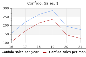
Confido 60 caps discount mastercard
Mnemonic Traces of Concepts 233 In a subsequent report involving a large affected person pattern (Damasio prostate cancer 7 stage 60 caps confido order mastercard, Tranel mens health 2012 confido 60 caps lowest price, Grabowski, Adolphs, & Damasio, 2004), a considerably totally different image emerged: on the idea of the effects of focal lesions, it appeared that left hemisphere lesions produce deficits in naming, whereas bilateral lesions produce deficits in object recognition. That is, it appeared as if focal lesions interfere with name circuits, whereas bilateral lesions interfere with circuits representing basic knowledge concerning the objects. Moreover, it appeared as if mnemonic traces of each kinds are additional segregated in that the naming of well-known individuals was affected by lesions within the left temporal pole; that of animals was affected by lesions within the anterior a half of the left inferior temporal gyrus, the anterior insula, and the dorsal temporo-occipital junction; and the naming of instruments by lesions in the posterior lateral temporaloccipital-parietal junction, the inferior sector of the pre- and post-central gyri and the insula, whereas the naming of musical instruments was compromised by lesions within the temporal pole, the anterior part of the left inferior temporal gyrus, the posterior part of the lateral temporal cortex, the insula, and the inferior a half of the pre- and post-central gyri. On the other hand, semantic circuits appeared to be compromised by bilateral lesions, however principally by lesions in the proper hemisphere. These lesions also selectively affected knowledge of objects belonging to the aforementioned categories relying on their precise location, thus pointing, again, to a spatial segregation of semantic circuits by category. However, using correlations of grey mater quantity and the naming accuracy of line drawings of residing and non-living objects, in a sequence of 152 patients, Brambati et al. Namely, these for living issues (animals and fruits) appeared to be located in the anterior and medial a part of the right temporal lobe and those for non-living things in the posterior part of the left middle temporal gyrus. Evidently, variability within the exact cortical loci of the putative name and semantic circuits of objects belonging to completely different classes is considerable from one research to the following, as is the variability of the selective deficits produced by particular lesions from which the categories of semantic and name circuits are deduced. The issue with deciphering them is due to a quantity of components, the detailed examination of which is past the scope of this chapter. But, variability in the specification of the type of classes of ideas or cardinal concept options associated with particular lesion places aside, the central query here is the soundness of decoding the selectivity of any such deficit as interference with mnemonic traces. Inability or difficulty in naming objects and incapability or difficulty in identifying the nature of objects seen or described may be because of a number of factors along with interference with the concept-specific circuits themselves. The variety of these factors is determined by the particular mannequin of recognition one is implicitly or explicitly adopting. In the case of the final "embellished" mannequin sketched earlier, the deficits may be attributed to (1) the degradation of the network of the cognitive operation of retrieval of the semantic circuits representing entire concepts, (2) interference with the retrieval operation of feature-specific circuits, and/ or (3) interference with the operation of binding the sensory features right into a pictorial representation or mental picture of the object, along with or as a substitute of (4) the degradation of the concept-specific circuits themselves. As talked about previously, of these fundamental alternatives, the first three might conceivably be eradicated on the assumption that the mechanisms of retrieval, binding, and integration are intact for the reason that patients have problem in retrieving, binding, and reconstructing only some specific concepts. It can be possible that retrieval, binding, or integration mechanisms are specialised and hooked up to each of the a quantity of completely different classes of objects. For example, lesions in the anterior part of the temporal lobes are stated to degrade the semantic circuits of living things (Gainotti, 2000) or solely the name circuits of objects (Damasio et al. Accordingly, to decide among the many alternatives, one should search the best-fitting interpretation of regional mind specialization that the empirical information of lesion and functional neuroimaging studies allow-provided, in fact, that the belief that mnemonic traces do exist is true. One such that involves mind is if specific objects or narrowly outlined classes were irrevocably lost in associative agnosia or if specific idea names were similarly misplaced in circumstances of anomia. Instead, what appears to be a permanently eradicated memory is only inaccessible on the time of testing, Accordingly, not having proof that specific recollections are ever eradicated one has to conclude that nothing certain in regards to the traces has been found, and but traces are still assumed to be there! At this juncture, the potential of functional neuroimaging for resolving a variety of the ambiguity turns into obvious. By using normal people with uncompromised brains, with the ability to visualize focal activation with a considerably higher diploma of spatial resolution than that afforded by lesions and in addition having the potential of experimenting with massive numbers of different stimulus objects or object options, it appears possible that an answer as to which ones a specific cortical patch responds selectively can be approximated. Category-Selective Brain Areas Although the early lesion literature would recommend that there are several brain areas specialized for recognizing objects belonging to different classes, few such areas have been identified through functional neuroimaging. The proof for the specialization of these areas might be briefly reviewed to find a way to address the query of whether-whatever else specialization could mean-it may be interpreted as evidence that these areas contain mnemonic traces of ideas or traces of cardinal concept options. The Face Area Early hints for the existence of a mind space specialized for the recognition of faces got here from observations of the selective effects of lesions in the ventral facet of the occipito-temporal area of each hemispheres (or of only the best hemisphere) that encompass the fusiform gyrus (Benton, 1980; Damasio, Damasio, & Van Hoesen, 1982; Damasio, Tranel, & Damasio, 1990a; Lhermitte, Chain, Escourolle, Ducarne, & Pillon, 1972; Meadows, 1974a; Newcombe, 1979; Wilbrand, 1892). These lesions end in prosopagnosia, which is a specific case of object agnosia. But, in any case, on the basis of lesions, this could be very troublesome or impossible to determine the exact regions that specialize for the recognition of individual faces or different regions that will specialize for the popularity of particular person instances of different object categories. As talked about earlier, in the context of empirical research, nevertheless intensive they might be, one may never assert with certitude that an area responds selectively to a particular object or particular characteristic. One might solely cut back incrementally by way of successive research the likelihood of misattribution of the specialization of an area for a particular object or function. To tackle that issue, a condition was included in which footage of individual houses and of particular person faces had been passively seen. It might very well be the case that, whereas we habitually view faces to be able to set up their identification and their distinctions from all different faces, we habitually view all different stimuli, including houses, only aiming to set up their class membership. We are led to the identical conclusion by way of the examination of neuroimaging proof relating to the function of other "specialised" mind regions, and that is introduced subsequent. Rather, they lead to a discount of the efficiency of discriminating photos of our bodies or physique components, a phenomenon reported solely once within the medical literature (Moro et al. Usually, they affect a broader area and end in object agnosia or prosopagnosia (see Moro et al. But our bodies and physique components are rarely if ever static figures within the natural setting. Yet they simply reduce the effectivity of nice discrimination, which is indicative of interference with the mechanisms processing the visible enter. The existence of such an space specialised for scenes was first reported by Epstein and Kanwisher (1998). Failure to recognize landmarks is referred to as topographagnosia, and failure in utilizing cognitive maps is called topographic amnesia (De Renzi, Faglioni, & Villa, 1977; Hecaen, Tzortzis, & Rondot, 1980; Maguire, Burke, Phillips, & Staunton, 1996; McCarthy, Evans, & Hodges, 1996). Lesions in the parahippocampal gyri-especially the right-like strokes of the posterior cerebral artery, produce amongst other visible deficits similar to hemianopia two variants of topographic amnesia: first, they produce a profound deficit in the capability of patients to learn their method round new environments (Barrash, Damasio, Adolphs, & Tranel, 2000; Habib & Sirigu, 1987; Ross, 1980; Teng & Squire, 1999). Second, they produce in some sufferers extra difficulties in finding their way in acquainted environments as well (della Rocchetta, Cipolotti, & Warrington, 1996; Habib & Sirigu, 1987; Landis, Cummings, Benson, & Palmer, 1986; McCarthy et al. The deficit in studying to navigate in new environments is clearly demonstrated in a affected person described by Epstein et al. This patient had sustained damage to the proper medial temporal and occipital cortex and later to the homotopic area within the left hemisphere that resulted in peripheral vision deficits, in difficulties in recognizing colors (cerebral dyschromatopsia), and in topographical orientation. Otherwise, the patient behaved fairly usually, exhibiting no linguistic or affective problems. His most conspicuous deficit was his incapability to learn to orient himself to area. According to the authors, "even after a number of days of testing, he never learned the relative locations of the completely different rooms of the lab. Soon after his harm, he moved to a neighborhood with many similarlooking houses. Clearly, this patient had misplaced his capacity to be taught new spatial data, although he did perceive and will recognize individual objects that made up the scene, such as individual homes. That is to say, the deficit is instantly attributable to degraded cerebral networks for processing topographical material and to not degraded traces. But, even in those instances in which patients present with difficulties in navigating acquainted environments regardless of recognizing individual landmarks, the deficit hardly constitutes evidence for the existence of scene-specific traces within the space that has been degraded by the lesion. For instance, in a single such case (Mendez & Cherrier, 2003), the authors concluded that the deficit was as a outcome of the compromised mechanics of the processing operation for retrieval of saved representations of scenes rather than because of compromised stored representations. It might, of course, be debated whether the chance that mnemonic hint degradations can be excluded as a proof.
Confido 60 caps buy cheap on-line
A lateral insertion of the recti muscles could be palpated in the supraumbilical space (type C) prostate over the counter 60 caps confido buy. In this case mens health 062012 confido 60 caps discount visa, only correction of the myoaponeurotic layer was performed by a direct access, advancing the recti sheaths. She is shown 1 year postoperatively demonstrating improvement of the skin redundancy and better rigidity of the stomach wall, obtained by the plication of the anterior rectus sheath. The solely myoaponeurotic process was plication of the anterior rectus sheath (type A). She is shown 2 years postoperatively demonstrating enchancment of the anterior projection of her stomach and enhanced contours the place liposuction was carried out. She had a 2 cm diastasis with an excellent muscular tonus and had a great amount of extra infraumbilical pores and skin. The diastasis was corrected and the excess skin between the pubic area and the umbilicus was removed. Two years postoperatively, the development of the muscular pressure of the stomach wall and her stomach contour is clear. She underwent an abdominoplasty with correction of the rectus diastasis, plus liposuction of the higher thighs, flanks, lumbar and sacral area, and posterior abdomen. Note the sleek dimple created by the plication of the recti muscle tissue and the fragile contour created by liposculpture. Although she had a considerable amount of intraabdominal fat, the main adipose quantity was situated in the subcutaneous tissue. She also had a rectus diastasis and a common laxity of the anterior abdominal wall. Plication of the anterior rectus sheath and of the exterior oblique aponeurosis had been carried out. She is proven 6 months postoperatively; note the numerous discount of the subcutaneous fat and the increase in tension of the myoaponeurotic layer. Note her improved waistline and that the scar was positioned within the lower abdominal fold, between the belly anatomic items. However, this operation still has a substantial range of related complications. Scar widening, scars that are hypertrophic and dyschromic, and dog-ears are widespread. Specific therapies could be applied to control these conditions, corresponding to the use of chemical peels for hyperchromic scars, intralesional steroid injections for hypertrophic scars, removal of extra pores and skin in dog-ears, and scar excision to appropriate extensive scars. The high quality of the scar may be improved with good surgical planning by reducing the tension on the belly flap. The use of Micropore strips or a silicone sheath over the scar can even improve scar high quality. Sitting the affected person upright in the course of the operation after skin closure permits the surgeon to determine and treat dog-ears. Loss of Sensitivity Flap undermining and traction after abdominoplasty may result in decreased sensitivity of the belly skin. Therefore, these patients are extra exposed to contact burn accidents on sizzling surfaces such as hot ovens and barbecue grills. On the other hand, when lipoabdominoplasty is carried out, the innervation of the belly flap is stored intact, thus preserving pores and skin sensibility in these areas. The burn was triggered by means of a hot towel applied over the supraumbilical space to enhance what she thought was an inflammatory lymph node, however it was actually a small epigastric hernia. When trying to acquire a younger and exquisite umbilicus, some plastic surgeons are inclined to lower the quantity of pores and skin left within the umbilical pedicle. The umbilical pedicle fixation to the anterior rectus fascia permits the surgeon to management the pedicle top and consequently the tension at the pores and skin edges through the suture. Therefore, in some circumstances, the skin pedicle must be advanced superficially utilizing the aponeurosis as a step to get hold of tension decrease of the skin suture between the umbilical skin and the belly flap. The umbilicus ought to match within the gap created on the abdominal flap by skin incisions so that a damaged line is formed when sutured, to keep away from scar constriction. Another way to decrease skin rigidity of the umbilical skin is to fix the dermis of the abdominal flap in two or three points across the umbilicus after which suture the pores and skin with a lot less tension. It has been considered as an inevitable complication until Baroudi and Ferreira in 1998 described using quilting sutures to forestall a seroma. A deformity of the abdomen will happen from the contraction of this capsule around the fluid assortment, in the same way that it occurs round a breast prosthesis. Extensive undermining is usually necessary to appropriate this secondary complication. Note the pseudobursa, which is similar to a capsule formed round an implant. The capsule was eliminated and the tissue was closed with quilting sutures to avoid recurrence. Flap Necrosis Although flap necrosis is a multifactorial complication, extreme pressure and undermining are two frequent technical causes of necrosis after abdominoplasty. Abdominal flap necrosis is often a dramatic complication, relying on its extension. Making a great preoperative plan primarily based on the sort of pores and skin and subcutaneous deformity can forestall extreme tension of the stomach flap. A conservative undermining must also be carried out at the upper stomach to lower the number of vessels severed. A narrow tunnel above the umbilicus can protect some of the rectus perforator arteries, thus growing the arterial flow of the flap. For this reason, surgeons should be careful when performing liposuction of the undermined flap. These sutures are progressively positioned attaching the belly flap to the aponeurosis, advancing the flap, thus decreasing pressure on the skin edge. Hematoma Many components can improve the incidence of hematoma after abdominoplasty. The surgeon should be aware that the area between the abdominal pores and skin flap and the aponeurosis can increase when a hematoma occurs. Therefore, to keep away from this immense lifeless space, quilting sutures can be utilized when this complication happens to restrict a doubtlessly large hematoma, and a small hematoma could additionally be shaped. A small hematoma is treated conservatively, whereas a big hematoma must be surgically eliminated to avoid secondary fibrosis. In such cases, a seroma could occur secondarily, which could be evaluated by ultrasonography. After elimination of the stays of the bloody content material, it was possible to determine a thick capsule that was incised and removed. Quilting sutures were used, just like the correction of a pseudobursa resulting from a seroma. A local swab can also help in the identification of the microorganism liable for this condition; nonetheless, an isolated constructive culture may point out colonization solely. Some ischemic flaps with necrosis could distort the diagnosis, giving the impression that the infection is local.
Confido 60 caps cheap online
There is the added danger of screw cut-out through the concave volar floor of the scaphoid or through the dorsoulnar side of the proximal pole prostate month generic 60 caps confido with mastercard. The hand is suspended by the thumb alone in a single Chinese finger trap with no countertraction prostate cancer 78 years old confido 60 caps buy discount line. This position extends the scaphoid and ulnarly deviates the wrist to improve entry to the distal pole of the scaphoid. Most importantly, this position permits free rotation of the hand all through the operation and the scaphoid stays within the heart of the x-ray subject throughout the procedure. The distinction in size between the trailing end of each wire is the scaphoid size. This permits 2 mm of clearance of the screw at each finish of the scaphoid, thus ensuring full implantation with out screw exposure. This may be doublechecked by holding the screw over the snuffbox within the desired angle of insertion whereas performing a lateral fluoroscopic view. Note how the fracture line is reduced (arrows) and there are a minimum of four screw threads within the proximal pole. The picture intensifier C-arm is turned to a horizontal place and positioned so that the wrist is in the central axis. K-wires may be inserted and used as joysticks to manipulate the fragments into position as needed. The quality of the reduction can then be checked radiographically and, if essential, arthroscopically, without disturbing the overall setup. The length of the screw is determined utilizing a second guide wire of the identical length inserted in the distal cortex of the scaphoid and undersized by 4 mm. The information wire is then advanced by way of the proximal pole to exit on the dorsal facet of the wrist to reduce the chance of inadvertent withdrawal, adopted by reaming and screw insertion. Although some authors permit quick and unrestricted motion, I favor to immobilize the wrist in a short-arm thumb spica solid for 6 to 8 weeks. The authors have been normally in a position to get hold of a passable dorsal-central position without drilling by way of the trapezium. The wrist is then placed into extension and ulnar deviation and the information wire is advanced into the radius to forestall dislodgement after the length of the screw is determined. The scaphoid is reamed to inside 2 to 3 mm of the proximal pole and a screw is inserted beneath fluoroscopic control. Arthroscopic Bone Grafting Ho recently described a method of arthroscopic-assisted bone grafting of scaphoid nonunions. Loose implants (if present) are exchanged with a larger screw or multiple K-wires. Care is taken to preserve any intact cartilage or pseudocapsule over the nonunion website to keep away from subsequent graft protrusion into the radiocarpal joint. Cancellous bone graft is tightly impacted into the nonunion website through an arthroscopic cannula. His collection included 37 patients with established symptomatic nonunions and 6 sufferers with delayed unions with an average age of 28. Penetration of the dorsal-radial surface of the scaphoid, which is exacerbated by any angular or humpback deformity aspect of the proximal pole, is widespread and could be minimized by utilizing the pronated indirect views and undersizing the screw. Screw cut out of the tuberosity can typically be salvaged by using an open technique and creating a gliding trough within the trapezium, switching to a smaller diameter screw and levering the tuberosity volarly with an elevator. They incorrectly placed the screw above the subchondral bone regardless of live fluoroscopy in two specimens. They famous that these soft-tissue accidents could be avoided utilizing a miniopen dorsal strategy. The coronal fractures could be complete and involve the entire physique of the scaphoid. The scapholunate interosseous ligament could be utterly detached or break up into a dorsal and a volar half. The scapholunate ligament may be attached to either the volar or dorsal fragment as a result of a coronal fracture can split the ligament into dorsal and volar halves. In some cases, reduction of the fracture fragments restores the integrity of the ligament, whereas in different circumstances a scapholunate ligament restore may be essential. Arrows are highlighting the malunion site and the step off of the proximal articular floor. The scaphoid fracture is being pried apart and opened like a clam shell, demonstrating the dorsal (D) and volar (V) fracture fragments. Bony union was achieved in all the cases, within a median time of sixty two days (range, 45�80 days). The practical restoration of the operated wrists was reported to be good with an average return to work at 21 days following surgery. The study demonstrated the utility of arthroscopy, which aided the fracture reduction in 67 displaced fractures in addition to the 10 humpback deformities. Radiographic signs of arthritis within the radioscaphoid joint had been more widespread within the surgically treated group (3/14) than in the conservative group (2/21). The medical implication of this research is that their first selection of treatment for nondisplaced or minimally displaced scaphoid waist fractures is solid immobilization. For sufferers for whom a quick return to work or sport actions is of great importance, arthroscopic-assisted screw fixation was provided. Arthroscopic instability was identified if the fracture fragments could be moved with gentle manipulation of the bone by making use of external stress on the distal pole of the scaphoid, by deviating the wrist in radial and ulnar directions, or by inserting a probe between the fracture fragments. Arthroscopy revealed 38 unstable fractures (movement between fracture fragments; 66%), 27 of which have been also displaced. All arthroscopically determined displaced fractures had been unstable, and 11 of the 31 arthroscopically determined nondisplaced fractures had been unstable. There was a significant correlation between radiographic comminution (more than two fracture fragments) and arthroscopically determined displacement and instability. They additionally make the point, however, that given the fact that over 90% of radiologically nondisplaced scaphoid waist fractures heal with cast immobilization, instability is probably not a risk issue for nonunion. These soft-tissue injuries negatively impacted the outcomes following therapy for the scaphoid fracture. The elements affecting outcome after non-vascular bone grafting and inside fixation for nonunion of the scaphoid. Management of displaced fractures of the waist of the scaphoid: meta-analyses of comparative studies. The impact of delayed treatment on scientific and radiological effects of anterior wedge grafting for non-union of scaphoid fractures. Screw fixation of scaphoid fractures: a biomechanical assessment of screw length and screw augmentation. Comparison of screw trajectory on stability of oblique scaphoid fractures: a mechanical research. A comparative evaluation of the accuracy, diagnostic uncertainty and price of imaging modalities in suspected scaphoid fractures.
Bear Grass (Yucca). Confido.
- Dosing considerations for Yucca.
- Are there safety concerns?
- What is Yucca?
- How does Yucca work?
- Arthritis, migraines, digestive disorders, diabetes, high blood pressure, high cholesterol, high triglycerides, poor blood circulation, skin problems, and other conditions.
Source: http://www.rxlist.com/script/main/art.asp?articlekey=96718
60 caps confido cheap free shipping
The solely exception is when there is a sign that the operative time must be decreased prostate cancer robotic surgery buy confido 60 caps fast delivery. In chubby kind C sufferers mens health dwayne johnson supplements confido 60 caps amex, when plicating the posterior rectus sheath, it may be troublesome to keep away from passing the needle within the intestine wall. In such circumstances, a 2 cm incision within the posterior rectus sheath as properly as within the peritoneum will forestall bowel damage during the plication of the posterior rectus sheath. In type B patients, extreme tension in horizontal plications on the inguinal area must be avoided in the course of the L-plication of the exterior indirect aponeurosis. This might decrease the aponeurotic resistance within the groin area, leading to a hernia. In dissecting the posterior rectus sheath in sort C patients, care have to be exercised with the inferior epigastric vessels that run on the posterior surface of the recti muscles, penetrating the muscle tissue above the arcuate line. Benchmarking outcomes in plastic surgery: national complication charges for abdominoplasty and breast augmentation. Use of the, anterior rectus sheath for stomach wall reconstruction: a examine in cadavers. A, variation in the part separation approach that preserves linea semilunaris: a examine in cadavers and a scientific case. Does the use of compression garments increase venous stasis within the frequent femoral vein Body dysmorphic disorder in sufferers seeking abdominoplasty, rhinoplasty, and rhytidectomy. Should plastic surgeons operate on sufferers diagnosed with body dysmorphic disorder Reply: Should plastic surgeons operate on sufferers diagnosed with physique dysmorphic disorders Seroma in lipoabdominoplasty and abdominoplasty: a comparative research using ultrasound. Patients with mild to average physique dysmorphic disorder may profit from rhinoplasty. Abdominal-wall reconstruction with expanded musculofascial tissue in a posttraumatic defect. A method for repairing large ventral incisional hernias with out the use of a mesh. Effects of aesthetic abdominoplasty on stomach wall perfusion: a quantitative analysis. Advancement of the external oblique muscle flap to enhance waistline: a examine in cadavers. Commentary on: Improvements in vertebral-column angles and psychological metrics after abdominoplasty with rectus plication. Discussion: Evaluation of the long-term stability of sheath plication using absorbable sutures in fifty one patients with diastasis of the recti muscular tissues: an ultrasonographic research. A pragmatic approach to treat stomach deformities based on pores and skin and subcutaneous extra. Wide abdominal rectus plication abdominoplasty for the therapy of chronic intractable low back pain. Factors that will influence failure of the, correction of the musculoaponeurotic deformities of the stomach. Components separation, method with restricted subcutaneous undermining: a cadaver examine. Seroma after lipoabdominoplasty: fats thickness of the abdominal wall is probably a contributory issue. Ventilatory operate and intraabdominal stress in patients who underwent abdominoplasty with plication of the external indirect aponeurosis. Does diastasis width affect the variation of the intra-abdominal stress after correction of rectus diastasis Is it, possible to restore diastasis recti and shorten the aponeurosis on the same time Deos Ricardo Arnt Eduardo Gus the abdominal wall is likely certainly one of the areas that most incessantly has a variety of anatomic variations and deformities, including fat extra or misdistribution, pores and skin or muscular laxity, hernias, and other issues related to these traits. Traditional abdominoplasty and mini-abdominoplasty procedures, with or without liposuction, often handle most of these problems. However, abdominal pores and skin with or with out fats excess-especially in the supraumbilical abdominal region-still stays a challenge for most moldable surgeons. Mini-abdominoplasties produce inadequate results, and conventional abdominoplasties end in a cephalad-positioned scar or in a further infraumbilical vertical scar. Resection of pores and skin and adipose tissue in the higher stomach was first described by Thorek in 1942. In 1977 Rebello and Franco described and systematized the strategy to belly plastic surgery by way of the inframammary sulcus. Originally the technique involved fixation of the flap solely to the mammary sulcus, which resulted in its inferior dislocation or even in hypertrophic scarring, as a result of the burden of the flap constantly exerted rigidity on the sutures. Traction and fixation of the flap: the flap is put under in depth traction toward the mammary sulcus and is strongly fixated to the muscular belly aponeurosis, thereby minimizing and even eliminating the tension on the scar and preventing the previously mentioned problems. Extension of the inframammary incision and the amplitude of the undermining area: Patients with larger amounts of pores and skin to be resected require incision unification on the midline, which leads to a single U-shaped flap. It is bounded superiorly by the xiphoid course of and the costal margin, laterally by the iliac crest and the oblique muscle tissue, and inferiorly by the pubis and the inguinal ligament. It is of utmost necessary that surgeons perceive the vascularization sample of the belly wall. According to Huger, the primary blood provide of the anterior abdominal wall is provided by the deep epigastric arcade, which is shaped by the superior and inferior epigastric arteries (Huger zone I). The anastomoses between the superior and inferior methods occur mainly within the midline between the costal margin and the umbilicus. Indications and Contraindications As for all different surgical procedures, the right indications for the technique and acceptable patient choice are the cornerstones of successful outcomes. This leads to a a lot smaller space of undermining in contrast with that related to typical abdominoplasty. Conventional abdominoplasty and mini-abdominoplasty ought to all the time be considered first, because the scars are inclined to be coated by garments and favor gravitational forces. The patient is then asked to carry out a Valsalva maneuver in order that the surgeon could decide the presence of any muscle diastasis or hernias. The degree of lipodystrophy and the need for additional liposuction should also be assessed at this time. In these positions, the patient should bend forward so the surgeon can higher evaluate the amount of pores and skin extra and the placement in the stomach the place this excess predominates. Preoperative Planning and Preparation the inframammary scar extension and the dissection amplitude are determined by the depth of the supraumbilical skin excess.
Cheap 60 caps confido with amex
The mind is richly provided with blood vessels that carry oxygen-rich blood to the mind man health magazine garcinia test fixed confido 60 caps buy without prescription. These blood vessels additionally convey a unbroken supply of glucose to meet the huge vitality wants of the mind mens health 9 week plan confido 60 caps generic with amex. In fact, although the brain consists of just 2% of the weight of the body, it requires 20% of the glucose (energy) demands of the physique. Deprivation of either oxygen or glucose can end result in irreparable damage to the mind inside 10 minutes. Depriving the mind of a gentle stage of glucose leads to rising reactivity and ultimate instability of mobile functioning. For example, in fundamental mind functioning thirteen a diabetic, fluctuating blood glucose levels may end up in initial irritability and mood modifications. If a blood glucose degree drops too low without the administration of or intake of a supply of glucose, the brain will reply with eventual shutdown including seizure, coma, after which demise. The brain is covered with three layers of membranes referred to as meninges, which line the cranium and supply an additional layer of cushion between mind and skull. The mind could be thought to float on this upside-down, hardsided bowl of the skull. It is bathed in cerebrospinal fluid, which helps to cushion and protect the mind by absorbing shock from blows to the top. For instance, latest findings point to an association between head injury and a rise in dangerous behaviors in adolescents (Thompson, 2014). The frontal lobe of the mind seems to be notably weak to injury from head injury. Damage to the frontal lobe can negatively affect determination making and executive planning, with ensuing enhance in dangerous behaviors (McKee & Robinson, 2014). New info is on the market for clinicians supporting education for prevention of head injury and on assessment of, and therapy for, injury to the brain due to head trauma. Counselors and other psychological well being specialists ought to embrace in each 14 basic mind functioning consumption assessment an inquiry of historical past of head traumas and concussions. The brain has many convolutions and folds, principally seen throughout the outer area of the cerebrum. These folds permit for a bigger brain floor space than is quickly obvious on easy examination. These folds lengthen lengthwise from the pinnacle all the means down to the higher space of the spinal column. The mind accommodates inner ventricles or chambers which are filled with continuously replenished cerebral spinal fluid. Cerebral spinal fluid carries nutrients to the mind and removes waste products from the brain. Recent medical consideration is concentrated on the importance of identifying and treating head accidents. Many returning struggle veterans have suffered head accidents during their service, and increasing brain-related accidents are famous in sports activities and some office environments. There is a superb need for schooling, identification, and prevention of head harm and trauma. Counselors and trauma-focused therapists could help their shoppers to connect with medical resources for head trauma treatment and to develop collaborative therapy planning. A trauma-aware counselor will inquire about history of head or spinal injuries, gain a complete diagnostic picture, and assist in remedy planning and drawback fixing with head harm awareness. The brain sits at the high of the body, performing as a director and coordinator of activity and action throughout the physique. It does so via nerves positioned within the mind and through fundamental brain functioning 15 nerves that reach down through the spinal twine and outward to the physique. Within the head, there are additionally 12 pairs of cranial nerves, that are linked immediately inside the brain and the mind stem. The cranial nerves primarily hook up with the sensory organs (eye, ear, and nose) and provide enervation for necessary actions corresponding to chewing, talking, and eye actions. Cranial nerves are responsible for minute and complicated movements concerned in facial expressions, to which humans respond and with which they work together, fostering an all-important social connection. An understanding of vaso�vagal nerve responses and influences on body organs corresponding to coronary heart, lungs, muscular tissues, and digestive system leads to a discussion of sympathetic and parasympathetic nervous system responses. It is useful to contemplate the mind as having three functioning layers from the bottom of the mind to the top of the head. The brain is further divided into two hemispheres, one on the proper side of the skull and one on the left, and these hemispheres are linked by a construction referred to as the corpus callosum. The corpus callosum permits for communication between the neurons in both brain hemispheres. The cerebral cortex, with its many folds and convolutions, wraps across the sides and over the top space of the mind. For functions of location and identification, particular regions of the cerebral cortex are recognized as lobes, or specialised 16 fundamental brain functioning areas. The cerebellum is located underneath the occipital lobe, within the back decrease area of the top. The brain stem is located deep and center on the base of the mind, just above the spinal twine. Brain lobes correspond considerably loosely with completely different areas of performance in the brain. To illustrate a tough three-dimensional mannequin of the mind, the advisers can increase their hand with palm going through outward, inserting the thumb alongside the within of the palm. The counselor can level out that the thumb represents the inside limbic system of the brain. Then, upon closing the fingers over and making a fist, can explain how the cortex is wrapped excessive of the brain. The brain stem can be pointed to because the wrist space, and extending down the arm is the spinal wire (Siegel, 2010). While simplistic, such illustrations could be helpful for purchasers as they achieve primary understanding of brain responses to and restoration from trauma. Viewing the brain from underneath, one would see a portion of the frontal lobe and temporal lobe on the best and left facet, in addition to the olfactory bulb main from the nostril, the pons and cerebellum, and the 12 cranial nerves. The inner elements of the brain are often considered from a sagittal (or vertical) view, usually by dividing down the corpus callosum and looking from inside to out. Major constructions seen in this manner would include the cingulate sulcus, diencephalon, anterior commissure, and temporal lobe portions. From this viewpoint, one would additionally see the midbrain, containing the limbic system constructions of the amygdala, hypothalamus, and hippocampus, and below it the pons, cerebellum, and the poetically named medulla oblongata.
60 caps confido buy with amex
The interconnections of neurons to other neurons leads to the risk of an estimated 100 trillion connections androgen hormone treatment 60 caps confido order. The actions that happen within the synaptic area could be functionally divided into two ideas prostate cancer wristbands order confido 60 caps free shipping. During peak months of fetal growth, an estimated 250,000 neurons are being produced every minute (LeDoux, 2002). Initially, some of the divided cells located on the outside of the blastocyst (the bundle of cells growing just after fertilization) start to migrate toward an inside cluster of the developing cells, nearly like folding in on itself, moving to the inside of the ball of cells. These cells start to form one thing akin to a tube with the rudimentary formation of a future brain located on one end, and the tube extending the size to the other finish. Running alongside this length of neurons is the early formation of the spinal twine and peripheral nerves. From earliest moments after conception, cells are dividing and proceed to divide at a rapid and livid tempo. By the eighth week, the mind itself has developed into its three areas, the forebrain, midbrain, and hindbrain. In the early weeks there are an estimated 250,000 neuroblasts, the primitive precursors to neurons. Neural production begins around gestational day 42, and neurons instantly begin a strategy of migration to various areas of the brain. Neurons that Fire Together Wire Together From the earliest beginning, nerves cells are about connection and network. It is as if nerve cells reach out to each other from the beginning, with these cells that join with different neurons humming with life; those not capable of connect basic mind functioning 21 will die or be pruned away. Neurons begin the process of a great migration to align with and connect to other neurons. The glial cells additionally protect and nourish the neurons, with one type of glial cell that controls neuron cell metabolism and one other sort of glial cell that coats the neuron with a substance called myelin, forming a sheath-like cover surrounding the neuron, very related to the rubber coating round electrical wiring. The presence of myelin controls the pace of conduction of electrical impulses traveling via the axons of neurons. Continuing studies of fetal neuron migration have proven that there are chemical alerts known as trophic elements that seem to direct neural axons on the place to join. These chemical indicators, together with sustained electrical stimulation, determine if the connection between neurons will hold, and finally whether a given neuron will survive or die. These cells begin a migration outward to totally different parts of the brain; for instance, a visible neuron could migrate to the visual cortex area-located in the occipital lobe, or back decrease part of the head-or it could join different visual neurons to type the optic nerve. There is proof that some neurons travel faraway and arrange new clusters of neural neighborhoods that in flip open additional communications between varied neural sites. As neurons connect, the factor of environment comes into play within the type of studying. Neural connections which are repeatedly activated by sensory enter turn into stronger, with their connection community staying intact. The widespread saying that "neurons that fireplace collectively wire together" serves to explain this course of. It had lengthy been thought that the structure and functioning capacity of the brain was unchanging after early development was full. It is primary mind functioning 23 now recognized that brain tissue could be strengthened and might restructure itself in response to studying. There are many sensible implications of these findings resulting in new support and preventive efforts to promote wholesome growth within the early prenatal interval of life. This is pointing to renewed curiosity in and necessary emphasis upon good maternal and fetal well being within the prenatal, delivery, and perinatal intervals. Maternal state of health, use of gear, hormonal influences, and cellular metabolism are simply a quantity of attainable factors that can have profound results on neural migration and the following neural connections of the growing fetus. Disorders corresponding to autism, schizophrenia, epilepsy, and others are actually thought to have beginnings inside defective migration patterns of neurons within the brain (Ratey, 2001). The whole brain is itself aligned in a vertical style, starting at the base or mind stem, by way of the midbrain, and upward towards the cerebral cortex. For instance, the practical tasks of the decrease mind stem embrace fundamental physiological features such as respiration, while higher areas of the cerebral cortex are targeted on functions of studying and downside fixing. There is also side-to-side perform, with the proper hemisphere and left hemisphere linked through the corpus callosum. It is thought that the human brain organized bottom to prime throughout evolution similarly to what occurred with different animal species. Reptiles, fundamental brain functioning 25 birds, and fish, for instance, have rudimentary brain stems. The mind stem seems like a widening area at the finish of the stalk-like spinal cord. The decrease finish of the mind stem is situated via an opening within the base of the cranium, and its upper area sits at about eye degree within the center of the pinnacle. The brains of mammals advanced to embody midbrain areas such because the limbic system, with the cortex sitting above. Human mind evolution dramatically elevated the size, the density, and the convolutions throughout the cortex, particularly within the space of the frontal lobe of the mind. The brain stem accommodates the lower mind areas connecting with the spinal twine, the midbrain space, and the lower hindbrain space. At the bottom finish of the brain stem sits the medulla oblongata, additionally known as simply the medulla, with the pons and the midbrain area above. The brain stem performs critical but fundamental life-promoting functions, most of that are automatic and ongoing and usually exterior of acutely aware consciousness, corresponding to regulation of heart fee, respirations, blood strain, and scanning actions of the eye. Other mostly automated actions initiated within the mind stem include swallowing, coughing, and sneezing. In moments of imminent hazard, or with inside physiological disturbance, the mind stem response might include fainting or lack of consciousness, or autonomic responses of change in blood strain and coronary heart price. The brain stem plays a task within the circadian rhythm within the body, adjusting to a 24-hour cycle of waking and sleeping. Assisting and educating purchasers around the need for reestablishing normal sleep�wake cycles may be a really essential and important consideration for therapists working with a trauma focus. Chapter 9 will provide the counselor with further data on the neuroscience of sleep and implications for the treatment of trauma. Some contemplate the thalamus to be part of the brain stem whereas different sources point out its location in proximity to midbrain buildings such because the limbic system. Appropriately, the thalamus is functionally similar to a preprocessing or switch heart, relaying messages principally acquired from sensory organs or from the brain stem as a lot as cortical areas for interpretation and signal to action. Messages from every sensory organ, apart from the olfactory sense of smell, are processed through the thalamus. The thalamus is a crucial link for management and regulation of the motor methods located within the mind that affect voluntary movements and basic coordination of the physique.