

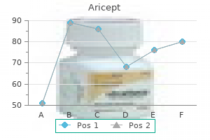
Aricept dosages: 10 mg, 5 mg
Aricept packs: 30 pills, 60 pills, 90 pills, 120 pills, 180 pills, 270 pills, 360 pills
Aricept 10 mg safe
This area known as the anal transitional zone is examined at greater magnification within the backside left figure treatment kennel cough aricept 5 mg mastercard. The right rectangular area contains the stratified squamous epithelium (StS) of the skin in the squamous zone of the anal canal and is examined at greater magnification in the bottom proper figure symptoms 1974 aricept 10 mg free shipping. Between the two diamonds within the rectangular areas proven is epithelium of the decrease a part of the anal canal. Characteristically, the lamina propria contains massive numbers of lymphocytes (Lym), particularly so in the region marked. A larger magnification of the stratified columnar epithelium (StCol) and stratified cuboidal epithelium (StC) discovered in the transition zone is shown in the inset. The ultimate change in epithelial kind that occurs on the squamous zone of the anal canal is shown right here. Again, numerous lymphocytes (Lym) are in the underlying connective tissue, and heaps of have migrated into the epithelium in the nonkeratinized area. It is located within the upper proper and partially within the upper left quadrants of the abdominal cavity, protected by the rib cage. The liver is anatomically divided by deep grooves into two giant lobes (the right and left lobes) and two smaller lobes (the quadrate and caudate lobes;. This anatomic division has solely topographic significance as a result of it relates lobes of the liver to other stomach organs. Division into functional or surgical segments that correspond to the blood supply and bile drainage is more clinically important. In the embryo, the liver develops as an endodermal evagination from the wall of the foregut (specifically the location that may become the duodenum) to type the hepatic diverticulum. The diverticulum proliferates, giving rise to the hepatocytes, which become organized in mobile (liver) cords, thus forming the parenchyma of the liver. An outgrowth from the frequent bile duct types the cystic diverticulum that provides rise to the gallbladder and cystic duct. Liver Physiology Many circulating plasma proteins are produced and secreted by the liver. The liver performs an essential position in the uptake, storage, and distribution of both nutrients and nutritional vitamins from the bloodstream. In addition, the liver degrades or conjugates quite a few poisonous substances and drugs, however it may be overwhelmed by such substances and damaged. The liver is also an exocrine organ; it produces a bile secretion that incorporates bile salts, phospholipids, and ldl cholesterol. Several nutritional vitamins are taken up from the bloodstream and are then saved or biochemically modified by the liver. They embody: � falciform ligament ligamentum teres terminal hepatic venule (central vein) vitamin A (retinol), an necessary vitamin in imaginative and prescient. This diagram exhibits the gross view of the diaphragmatic and visceral surfaces of the liver, with labeled anatomic landmarks found on both surfaces. The enlarged cross-sectional space of the liver (bottom) exhibits the overall microscopic group of the liver into lobules. Note the presence of hepatic portal triads at the periphery of each lobule, with the terminal hepatic venule (central vein) in the middle of the lobule. The circulating plasma proteins produced by the liver embrace: � � albumins, which are involved in regulating plasma quantity and tissue fluid steadiness by maintaining the plasma colloid osmotic stress. The liver additionally produces small quantities of different plasma lipoproteins, similar to low-density Vitamin A is the precursor of retinal, which is required for the synthesis of rhodopsin in the eye. The liver performs a serious role in the uptake, storage, and upkeep of circulating levels of vitamin A. When the vitamin A levels in the blood decrease, the liver mobilizes its storage websites in the hepatic stellate cells (see pages 634�635). Vitamin D is acquired from dietary vitamin D3 and can also be produced in the skin throughout exposure to ultraviolet light by conversion of 7-dehydrocholesterol. The liver performs an necessary position in vitamin D metabolism by converting vitamin D3 to 25-hydroxycholecalciferol, the predominant form of circulating vitamin D. Further conversion takes place within the kidney to 1,25-hydroxycholecalciferol, which is 10 times more energetic than vitamin D3. Vitamin D is important for growth and development of the skeletal system and enamel. Deficiency of vitamin D is related to rickets and problems of bone mineralization. Vitamin K deficiency is associated with hypoprothrombinemia and bleeding disorders. It synthesizes almost all of the proteins concerned in iron transport and metabolism, together with transferrin, haptoglobin, and hemopexin. The association of the protein with the lipid-containing core makes the complicated sufficiently hydrophilic to stay suspended in the plasma. Lipoproteins serve a big selection of capabilities in mobile membranes and in the transport and metabolism of lipids. The lipoprotein complexes cross to the Golgi, where secretory vesicles containing electron-dense lipoprotein particles bud off and are then released at the cell surface bordering the perisinusoidal area to attain the bloodstream. Several hormones, similar to estrogen and thyroid hormones, regulate the secretion of lipoproteins. These lipoproteins differ in chemical composition and could be isolated from plasma according to their flotation properties, from largest and least dense to smallest and most dense. Chylomicrons, the lightest of all lipoproteins, are made solely within the small intestine. Their major operate is to transport the large amount of absorbed fats to the bloodstream. Their operate is to transport a lot of the triglycerides from the liver to other organs. In liver biopsy specimens from these people, massive lipid droplets occupy a lot of the hepatocyte cytoplasm. Iron is saved inside the hepatocyte cytoplasm within the form of ferritin or may be transformed to hemosiderin granules. Recent studies point out that hepatocytes are the main sites of long-term storage of iron. Iron overload (as in multiple blood transfusions) could lead to hemochromatosis, a type of liver harm characterized by extreme amounts of hemosiderin in hepatocytes. Hepatocytes are involved in degradation of medication, toxins, and other proteins overseas to the physique (xenobiotics). It involves a series of biochemical reactions with proteins collectively named cytochrome P450. This process makes the product of phase I even more water-soluble in order that it can be simply eliminated by the kidney. The liver is important in carbohydrate metabolism as it maintains an sufficient provide of vitamins for cell processes. In glucose metabolism, the liver phosphorylates take up glucose from the gastrointestinal tract to glucose-6-phosphate. Depending on energy necessities, glucose-6-phosphate is either saved within the liver within the type of glycogen or used in the glycolytic pathways.
Diseases
- Epidermolysis bullosa, generalized atrophic benign
- Finnish lethal neonatal metabolic syndrome
- Congenital hemidysplasia with ichtyosiform erythroderma and limbs defects
- Cardiac valvular dysplasia, X-linked
- Muscular dystrophy congenital, merosin negative
- Haspeslagh Fryns Muelenaere syndrome
- Graham Boyle Troxell syndrome
- Respiratory distress syndrome, infant
Purchase aricept 10 mg line
In any section through the postpubertal ovary treatment of hemorrhoids cheap aricept 10 mg without a prescription, follicles of various levels may be seen present process atresia medications zovirax generic aricept 5 mg with mastercard. In atresia, the initial adjustments involve pyknosis of the nuclei of the follicular cells and dissolution of their cytoplasm. This might fold inward or collapse, however it usually retains its thickness and staining traits. When included in the airplane of part, a distorted zona pellucida serves as a dependable diagnostic characteristic of an atretic follicle. In atresia of large, nearly mature follicles, cells of the theca interna remain to form clusters of epithelioid cells in the ovarian cortex. These are referred to collectively as interstitial glands and continue to secrete steroid hormones. When seen with the electron microscope, they show the characteristics of endocrine cells, significantly steroid-secreting cells. In figure on proper, a later stage within the development of the secondary follicle is shown. In atresia of a more superior follicle, the follicular cells tend to degenerate extra quickly than the cells of the theca interna, and the basement membrane separating the two becomes thickened to kind a hyalinized membrane, the glassy membrane. Thus, the glassy membrane (arrows) separates an outer layer of remaining theca interna cells from the degenerating inner follicular cells. Note that even though the atresia in these follicles is well-advanced, a number of the cells exterior to one of the glassy membranes nonetheless retain their epithelioid character (arrowhead). The cells of the corpus luteum, luteal cells, quickly increase in dimension and turn out to be full of lipid droplets. A lipid-soluble pigment in the cytoplasm of the cells, lipochrome, gives them their yellow appearance in fresh tissue. Two forms of luteal cells are identified: Large, centrally located granulosa lutein cells are derived from the granulosa cells; smaller, peripherally situated theca lutein cells are derived from the theca interna. A rich vascular community is established in the corpus luteum into which progesterone and estrogen are secreted by the lutein cells. These hormones stimulate growth and differentiation of the uterine endometrium to put together it for implantation of a fertilized ovum. The plication of the membrana granulosa begins simply earlier than ovulation and continues because the corpus luteum develops. As the corpus luteum becomes extra plicated, the previous follicular cavity turns into shrunk. Cells of the theca interna observe the blood vessels into the outermost depressions of the plicated construction. These theca interna cells turn out to be transformed into cells of the corpus luteum called theca lutein cells. Keep in mind that the theca interna was derived from the connective tissue stroma of the ovary. The similar arrangement of cells is shown in determine on the proper at much higher magnification. The cytoplasm contains yellow pigment (usually not evident in routine H&E sections), hence the name, corpus luteum. Thus, when figuring out the two cell sorts, aside from location, observe that the nuclei of adjoining theca lutein cells usually appear to be closer to one another than nuclei of adjoining granulosa lutein cells. The adjustments whereby the ruptured ovarian follicle is reworked into a corpus luteum occur underneath the affect of pituitary luteinizing hormone. In turn, the corpus luteum itself secretes progesterone, which has a profound effect on the estrogen-primed uterus. The regressing mobile components of the corpus luteum are changed by fibrous connective tissue, and the construction is then known as a corpus albicans. The epithelial cells enhance in peak in the course of the center of the cycle, simply about the time the ovum shall be passing through the tube, and turn into lowered through the premenstrual interval. Not only does the number of ciliated cells improve during the follicular phase of the ovarian cycle but in addition removing of the ovaries leads to atrophy of the epithelium and lack of ciliated cells. The mucosal folds are evident in its distal portion, the infundibulum, as it nears the open end. The infundibulum leads proximally to the ampulla, which constitutes about two-thirds of the size of the oviduct, has essentially the most quite a few and sophisticated mucosal folds, and is the positioning of fertilization. Mucosal folds are least quite a few on the proximal finish of the oviduct, near the uterus, the place the tube is slender and referred to because the isthmus. A uterine or intramural portion measures about 1 cm in size and passes via the uterine wall to empty into the uterine cavity. For the first a number of days of development, as it navigates the complicated pathway created by the mucosal folds, the embryo is transported proximally by the beating of the cilia of the ciliated epithelial cells and by peristaltic contractions of the well-developed muscularis layer that underlies the mucosa. In addition to the mucosa (Muc), the rest of the wall consists of a muscularis (Mus) and connective tissue. The muscularis consists of clean muscle that types a relatively thick layer of circular fibers and a thinner outer layer of longitudinal fibers. The area enclosed by the rectangle in figure above is shown here at greater magnification. The ciliated cells are readily recognized by the presence of well-formed cilia (C). The character of the connective tissue is actually the same from the epithelium to the muscularis, and because of this, no submucosa is described. The uterine wall consists of a mucosa, referred to as the endometrium; a muscularis, referred to as a myometrium; and, externally, a serosal cover, the perimetrium. The myometrium consists of smooth muscle and connective tissue and incorporates the large blood vessels that give rise to the vessels that offer the endometrium. The uterus undergoes cyclical modifications that are largely manifested by adjustments that happen in the endometrium. The stratum basale is the deeper part of the endometrium and adjoins the myometrium. In the nonpregnant uterus, the sleek muscle cells are about 50 m in length; during pregnancy, they undergo enormous hypertrophy, often reaching more than 500 m in size. In addition, new muscle fibers develop after division of present muscle cells and division and differentiation of undifferentiated mesenchymal cells. Fibroblasts increase by division and secrete further collagen and elastic fibers. Collagen secreted during being pregnant is digested by the very cells that secreted it, the fibroblasts. Similar, however much less pronounced, proliferation and degeneration of fibroblasts and collagen happen in every menstrual cycle. The area inscribed in the upper small rectangle is shown at greater magnification in the inset on the right.
Discount aricept 10 mg with mastercard
Bronchiolar Function Respiratory bronchioles are the first part of the bronchial tree that permits gas trade medicine 8 - love shadow 5 mg aricept proven. It is characterized by recurrent obstruction of airflow attributable to a combination of inflammation of the bronchioles and constriction of their easy muscles (bronchospasm) symptoms 2015 flu order aricept 10 mg mastercard. Obstruction of airways makes it difficult for air to transfer in and out of the lung alveoli, causing symptoms similar to wheezing, coughing, shortness of breath, and chest tightness. Inflammation of the respiratory mucosa, underlying connective tissue, and easy muscle tissue of bronchioles is found in patients with bronchial asthma. It is characterised by infiltration of the bronchiolar wall by eosinophils (in some circumstances, neutrophils), lymphocytes (mostly activated helper T cells), and mast cells. Bronchiolar epithelium is thick, incorporates an increased number of goblet cells (thus produces extra mucus), and has a thick basement membrane as a end result of an elevated deposition of collagen fibers in the reticular lamina. The smooth muscle layer can be extra pronounced and incorporates a quantity of layers of hyperplastic smooth muscle cells. Traditionally, medications used to treat patients with bronchial asthma have been categorised as bronchodilators (cause rest of clean muscle) or anti-inflammatory medications (suppress inflammatory reactions). Currently, asthma drugs are categorized in accordance with their time of motion within the overall management of this illness. They are either quick-relief drugs, corresponding to -adrenergic agonist bronchodilators to reverse smooth-muscle constriction, or long-term management drugs, similar to inhaled corticosteroids, long-acting -agonist bronchodilators, and leukotriene modifiers. This part of the lung from a affected person with asthma reveals a bronchiole in the middle with surrounding alveoli. This high-magnification photomicrograph exhibits the structure of a bronchiolar pseudostratified columnar epithelium containing a lot of goblet cells. Note the presence of a lot of eosinophils (cells with pink cytoplasm), lymphocytes, and different connective tissue cells that infiltrated the lamina propria and submucosa of the bronchiole. The smooth muscle layer can additionally be thick and the underlying adventitia accommodates enlarged blood vessels. The epithelium of the initial segments of the respiratory bronchioles contains both ciliated cells and Clara cells. Occasional brush cells and densecore granule cells are additionally current along the size of the respiratory bronchiole. Scattered, thin-walled outpocketings, alveoli, extend from the lumen of the respiratory bronchioles. Alveoli are the websites at which air leaves and enters the bronchiole to enable fuel change. Alveoli are the terminal air spaces of the respiratory system and are the precise websites of gas trade between the air and the blood. Each alveolus is surrounded by a community of capillaries that brings blood into shut proximity to inhaled air contained in the alveolus. The alveoli are surrounded and separated from each other by a skinny connective tissue layer, the interalveolar septa, containing blood capillaries. On the proper is the lung surface, which is covered by visceral pleura containing simple squamous epithelium and an underlying layer of connective tissue. The junctions kind an effective barrier between the air area and the parts of the septal wall. These cuboidal cells are interspersed among the many sort I cells but are inclined to congregate at septal junctions. After lung damage, they proliferate and restore both kinds of alveolar cells throughout the alveolus. Surfactant decreases the alveolar surface rigidity and actively participates in the clearance of international supplies. Rings of smooth muscle are current within the knob-like interalveolar septa (see the next paragraph). Alveolar sacs normally occur at the termination of an alveolar duct however may occur anywhere along its length. Alveoli are surrounded and separated from each other by an exceedingly skinny connective tissue layer that contains blood capillaries. The tissue between adjacent alveolar air areas is known as the alveolar septum or septal wall. Surfactant synthesis within the fetus happens after the thirty fifth week of gestation and is modulated by a selection of hormones, including cortisol, insulin, prolactin, and thyroxine. Without adequate secretion of surfactant, the alveoli would collapse on each successive exhalation. In addition, administration of cortisol to mothers with threatened premature delivery decreases neonatal mortality. Surfactant proteins help arrange the surfactant layer and modulate alveolar immune responses. The alveolar epithelium consists of a quantity of specialised cells and their products, some of which play defensive and protecting roles: In addition to phospholipids, hydrophobic proteins are needed for the construction and function of surfactant. They are extremely skinny squamous cells; they line most (95%) of the floor of the alveoli. This electron micrograph reveals two alveolar areas separated by an alveolar septum containing capillaries, a few of which contain purple blood cells. Photomicrograph of an alveolus for comparison with the alveolar wall as seen in an electron micrograph. Connective tissue cells and fibers which could be present between the 2 basal laminae widen the air�blood barrier. It is assumed that nearly all gasoline exchange occurs throughout the thin portion of the barrier. The thick portion is assumed to be a web site by which tissue fluid can accumulate and even cross into the alveolus. Lymphatic vessels in the connective tissue of the terminal bronchioles drain fluid that accumulates in the thick portion of the septum. Alveolar macrophages remove inhaled particulate matter from the air spaces and red blood cells from the septum. Alveolar macrophages are unusual in that they func- the alveolar septum is the positioning of the air�blood barrier. The air�blood barrier refers to the cells and cell products across which gases should diffuse between the alveolar and capillary compartments. The thinnest air�blood barrier consists of a skinny layer of surfactant, a sort I epithelial tion both within the connective tissue of the septum and within the air house of the alveolus. Alveolar macrophages are derived from blood monocytes and belong to the mononuclear phagocyte system (see web page 181). Some engorged macrophages move up the bronchial tree in the mucus and are disposed of by swallowing or expectoration once they attain the pharynx. Thus, at autopsy, the lungs of city dwellers and smokers normally show many alveolar and septal macrophages filled with carbon particles, anthracotic pigment, and birefringent needle-like particles of silica. Alveolar macrophages also phagocytose infectious organisms such as Mycobacterium tuberculosis, which could be acknowledged within the cells in appropriately stained specimens. In addition, latest evidence means that apoptosis of septal macrophages contributes to the event of emphysema. Collateral air circulation via alveolar pores allows air to pass between alveoli.
Cheap aricept 10 mg amex
In addition medications depression purchase 10 mg aricept mastercard, the connective tissue of the higher eyelid incorporates tendon fibers of the levator palpebrae superioris muscle that open the eyelid keratin treatment aricept 10 mg buy cheap on line. In addition to eccrine sweat glands, which discharge their secretions immediately onto the pores and skin, the eyelid accommodates 4 other major kinds of glands. The vitreous body is loosely hooked up to the encircling structures, together with the inner limiting membrane of the retina. The main portion of the vitreous body is a homogeneous gel containing roughly 99% water (the vitreous humor), collagen, glycosaminoglycans (principally hyaluronan), and a small population of cells referred to as hyalocytes. These cells are believed to be responsible for synthesis of collagen fibrils and glycosaminoglycans. Hyalocytes in routine hematoxylin and eosin (H&E) preparation are troublesome to visualize. Fibroblasts and tissue macrophages are sometimes seen in the periphery of the vitreous body. The tarsal glands (Meibomian glands), long sebaceous glands embedded within the tarsal plates, appear as vertical yellow streaks within the tissue deep in the conjunctiva. About 25 tarsal glands are present within the higher eyelid, and 20 are present in the lower eyelid. The sebaceous secretion of the tarsal glands produces an oily layer on the floor of the tear movie that retards the evaporation of the traditional tear layer. Blockage of the tarsal gland secretion results in chalazion (tarsal gland lipogranuloma), an inflammation of the tarsal gland. It presents as a painless cyst normally on the upper eyelid that disappears after a couple of months without any therapeutic intervention. Sebaceous glands of eyelashes (glands of Zeis) are small, modified sebaceous glands which may be connected with and empty their secretion into the follicles of the eyelashes. Bacterial an infection of those sebaceous glands causes a stye (also referred to as hordeolum), a painful tenderness and redness of the affected space of the eyelid. Apocrine glands of eyelashes (glands of Moll) are small sweat glands with unbranched sinuous tubules that begin as a simple spiral. Accessory lacrimal glands are compound serous tubuloalveolar glands that have distended lumina. They are situated on the inner floor of the higher eyelids (glands of Wolfring) and within the fornix of the lacrimal sac (glands of Krause). It consists of a stratified columnar epithelium containing numerous goblet cells and rests on a lamina propria composed of loose connective tissue. The eyelashes emerge from probably the most anterior fringe of the lid margin, in entrance of the openings of the Meibomian glands. The lacrimal gland produces tears that moisten the cornea and pass to the nasolacrimal duct. Within each eyelid is a flexible help, the tarsal plate, consisting of dense fibrous and elastic tissue. Its lower free margin extends to the lid margin, and its superior border serves for the attachment of easy muscle fibers of the superior tarsal muscle (of M�ller). The undersurface Tears are produced by the lacrimal glands and to a lesser diploma by the accessory lacrimal glands. The lacrimal gland is positioned beneath the conjunctiva on the upper lateral facet of the orbit. The lacrimal gland consists of a quantity of separate lobules of tubuloacinar serous glands. Myoepithelial cells, situated below the epithelial cells throughout the basal lamina, aid within the launch of tears. Approximately 12 ducts drain from the lacrimal gland into the reflection of conjunctiva simply beneath the upper eyelid, generally identified as the fornix of the lacrimal sac. This schematic drawing of the eyelid shows the skin, associated skin appendages, muscle tissue, tendons, connective tissue, and conjunctiva. Note the distribution of a number of small glands associated with the eyelid and observe the reflection of the palpebral conjunctiva in the fornix of the lacrimal sac to become the bulbar conjunctiva. Photomicrograph of a sagittal part of the eyelid stained with picric acid for better visualization of epithelial parts of the pores and skin and the numerous glands. Higher magnification of a tarsal gland from the boxed area, displaying the standard structure of a holocrine gland. The higher and decrease canaliculi be part of to kind the common canaliculus, which opens into the lacrimal sac. The sac is steady with the nasolacrimal duct, which opens into the nasal cavity under the inferior turbinate. A pseudostratified ciliated epithelium strains the lacrimal sac and the nasolacrimal duct. It usually affects older individuals and is most frequently secondary to stenosis of the lacrimal canaliculi. This drawing reveals the location of the lacrimal gland and parts of the lacrimal apparatus, which drains the lacrimal fluid into the nasal cavity. Tears maintain the conjunctiva and corneal epithelium moist and wash foreign materials from the attention as they flow across the corneal floor towards the medial angle of the attention. The tear film contains proteins (tear albumins, lactoferrin), enzymes (lysozyme), lipids, metabolites, electrolytes, and medicines, the latter secreted during remedy. The tear cationic protein lactoferrin increases the activity of assorted antimicrobial brokers such as lysozyme. The eye is moved within the orbit by coordinated contraction of extraocular muscle tissue. Six muscle tissue of the eyeball (also called extraocular or extrinsic muscles) attach to every eye. These are the medial, lateral, superior, and inferior rectus muscular tissues and the superior and inferior indirect muscles. The mixed, exactly managed action of those muscular tissues allows vertical, lateral, and rotational motion of the eye. Normally, the actions of the muscle tissue of each eyes are coordinated in order that the eyes transfer in parallel (called conjugate gaze). The tissues of the eye are derived from neuroectoderm (retina), floor ectoderm (lens, corneal epithelium), and mesoderm (sclera, corneal stroma, vascular coat). The eyeball consists of three structural layers: the outer corneoscleral (fibrous) coat consisting of the clear cornea and the white opaque sclera; the middle vascular coat consisting of the choroid, ciliary body, and iris; and the inside layer, the retina. The layers of the eye and the lens function boundaries for three chambers: the anterior chamber and posterior chamber, that are full of aqueous humor, and the vitreous chamber, which is occupied by a transparent gel, the vitreous body. It communicates with the cornea at the corneoscleral limbus, which incorporates corneolimbal stem cells.
May Lily (Lily-Of-The-Valley). Aricept.
- What is Lily-of-the-valley?
- Are there any interactions with medications?
- Dosing considerations for Lily-of-the-valley.
- How does Lily-of-the-valley work?
- Are there safety concerns?
- Heart arrhythmias and other heart problems, urinary tract infections (UTIs), kidney stones, weak contractions in labor, epilepsy, fluid retention, strokes, paralysis, infection of eye (conjunctivitis), and leprosy.
Source: http://www.rxlist.com/script/main/art.asp?articlekey=96312
5 mg aricept purchase amex
The most immature spermatogenic cells medications for depression 10 mg aricept purchase with amex, known as spermatogonia treatment erectile dysfunction trusted aricept 10 mg, relaxation on the basal lamina. The most mature cells, referred to as spermatids, are attached to the apical portion of the Sertoli cell, where they border the lumen of the tubule. Leydig Cells Leydig cells (interstitial cells) are massive, polygonal, eosinophilic cells that typically comprise lipid droplets. Lipofuscin pigment can be regularly present in these cells as well as distinctive, rod-shaped cytoplasmic crystals, the crystals of Reinke. This low-magnification photomicrograph of an H&E�stained part of a human testis reveals seminiferous tubules and the tunica albuginea. The seminiferous tubules are extremely convoluted; thus, the profiles that they present within the part are variable in look. A greater magnification of the earlier specimen exhibits several seminiferous tubules. Note the population of Leydig (interstitial) cells that happen in small clusters within the area between adjoining tubules. This drawing exhibits the relationship of the Sertoli cells to the spermatogenic cells. The seminiferous epithelium rests on a basal lamina, and a layer of peritubular cells surrounds the seminiferous tubule. The spermatogonia-type A pale, sort A dark, and sort B pale-and preleptotene spermatocytes are located within the basal compartment of the seminiferous epithelium under the junctional complicated, between adjacent Sertoli cells. Pachytene major spermatocytes, early spermatids, and late spermatids, with partitioning residual cytoplasm that turns into the residual body, are seen above the junctional complex within the abluminal compartment. Other features characteristic of the Leydig cell seen within the decrease energy micrograph are the quite a few lipid droplets (L), the segmented profiles of the Golgi equipment (G), and the presence of variable numbers of lysosomes (Ly). Although their exact nature and function stay unknown, they in all probability characterize a protein product of the cell. Mitochondria with tubulovesicular cristae, one other attribute of steroid-secreting cells, are additionally present in Leydig cells. Secretion of testosterone is required during embryonic development, sexual maturation, and reproductive operate: � secretion, and development of secondary sex traits. In the adult, secretion of testosterone is essential for the maintenance of spermatogenesis and of secondary sex traits, genital excurrent ducts, and accent intercourse glands. Testicular oxytocin stimulates contraction of myoid cells that encompass the seminiferous tubules, transferring the spermatozoa toward the efferent ductules. At puberty, secretion of testosterone is liable for the initiation of sperm production, accessory intercourse gland the Leydig cells are energetic within the early differentiation of the male fetus and then undergo a period of inactivity starting at about 5 months of fetal life. When Leydig cells are uncovered to gonadotropic stimulation at puberty, they once more become androgen-secreting cells and remain lively throughout life. Leydig cell tumors represent predominately benign tumors and happen during two distinct intervals (in childhood and in adults between 20 and 60 years old). They are hormonally active and secrete androgens or a combination of androgens and estrogens. The first symptom of these benign tumors, apart from testicular enlargement, usually is expounded to irregular stage of hormone manufacturing. In prepubertal boys, this results in sexual precocity (unexpected pubertal adjustments in early age), whereas in adults it may be noticed as feminization (development of female sexual characteristics) and gynecomastia (development of breast in males). Spermatogenesis, the method by which sperm are pro- duced, includes a fancy and unique series of events. It begins shortly earlier than puberty, beneath the influence of rising ranges of pituitary gonadotropins, and continues all through life. This electron micrograph reveals the internal construction of a Reinke crystal within the cytoplasm of a human Leydig cell. In early clinical research, these drugs have been shown to cause a big lower within the testicular testosterone concentration and inhibition of spermatogenesis. The endocrine perform of the testis resides primarily in the Leydig cell population that synthesizes and secretes the principal circulating androgen, testosterone. Nearly all of the testosterone is produced by the testis; less than 5% is produced by the adrenal glands. It is estimated in people that the entire Leydig cell population produces about 7 mg of testosterone per day. As testosterone leaves the Leydig cells, it passes into blood and lymphatic capillaries and across the peritubular tissue to attain the seminiferous epithelium. High native ranges of testosterone throughout the testis (estimated to be as much as 200 instances the circulating levels) are essential for the proliferation and differentiation of spermatogenic cells. This excessive testicular stage of testosterone could be significantly decreased by unfavorable suggestions from exogenous testosterone. Male Reproductive System At the tip of spermatogenesis, spermatids undergo their last maturation and are launched during a course of called spermiation from the supporting Sertoli cells into the lumen of the seminiferous tubule. Human spermatogonia are categorized into three varieties based on the appearance of the nuclei in routine histologic preparations: � Type A darkish (Ad) spermatogonia have ovoid nuclei Spermatogonial Phase In the spermatogonial section, stem cells divide to exchange themselves and provide a inhabitants of committed spermatogonia. Spermatogonial stem cells undergo a quantity of divisions and produce spermatogonial progeny that show differences � with intensely basophilic, finely granular chromatin. These spermatogonia are thought to be the stem cells of the seminiferous epithelium. They divide at irregular intervals to give rise to both a pair of sort Ad spermatogonia that remain as reserve stem cells or to a pair of sort Ap spermatogonia. Type A pale (Ap) spermatogonia have ovoid nuclei with flippantly staining, finely granular chromatin. ExpoDegenerative modifications, similar to apoptosis, premature sloughing of cells, or formation of multinucleated giant cells, are readily obvious after exposure to such agents. Factors that negatively affect spermatogenesis include these: � Dietary deficiencies. Vitamins, coenzymes, and microelements similar to vitamin A, B12, C, E, -carotenes, zinc, and selenium have been shown to have an result on sperm formation. A recent examine carried out in Denmark in contrast the sperm depend in two teams of young males from rural and urban populations. A larger median sperm count (24%) was discovered in the men from the rural group in contrast with these from the city group. Cryptorchidism, hypospadias, and components similar to low start weight have been found to be essential danger components for testicular most cancers related to decreased semen quality and reduced fertility. A sedentary lifestyle may impair the flexibility to keep the decrease temperature of the testis within the scrotum. A higher than common scrotal temperature has been linked to failure of spermatogenesis. Prenatal exposure to estrogens can potentially inhibit fetal gonadotropin secretion and inhibit Sertoli cell proliferation.
Discount aricept 5 mg amex
Hence symptoms 8 days post 5 day transfer buy aricept 10 mg overnight delivery, in periods of high salivary move medicine 1700s 5 mg aricept cheap free shipping, a larger release of drug from oral Pharmacokinetics the rate of clearance of chlorhexidine from the mouth after one mouth rinse with 10 mL of a 0. The pronounced substantivity, together with the relative susceptibility of oral streptococci, could account for the good effectiveness of chlorhexidine in inhibiting supragingival plaque formation. Ingestion (swallow) or Expectoration e nc ara cle Tissue Reservoirs (tooth, oral mucosa, tongue, etc. Furthermore, Streptococcus mutans and Antinomies viscosus appear to be significantly delicate. Low concentrations of chlorhexidine are bacteriostatic, whereas high concentrations are bactericidal. Bacteriostasis is the end result of chlorhexidine binding to the negatively charged bacterial cell wall. High chlorhexidine concentrations trigger intracellular protein precipitation and cell demise. Despite its pronounced effect on plaque formation, no detectable modifications in resistance of plaque bacteria were found in a 6-month longitudinal examine of mouth rinses. Desquamative gentle tissue lesions have additionally been reported with use of drug concentrations exceeding 0. In vitro, chlorhexidine can adversely have an effect on gingival fibroblast attachment to root surfaces. Furthermore, protein production in human gingival fibroblasts is decreased at chlorhexidine concentrations that might not have an result on cell proliferation. Such findings corroborate earlier studies displaying delayed wound healing in standardized mucosal wounds after rinsing with 0. As an oral rinsing agent, to date chlorhexidine has not been reported to produce any toxic systemic effects. Since chlorhexidine is poorly absorbed within the oral cavity and gastrointestinal tract, little if any enters the bloodstream. It was initially used in soaps, antiperspirants, and beauty toiletries as a germicide. Today, triclosan is incorporated into toothpaste due to its broad spectrum of antimicrobial activities and low toxicity. Clinical Uses the earlier routine remedy for instances of extreme gingival illness consisted of calculus and plaque removing and oral hygiene instructions. Subsequent resolution of the gingival inflammation was largely depending on day by day plaque control by the patient. Consequently, use of chlorhexidine is indicated within the following situations: in disinfection of the oral cavity earlier than dental treatment; as an adjunct throughout initial remedy, particularly in circumstances of local and basic aggressive periodontitis; and in handicapped patients. Pharmacokinetics Triclosan is retained in dental plaque for a minimal of 8 hours, which in addition to its broad antibacterial property might make it appropriate to be used as an antiplaque agent in oral care preparations. However, the compound is quickly released from oral tissues, resulting in relatively poor antiplaque properties as assessed in medical research of plaque formation. This observation is further corroborated by a poor correlation between minimal inhibitory focus values generated in vitro and scientific plaque inhibitory properties of triclosan. Adverse Effects and Toxicity essentially the most conspicuous facet impact of chlorhexidine is the event of a yellow to brownish extrinsic stain on the tooth and soft tissues of some sufferers. The discoloration on tooth surfaces is extraordinarily tenacious, and a professional tooth cleaning utilizing abrasives is important to remove it fully. The staining is dose dependent, and variation in severity is pronounced between individuals. This facet effect is attributed to the cationic na- Mechanism of Action Triclosan is active towards a broad range of oral grampositive and gram-negative bacteria. High concentrations trigger membrane leakage and in the end lysis of the bacterial cell. Triclosan has been shown to bind to cell membrane targets and inhibit enzymes associated with the phosphotransferase and proton motive force systems. This product (Total) was examined in a lot of short-term managed scientific trials, from which a statistically important however clinically modest 15 to 20% plaque discount was reported. The similar toothpaste composition additionally exhibited significant anticalculus properties. Finally, of considerable interest is the observation that triclosan inhibits gingivitis by a mechanism impartial of its antiplaque activity. In a clinical research, minimal plaque results accompanied an average 50% reduction in gingivitis. An rationalization of this stunning effect stems from analysis performed utilizing a gingival fibroblast cell culture mannequin. In distinction to the efficacy of fluorides in preventing carious lesions, these formulations have relatively poor antibacterial properties (Table 42. The weak therapeutic good thing about fluorides on gingivitis is because of a modest inhibition of glycolysis in plaque micro organism. Sodium fluoride, monofluorophosphate, and stannous fluoride are the compounds used in topically utilized agents. A few well-controlled scientific studies suggested a possible plaque-inhibiting effect for dentifrices containing stannous fluoride. However, these outcomes have been most likely as a end result of the stannous ion quite than to fluoride; the optimistic charge of the stannous ion could interfere with bacterial membrane operate, bacterial adhesion, and glucose uptake, thereby inhibiting the formation of plaque. Prebrushing Rinses Essential Oils A mixture of important oils consisting of thymol zero. Essential oils may reduce plaque levels by inhibiting bacterial enzymes and by lowering pathogenicity of plaque through discount of the quantity of endotoxin; the alcohol might be responsible for denaturing bacterial cell walls. The substantivity of Listerine seems to be fairly low, and subsequently, it should be used at least twice a day to be effective. A number of clinical studies have demonstrated that Listerine is able to decreasing plaque and gingivitis over extended durations; nonetheless, the degree of discount is variable. Adverse reactions embody a bitter taste and burning sensation within the oral cavity. Regular use of high-alcohol rinses can aggravate present oral lesions and desiccate mucous membranes. The topical software of a liquid rinse earlier than brushing as an assist in the mechanical removing of supragingival plaque is a novel concept. Since the introduction of the primary prebrushing rinse there has been a speedy improve within the number of generic products that declare to physically loosen or remove plaque. It has been instructed that sodium lauryl sulfate acts as a detergent to dislodge or loosen the plaque on tooth (Table forty two. On an annual basis, Americans spend more than $750 million on oral rinsing agents, though few effective plaque-inhibiting oral rinses are available and plenty of are associated with side effects that prohibit long-term use. Systemic administration of fluorides for caries prevention is on the market via drink- forty two Drugs for the Control of Supragingival Plaque 505 pounds with well-established plaque-inhibiting properties are beneath investigation. Among the most promising products are amine fluoride plus stannous fluoride and copper sulfate plus hexetidine. In the future, chemopre- vention of supragingival plaque will depend upon merchandise that are efficient, substantive, and secure.
Aricept 5 mg buy mastercard
Despite a standard mechanism of action symptoms joint pain fatigue 5 mg aricept overnight delivery, however medicine urinary tract infection aricept 5 mg fast delivery, retinoids vary broadly of their physiological effects. Isotretinoin Isotretinoin (Accutane) alters keratinization within the acroinfundibulum of sebaceous glands and shrinks them, thereby decreasing sebum excretion and comedogenesis. These features underlie its usefulness in zits vulgaris, since sebum secretion is a trademark of acneprone pores and skin. Isotretinoin is quickly absorbed orally, with peak blood concentrations 3 hours after ingestion. Isotretinoin is most useful for the treatment of severe recalcitrant nodular pimples vulgaris. It may also be 5 6 7 a Using the vasoconstrictor bioassay, class 1 is most potent; class 7 is least potent. Other toxicities: � Skin complaints, particularly xerosis, conjunctivitis, and cheilitis. Because of the much longer half-life of etretinate, which can be formed when ethanol is ingested with acitretin, female sufferers of childbearing potential should additionally agree to not ingest alcohol during remedy and for two months following its discontinuation. Other toxicities are much like those of isotretinoin; they embody cutaneous irritation and irritation, bone and joint ache, hyperlipidemia, hepatic enzyme elevation, and tendinous and ligamentous calcifications. Tretinoin Topical tretinoin (Retin-A, Renova, Avita), like isotretinoin, alters keratinization in the acroinfundibulum. In addition, it reverses certain premalignant and other histological modifications associated with the photoaging changes that accompany persistent publicity to ultraviolet radiation. Topically applied tretinoin is indicated in comedogenic and papulopustular acne vulgaris, and its mild exfoliative effects make it typically useful in molluscum contagiosum, flat warts, and some ichthyotic problems. It is commonly prescribed to reduce the medical signs of photoaging (wrinkling and hyperpigmented macules). Rather, it promotes normalization of dysregulated keratinocyte proliferative activity within the dermis and can also be antiinflammatory. Oral absorption is perfect when acitretin is taken with a fatty meal; peak levels are reached approximately three hours after ingestion, while steady-state plasma ranges are achieved after approximately 3 weeks of every day dosing. Acitretin is most useful for the remedy of extreme psoriasis, significantly the pustular and erythrodermic variants. Other situations for which the drug could additionally be especially helpful embody congenital and purchased hyperkeratotic disorders, such because the ichthyoses and palmoplantar keratodermas, and extreme lichen planus. Adapalene Adapalene (Differin) is a polyaromatic retinoidlike compound that binds to specific retinoic acid nuclear receptors and is assumed to normalize the differentiation of keratinocytes within the sebaceous acroinfundibulum. In contrast to other medicine of the retinoid group, adapalene has not been shown to be teratogenic in rodents. However, since adequate human research are lacking, its use in pregnant women should be discouraged till further data is on the market. Application web site burning, stinging, and desquamation are common unwanted effects, particularly with pimples. Its major use in dermatology is for lowering skin photosensitivity in sufferers with erythropoietic protoporphyria. The commonest photosensitizing medicine used in dermatology are synthetic psoralens; psoralens additionally occur naturally in lots of crops, similar to citrus fruits and celery). In vitro, bexarotene exerts antiproliferative effects on some tumor strains of hematopoietic and squamous cell origin. Peak plasma levels are achieved within 2 hours of oral administration, although higher levels are obtained when the drug is ingested with a fatty meal. It is assumed to be metabolized primarily by the hepatobiliary system, with a terminal half-life of roughly 7 hours. Oral bexarotene can also be accredited for refractory circumstances of advanced disease; nevertheless, the best response has been famous in early disease. Local irritation, similar to burning, pruritus, and irritant contact dermatitis, is widespread following topical application. Major unwanted facet effects seen after systemic administration embrace dyslipidemia, leukopenia, liver perform take a look at abnormalities, and possibly development of cataracts. Unlike other systemic retinoids, oral bexarotene causes thyroid abnormalities in approximately half of patients, which may necessitate therapy for hypothyroidism. It suppresses contact hypersensitivity and should evoke different immunological adjustments by affecting T lymphocytes and epidermal Langerhans cells. Orally administered psoralens are quickly absorbed (maximum photosensitivity for the commonest preparation, 8-methoxypsoralen [Oxsoralen Ultra], is 1�1. It may be absorbed if applied topically, and after application to the whole physique, therapeutic plasma levels can be detected. Lymphocytes are altered or destroyed by the treatment, and theoretically, the return of those abnormal cells triggers an immune response directed against sure lymphocyte floor antigens. Most sufferers have local irritation whereas using alitretinoin gel; however, the irritation hardly ever necessitates discontinuation of remedy. Long-term toxicities embody the next: � Squamous cell carcinoma of the pores and skin (especially of the male genitalia). This danger is increased in sufferers already in danger due to honest pores and skin, a history of skin cancer, and a history of publicity to different cutaneous carcinogens. Dapsone is permitted for the remedy of an autoimmune blistering pores and skin disease, dermatitis herpetiformis. This intensely pruritic eruption is characterised histologically by a dense dermal infiltration of neutrophils and subepidermal blisters. Other pores and skin diseases during which dapsone is useful are linear immunoglobulin A (IgA) dermatosis, subcorneal pustular dermatosis, leukocytoclastic vasculitis, and a big selection of rarer eruptions by which neutrophils predominate, together with some forms of cutaneous lupus erythematosus. This feature is used in photodynamic therapy, by which an artificial porphyrin is run and the patient is exposed to seen light. This modality has been proven to be effective in treating basal cell and squamous cell skin cancers, although a limiting toxicity has been that sufferers stay extraordinarily photosensitive for weeks after therapy due to the lengthy elimination half-life of the porphyrin analogues. Its absorption from the gastrointestinal tract is sluggish, with peak plasma ranges being reached after three to 6 hours. It appears to bear nonenzymatic hydrolysis in the plasma to a giant quantity of metabolites. Thalidomide is accredited for use in the United States for the remedy of cutaneous manifestations of erythema nodosum leprosum, a doubtlessly lifethreatening systemic vasculitis that happens in some sufferers with leprosy. Thalidomide is a highly teratogenic drug, characteristically causing phocomelia (aplasia of the midportions of the limbs). Thalidomide must be prescribed to ladies of childbearing potential solely when no acceptable alternative exists. Local burning and stinging of treated areas of pores and skin because of photosensitization can occur.
5 mg aricept proven
After the lens vesicle detaches from the floor ectoderm medicine in spanish aricept 10 mg visa, this similar web site again thickens to type the corneal epithelium 7 medications emts can give aricept 10 mg order on line. Mesenchymal cells from the periphery then give rise to the corneal endothelium and the corneal stroma. Grooves containing blood vessels derived from mesenchyme develop alongside the inferior surface of every optic cup and stalk. Called the choroid fissures, the grooves allow the hyaloid artery to attain the inside chamber of the eye. This artery and its branches provide the inside chamber of the optic cup, lens vesicle, and mesenchyme within the optic cup. The distal portions of the hyaloid vessels degenerate, but the proximal parts stay as the central retinal artery and central retinal vein. By the end of the seventh week, the perimeters of the choroid fissure fuse, and a spherical opening, the future pupil, forms over the lens vesicle. The inside layer undergoes a posh differentiation into the nine layers of the neural retina. The photoreceptor cells (rods and cones) in addition to the bipolar, amacrine, and ganglion cells and nerve fibers are present by the seventh month. During the third month, growth of the optic cup provides rise to the ciliary body and the lengthy run iris, which forms a double row of epithelium in entrance of the lens. The mesoderm situated external to this area becomes the stroma of the ciliary body and iris. At birth, the iris is mild blue in fair-skinned people as a result of pigment is often not present. The dilator and sphincter pupillary muscles develop in the course of the sixth month as derivatives of the neuroectoderm of the outer layer of the optic cup. The embryonic origins of the person eye structures are summarized in Table 24. All the layers of the attention are established, and the hyaloid artery traverses the vitreous physique from the optic disc to the posterior floor of the lens. As every optic vesicle grows lat- erally, the connection to the forebrain becomes constricted into an optic stalk, and the overlying floor ectoderm thickens and forms a lens placode. These events are followed by concomitant invagination of the optic vesicles and the lens the transparent cornea. It consists of three cellular layers that are distinct in both look and origin. Thus, the 5 layers of the cornea seen in a transverse part are the next: the corneal epithelium is a nonkeratinized stratified squamous epithelium. It is steady with the conjunctival epithelium that overlies the adjacent sclera. This photomicrograph of a section via the complete thickness of the cornea exhibits the corneal stroma and the two corneal surfaces lined by different sorts of epithelia. A greater magnification of the anterior surface of the cornea displaying the corneal stroma lined by a stratified squamous (corneal) epithelium. A larger magnification photomicrograph of the posterior surface of the cornea lined by a skinny layer of straightforward squamous epithelium (corneal endothelium). These cells are in direct contact with the aqueous humor of the anterior chamber of the attention. Like other stratified epithelium, corresponding to that of the skin, the cells proliferate from a basal layer and turn out to be squamous at the surface. The basal cells are low columnar with spherical, ovoid nuclei; the surface cells acquire a squamous or discoid shape, and their nuclei are flattened and pyknotic. As the cells migrate to the surface, the cytoplasmic organelles steadily disappear, indicating a progressive decline in metabolic activity. The corneal epithelium has a exceptional regenerative capacity with a turnover time of approximately 7 days. The actual stem cells for the corneal epithelium reside at the corneoscleral limbus, the junction of the cornea and sclera. The microenvironment of this stem cell area of interest is essential in maintaining the inhabitants of corneolimbal stem cells that additionally act as a "barrier" to conjunctival epithelial cells and usually forestall their migration to the corneal floor. The corneolimbal stem cells could additionally be partially or totally depleted by illness or in depth harm, leading to abnormalities of the corneal surface that result in conjunctivalization of the cornea, which is characterized by vascularization, look of goblet cells, and an irregular and unstable epithelium. Minor injuries of the corneal surface heal rapidly by inducing stem cell proliferation and migration of cells from the corneoscleral limbus to fill the defect. Numerous free nerve endings in the corneal epithelium provide it with excessive sensitivity to contact. Microvilli current on the floor epithelial cells help retain the tear film over the whole corneal floor. The corneal stroma, additionally referred to as substantia propria, consists of about 60 skinny lamellae. Located between lamellae are nearly full sheets of slender, flattened fibroblasts. The collagen fibrils in every lamella are arranged at roughly right angles to those in the adjacent lamellae. The floor substance incorporates corneal proteoglycans, that are sulfated glycosaminoglycans-chiefly, keratan sulfate (lumican) and chondroitin sulfate covalently bound to protein (decorin). Lumican regulates regular collagen fibril meeting in the cornea and is crucial within the growth of a extremely organized collagenous matrix. It is believed that the uniform spacing of collagen fibrils and lamellae, as nicely as the orthogonal array of the lamellae (alternating layers at proper angles), is answerable for the transparency of the cornea. Proteoglycans (lumican), together with type V collagen, regulate the exact diameter and spacing of the collagen fibrils. Swelling of the cornea after harm to the epithelium or endothelium disrupts this precise array and results in translucency or opacity of the cornea. Lumican is overexpressed through the wound-healing process following corneal damage. During an inflammatory response involving the cornea, giant numbers of neutrophilic leukocytes and lymphocytes migrate from blood vessels of the corneoscleral limbus and penetrate the stromal lamellae. Instead, it has lately been shown that corneal epithelial cell nuclei contain ferritin, an iron-storage protein. It lies between the corneal epithelium and the underlying corneal stroma and ends abruptly on the corneoscleral limbus. Note that the collagen fibrils in adjoining lamellae are oriented at proper angles to one another. The corneal endothelium supplies for metabolic trade between the cornea and aqueous humor. The cells are joined by welldeveloped zonulae adherentes, comparatively leaky zonulae occludentes, and desmosomes. Virtually all of the metabolic exchanges of the cornea occur across the endothelium. Transparency of the cornea requires exact regulation of the water content of the stroma.
Buy 10 mg aricept otc
Placenta increta (about 15% of all cases) occurs when the placental villi penetrate deep into the muscular layer of the myometrium treatment vitiligo purchase aricept 5 mg mastercard. In the remaining 10% of all cases medicine xl3 purchase aricept 10 mg line, placenta percreta penetrates via the entire uterine wall and attaches to one other organ such because the bladder, rectum, intestines, or giant blood vessels. It is probably the most critical complication of placentation and will cause rupture of the uterus and other problems associated to its attachment. A retained abnormal placenta or placental fragments could trigger massive postpartum bleeding and must be manually removed. After physiologic supply of the placenta, the endometrial glands and stroma of the decidua basalis regenerate. Endometrial regeneration is accomplished by the end of the third week postpartum besides on the placental site, the place regeneration often extends for one more three weeks. During the first week after delivery, remnants of the decidua are shed and represent the red-brown uterine discharge known as lochia rubra. Synthesized in the syncytiotrophoblast, it promotes general growth, regulates glucose metabolism, and stimulates mammary duct proliferation within the maternal breast. Relaxin is synthesized by decidual cells and is involved within the "softening" of the cervix and the pelvic ligaments in preparation for parturition. Leptin is synthesized by the syncytiotrophoblast, notably during the last month of gestation. Leptin seems to regulate maternal nutrient storage to the nutrient requirements of the fetus. It can be involved in transporting nutrients across the placental barrier from mother to the fetus. The mucosal layer consists of a stratified squamous epithelium and the underlying connective tissue. The epithelial connective tissue boundary is usually very irregular, with prominent papillae projecting into the undersurface of the epithelium. The muscular layer is seen solely partly; it consists of irregularly organized bundles of easy muscle cells. Also, the deep region of the connective tissue incorporates a wealthy supply of blood vessels that supply the various layers of the vaginal wall. The vagina is a fibromuscular sheath extending from the cervix to the vestibule, which is the area between the labia minora. In a virgin, the opening into the vagina could also be surrounded by the hymen, folds of mucous membrane extending into the vaginal lumen. The hymen or its remnants are derived from the endodermal membrane that separated the growing vagina from the cavity of the definitive urogenital sinus in the embryo. Connective tissue papillae from the underlying lamina propria project into the epithelial layer. Therefore, nuclei may be seen in epithelial cells throughout the thickness of the epithelium. An intermediate muscular layer is organized into two typically indistinct, intermingling easy muscle layers, an outer longitudinal layer, and an internal circular layer. The outer layer is steady with the corresponding layer within the uterus and is far thicker than the inner layer. Striated muscle fibers of the bulbospongiosus muscle are present on the vaginal opening (Plate 101, page 894). An outer adventitial layer is organized into an inside dense connective tissue layer adjacent to the muscularis and an outer free connective tissue layer that blends with the adventitia of the encompassing structures. The inner layer incorporates quite a few elastic fibers that contribute to the elasticity and strength of the vaginal wall. The number of lymphocytes and leukocytes within the mucosa and vaginal lumen dramatically will increase around the time of menstrual circulate. The sensory nerve endings which might be extra plentiful within the decrease third of the vagina are most likely associated primarily with ache and stretch sensations. Note a single layer of basal cells and two or three layers of cells undergoing differentiation (with eosinophilic cytoplasm). Projections of the connective tissue papillae into the epithelium give the connective tissue�epithelial junction an uneven look. The ideas of those projections usually seem as isolated constructions surrounded by epithelium (arrows). The mons pubis is the rounded prominence over the pubic symphysis shaped by subcutaneous adipose tissue. The labia majora are two large longitudinal folds of skin, homologous to the pores and skin of the scrotum, that extend from the mons pubis and kind the lateral boundaries of the urogenital cleft. They comprise a skinny layer of smooth muscle that resembles the dartos muscle of the scrotum and a large amount of subcutaneous adipose tissue. The lumen of the vagina is lined by stratified squamous, nonkeratinized epithelium. The greater and lesser vestibular glands located in the wall of the vaginal vestibule produce extra mucus that lubricates the vagina. Under the influence of estrogens, during the follicular part, the epithelial cells synthesize and accumulate glycogen as they migrate towards the floor. Cells are continuously desquamated, however near or through the menstrual phase, the superficial layer of the vaginal epithelium could also be shed. The outer area instantly below the epithelium is a extremely cellular loose connective tissue. The deeper area, adjoining to the muscular layer, is denser and may be considered a submucosa. The deeper region incorporates many thin-walled veins that simulate erectile tissue throughout sexual arousal. Numerous elastic fibers are current instantly beneath the epithelium, and a few of the fibers extend into the muscular layer. Note the continuity of the duct epithelium with the epithelium of the skin and the sebaceous gland epithelium. At this magnification, a quantity of clean muscle bundles can just barely be discerned (arrows). The superficial epithelial cells are scraped from the mucosa, unfold on a glass slide, mounted, after which stained with the Papanicolaou stain (a mixture of hematoxylin, orange G, and eosin azure). Examination of the Pap smear provides priceless diagnostic information about the epithelium relating to pathologic modifications, response to hormonal changes through the menstrual cycle, and the microbial surroundings of the vagina. The synthesis and launch of glycogen by the epithelial cells of the uterus and vagina are instantly related to changes in the pH of vaginal fluid. The pH of the fluid, which is generally low, round pH four, turns into more acid close to midcycle as Lactobacillus acidophilus, a lactic acid� forming bacterium in the vagina, metabolizes the secreted glycogen. An alkaline environment can favor the expansion of infectious agents similar to Staphylococci, Corynebacterium vaginale, Trichomonas vaginalis, and Candida albicans, causing an abnormal improve in vaginal transexudates and inflammation of the vaginal mucosa and vulvar skin often known as vulvovaginitis. Specific antimicrobial agents (antibiotics, sulfonamides) are used together with nonspecific therapy (acidified 0.