

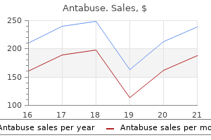
Antabuse dosages: 500 mg, 250 mg
Antabuse packs: 30 pills, 60 pills, 90 pills, 120 pills, 180 pills, 270 pills, 360 pills

500 mg antabuse cheap with amex
High-resolution black-blood contrast-enhanced T1 weighted photographs for the diagnosis and follow-up of intracranial arteritis treatment quotes buy 500 mg antabuse with amex. Diagnostic yield and security of brain biopsy for suspected primary central nervous system angiitis medicine 4212 antabuse 250 mg cheap online. The typical thunderclap headache of reversible cerebral vasoconstriction syndrome and its numerous triggers. Inflammatory cerebral amyloid angiopathy, amyloid-�related angiitis, and primary angiitis of the central nervous system similarities and differences. The scientific presentation is a combination of systemic manifestations (mostly spiking fever, evanescent rash, and lymphadenopathy) and joint symptoms ranging from arthralgia to aggressive arthritis. No specific diagnostic test is out there; the clinical analysis is predicated on sample recognition and exclusion of different ailments. The outcome is unfavorable in one third of patients with the event of a chronic arthritis. Therapeutic selections ought to be primarily based on the extent and severity of organ involvement. Nonsteroidal antiinflammatory medicine, glucocorticoids, methotrexate, and organic therapies are good choices. Many viruses, as nicely as some bacterial infections, have been reported to work together with the genetic background of the host,12 however there remains no proof of an infectious etiology. It occurs worldwide and affects girls slightly more often than males and younger people with 75% of the patients reporting disease onset between 16 and 35 years of age. The annual incidence estimated in retrospective studies of European international locations is 0. Not all three symptoms must be present at illness onset, and atypical displays might happen; subsequently, the pleiotropic presentation often leads to diagnostic and treatment delays. In general, a shared constellation of scientific options may be present in most cohort research reported over the past years from varied elements of the world (Table 173. It is proposed that environmental components can trigger deregulated immune pathways in a genetically predisposed host. These manifestations may evolve over a period of months into a more severe, harmful symmetric polyarthritis. Joints affected most frequently are the knees, wrists, and ankles, although involvement of the elbow, shoulder, proximal and distal interphalangeal joints, metacarpophalangeal joints, metatarsophalangeal joints, temporomandibular joints, and hip have been described as nicely (Table 173. The histopathology is nonspecific, displaying persistent synovitis with slight cell proliferation within the synovial lining layers, variable levels of vascular engorgement, and moderate infiltrates with mononuclear cells. Lymphadenopathy is detected in 33% to 73% of the patients with the cervical chain most commonly concerned. The exact relationship between lymphadenopathy and different reticuloendothelial system manifestations similar to hepatomegaly or splenomegaly is unclear; its mixed presence with constitutional signs may result in diagnostic confusion with lymphoma and even lymph node biopsy. Histopathology exhibits a benign reactive hyperplasia with plasma cells and polymorphonuclear cell infiltration. However, severe interstitial lung illness has been described, and some sufferers progress to acute respiratory distress syndrome. However, a low titer of both test happens in less than 10% of patients with out medical implications and may reflect their presence within the common population. Complement consumption is uncommon, with C3 ranges normally regular to slightly elevated in the acute part. The Fautrel criteria,42 that are more specific, require the not readily available measurement of glycosylated ferritin. The laboratory profile of the illness is a reflection of the systemic inflammation and cytokine cascade current. The erythrocyte sedimentation rate and C-reactive protein are nearly invariably elevated. These hematologic abnormalities could generally attain extreme ranges and mimic major hematologic disease. Bone marrow examinations have been reported to present hyperplasia of granulocytic precursors in all sufferers and hypercellularity and hemophagocytosis in some. Abnormal liver operate is thought to be a part of the inflammatory means of the disease itself and tends to normalize with disease remission, although concomitant use of probably hepatotoxic treatments often complicates interpretation. Nonsteroidal antiinflammatory medication should be the preliminary therapy in sufferers with mild illness. Most sufferers require treatment with glucocorticoids, particularly these with fever, joint symptoms, or internal organ involvement. Aspirin (3�4 g/day), ibuprofen (800 mg 4 instances daily), and naproxen (500 mg/twice daily) are choices for initial therapy. Patients with severe symptoms ought to be handled with glucocorticoids from the outset of therapy. No consensus has been reached on a therapeutic tapering scheme as quickly as scientific remission is achieved. Slow discount over a 6- to 12-month interval is recommended to maintain response and avoid relapse. At the time of the survey, 10 of the 15 patients treated with infliximab had discontinued the treatment after a imply remedy length of 9 months because of lack of efficacy (6 patients) or a aspect effect (3 patients). In the identical research, among the many 10 sufferers who were handled with etanercept, just one achieved a whole response and 7 of 10 a partial response. In some patients, a discount within the frequency of anakinra injections was attainable. Promising results had been reported with infliximab and etanercept, but treatment failures and loss of efficacy have also been described. Recent work showed a marked medical and laboratory enchancment in an open research of 34 patients. However, the looks of this complication during the diagnostic process or evolution of the disease is most typical. Clues to the analysis are fever, cytopenia, hypertriglyceridemia, and an increase in ferritin and lactate dehydrogenase. Treatment with biologic drugs is really helpful to halt the evolution to renal failure. Approximately one third of sufferers fall into every sample: Monophasic sample: Patients with the monophasic sample have a disease course that often lasts less than 1 year, with complete resolution of symptoms. Intermittent sample: Patients have two or more disease flares with complete remission between episodes. Subsequent flares tend to be much less extreme and of shorter period than the initial episode. In one series of 62 patients, 90% were American College of Rheumatology useful class I.
Diseases
- Erythroderma desquamativa of Leiner
- Cleft lip palate deafness sacral lipoma
- Tuberous Sclerosis
- Occupational asthma - wood
- Fetal akinesia syndrome X linked
- Ceroid lipofuscinois, neuronal 6, late infantile
- Faciocardiomelic dysplasia lethal
- Encephalomyelitis, myalgic
Generic antabuse 500 mg without prescription
Several bigger radicular arteries could also be discerned in the cervical and upper thoracic areas treatment resistant anxiety discount 500 mg antabuse with visa, however the largest treatment ketoacidosis 500 mg antabuse generic, the arteria radicularis magna (artery of Adamkiewicz76), is an asymmetrical contribution from one of many higher lumbar, or lower thoracic, segmental arteries. It travels obliquely upward with a ventral spinal root to be part of the anterior spinal artery in the region of the conus medullaris. Immediately distal to this point, it divides into dorsal and medial branches; the larger, dorsal branch ramiies within the larger muscle mass of the erector spinae, whereas the medial branch follows the external contours of the lamina and the spinous course of. An intermediate space that normally contains the lower two cervical and upper two thoracic vertebrae is supplied by costocervical branches of the subclavian artery that are of variable pattern and oten bilaterally dissimilar. From T2 to L3, the typical segmental association prevails, however in the sacral space lateral sacral branches of the hypogastric artery and middle sacral branches assume the operate of supporting the nutritional vasculature to the vertebral parts. Atlantoaxial Complex With their complex phyletic and developmental history, the components of the atlantoaxial articulation show probably the most atypical vascular sample of all of the vertebrae. Its ixed position relative to the rotation of the atlas and the adjacent sections of the vertebral arteries prevents formation of major vascularization by direct branches at its corresponding segmental stage. One would possibly assume that the vitamin of the dens would easily be completed by interosseous vessels derived from the spongiosa within the supporting body of the axis. It is axiomatic, nevertheless, that the vascular patterns of bones were developmentally established to supply the original ossiication centers inside the nonvascular cartilage matrices, and despite the eventual obliteration of the separating cartilage, the original patterns of vascularity generally prevail all through life. Occasionally, noncalciied remnants of this plate might persist in adults; although there could also be a secure union between the 2 components, a radiolucent space may suggest a fracture nonunion or a "false" os odontoideum. In gentle of the foregoing information, it was not sudden that the investigations of Schif and Parke78 revealed that the odontoid process was supplied primarily by pairs of anterior and posterior central branches that coursed upward from the surfaces of the body of the axis and had been derived from the vertebral arteries at the degree of the foramen of the third cervical nerve. A small descending branch anastomoses distally with vessels of the next decrease section. Dorsal to the alar ligament, it sends an anterior anastomotic department over the cranial edge of this ligament to form collateral connections with the anterior ascending artery. Fine medial branches ship perforators into the substance of the vertebral body and meet in a median anastomosis typical of the anterior central branches of the lower cervical region. Here every artery sends numerous ine perforators into the anterolateral surfaces of the neck of the odontoid process and terminates in a spray of vessels that provide the synovial capsule of the median atlantoaxial joint. Fine branches from the anterior and posterior ascending arteries additionally help within the nutrition of the syndesmotic relations of the atlantoaxial and craniovertebral articulations. Collateral vessels cross over and under the anterior arch of the atlas to anastomose with the apical arcade and ascending arteries. Its descending branches supply the periforaminal dura, the tectorial membrane and alar and apical ligaments, and the ine anastomoses to the arcade. Because the aorta terminates in a bifurcation ventral to the fourth lumbar vertebral physique, the vertebrae and the related tissues caudad to this point depend on an arterial advanced derived mostly from the interior iliac (hypogastric) arteries. With the growing use of percutaneous approaches to the decrease lumbar discs, this infra-aortic system of vessels has assumed some surgical signiicance, significantly because, in distinction to the standard segmental supply to the more superior vertebrae, its major components are longitudinally associated to the dorsolateral surfaces of the discs most frequently concerned in these procedures. It then continues to provide the decrease posterolateral stomach wall as it programs superior to the crest of the ilium. These patterns of the vessels have been derived from radiographs of perinatal specimens and dissections of adults and drawn towards a tracing of the lumbosacral region taken from a left anterior oblique radiograph of a person. The aorta lies to the left of middle because it approaches the bifurcation ventral to the fourth lumbar vertebra. This schema shows the extra frequent association of the sacroiliolumbar system on the right facet of the illustration, where the iliolumbar vessel (7) has a single origin from the dorsum of the posterior division of the (removed) inside iliac artery. The left aspect reveals the frequent variation where the iliac artery and the lumbar artery (14) are derived separately. The center sacral artery (16) is in its typical place, and the anastomotic contribution from the fourth lumbar artery (4) reveals its most frequent type. It normally has a medial branch that provides the exterior aspects of the facet joints and neural arch elements and the transversospinal group of muscles and a lateral department to the transversocostal group of the erector spinae. This specimen shows considerable variation between the 2 sides of the sacroiliolumbar system. The middle sacral artery can be absent, and different branches of the system supply its domain. As against the mostly visceral distribution of the anterior division of the interior iliac (hypogastric) artery, the posterior division is actually a somatic artery giving rise to gluteal, iliolumbar, and lateral sacral branches. It is directed dorsosuperiorly, passing near the ventrolateral floor of the irst sacral vertebral phase. It programs superiorly, dorsal to the obturator nerve and ventral to the lumbosacral trunk. Lateral to the inferior margin of the L5-S1 disc, the iliolumbar artery often divides right into a laterally directed iliac artery and an ascending lumbar artery. In most cases, a branch of this vessel continues rostrally to anastomose with the descending department of the fourth lumbar artery. Sacral Arteries Lateral Sacral Arteries Lateral sacral arteries usually type the second branch of the dorsal division of the internal iliac arteries and course down the pars lateralis on each side of the sacrum. Opposite the sacral foramina, they provide of medial branches that dorsally enter the foramina. Ater providing the everyday vertebromedullary derivatives, their dorsal muscular branches exit through the dorsal sacral foramina to supply the sacral origins of the erector spinae muscular tissues. In people, it is a variable vessel, being totally absent in some instances or changed by a branch of one of many lateral sacral arteries. It is a valveless venous complicated that receives the draining segmental tributaries of the interior veins through the intervertebral foramina and communicates in the end with the lumbar and intercostal tributaries of the caval and azygos system. The pure curvature of the sacrum offered oblique sections through segments 2, three, and 4. The dorsal branches cross into the anterior sacral foramina to present posterior, central, neural, and prelaminar branches. The dorsal branches go away through the posterior sacral foramina to provide the muscles and posterior laminar branches. Chapter 2 Applied Anatomy of the Spine 43 he internal venous plexus is of more useful and anatomic curiosity. When injected with a contrast medium, the primary channels might seem as a segmental chain of rhomboid beads. Chaynes and colleagues84 studied the interior venous plexus using silicon injection methods. In the cervical spine, the retrocorporeal vein was found deep to the posterior longitudinal ligament, whereas it was supericial to the ligament within the thoracic and lumbar regions. Where the primary anterior sinuses cross join, they obtain the large unpaired basivertebral sinus that arises inside the dorsal central concavity of the spongiosa and drains the intraosseous labyrinth of sinusoids. Regional visualization of the epidural plexus could be accomplished by introducing a radiopaque medium instantly into the spongiosa or the cancellous bone of the spinous course of (intraosseous venography). Breschet81 surmised that the epidural plexus served as a collateral route for the valveless caval and azygos techniques.
500 mg antabuse order visa
The attack is usually monoarticular in men and oligoarticular or polyarticular during later attacks in males and initial attacks in postmenopausal girls medications made from plasma buy antabuse 250 mg on line, lasting from days to weeks 5 medications cheap antabuse 250 mg visa. Attacks are initially separated by intervals of complete freedom from all signs (intercritical gout); however, because the disease progresses, the intervals between assaults could shorten, and the assaults might lengthen, leaving evidence of chronic arthritis with some patients having persistence of ache and inflammation during the intervals between assaults. Approximately 60% will expertise a second assault within 1 yr and 80% within three years. In addition, seasonal factors, similar to elevated attacks of gout in the spring, have been reported. The word tophus comes from the Greek word tophos, which means a porous volcanic stone or "chalk stone. Tophi of the helix or antihelix of the ear are basic however not a typical website. The fourth Earl of Orford, Horace (Horatio) Walpole, developed continual tophaceous gout and stated that "his fingers have been swelled and deformed, having more chalk-stones than joints in them. Tophi are prone to develop at avascular tissues, which may clarify reviews of tophus deposition within the cornea. Rarely, tophi have been reported to seem as an asymptomatic cutaneous rash consisting of pustule-like lesions containing tophaceous material. Characterization of, the conventional temporal sample of plasma corticosteroid ranges. Efficacy of colchicine prophylaxis: prevention of recurrent gouty arthritis over a imply interval of five years in 208 gouty topics. Tophi because the preliminary manifestation of gout: report of six cases and evaluation of the literature. Joint and tendon subclinical involvement suggestive of gouty arthritis in asymptomatic hyperuricemia: an ultrasound controlled examine. Management of gout and hyperuricemia Robert Terkeltaub 192 Key Points Therapeutic methods for gout and hyperuricemia have been subjected to systematic and formal consensus review processes and disseminated in current pointers. Management strategies contain distinct however linked arms, with consideration to safety and improved high quality of life: � Antiinflammatory remedy and prophylaxis of gouty arthritis. Treatment options are designed to either stop acute gout flares or deal with active inflammation of acute and persistent gouty arthritis. Treatment choices purpose to lower serum urate ranges, in addition to to obtain final decision of tophi and stop the disabling tissue consequences of urate crystal deposition. The lipophilic nature of colchicine facilitates cell uptake by permitting colchicine to bind tubulin, its major goal. Long-term objectives embrace limiting recurrences of acute gouty arthritis and inhibiting chronic gouty synovitis and its associated connective tissue destruction. Treatment of each the pain and irritation related to acute gout is achieved with antiinflammatory agents. Mechanism of action Colchicine binds tightly to unpolymerized tubulin and forms a tubulin� colchicine advanced that regulates microtubule and cytoskeleton operate. Colchicine elimination driven by hepatic metabolism and intestinal excretion follows a first-order course of, with enterohepatic circulation playing a considerable function. Colchicine myopathy, which affects proximal greater than distal muscle tissue and is accompanied by elevated creatine kinase in the early part and by various neuropathy, can mimic inflammatory muscle disease (see Chapter 160). Severe cases of colchicine intoxication are treated by supportive care and can be deadly. Monotherapy with a potent uricosuric is an alternative first-line method in young patients with normal kidney function and no tophaceous depositis,21 and probenecid is probably the most extensively out there drug with uricosuric motion. Targeting the uric acid underexcretion that drives hyperuricemia in most patients can robustly decrease body urate stores. The algorithm, discussed within the textual content, summarizes the first-, second-, and third-line approaches to pharmacologic urate-lowering remedy, together with management of refractory hyperuricemia in troublesome gout. Racial disparities in the danger of Steven Johnson syndrome and poisonous epidermal necrolysis among us adults with gout or urate-lowering drug use. Subjects had been treated with allopurinol, the potent uricosuric benzbromarone (a drug not out there within the United States), or a mix of the 2. Effect of urate-lowering therapy on the speed of measurement discount of tophi in chronic gout. A serum urate goal of lower than 6 mg/dL (<360 mmol/L) is the minimal acceptable target stage, with a lower target degree of 5 mg/dL (<300 mmol/L) being acceptable for persistent tophaceous gouty arthritis. Concordantly, current tips for decreasing whole body uric acid shops, debulking and resolving tophi, reducing the last word frequency of gout flares, and reducing the danger for ongoing precipitation of urate crystals support persevering with (lifelong) discount in serum urate to lower than 6 mg/dL. Because of main renal clearance of oxypurinol, its half-life rises substantially in these with renal impairment. Allopurinol and oxypurinol decrease serum urate not only by inhibiting xanthine oxidase but in addition by competing for phosphoribosylpyrophosphate in the salvage pathway and by the suppressive results of drug nucleotides on amidotransferase activity, the rate-limiting step in purine synthesis. Pruritus alone is a classic premonitory sign of rash and by itself is a useful indication for a beforehand knowledgeable affected person to instantly cease taking allopurinol. Allopurinol has main drug interactions with azathioprine, 6-mercaptopurine, and theophylline, whose metabolism is mediated by xanthine oxidase. Patients taking warfarin need careful remark of their anticoagulation standing. In addition, ampicillin and amoxicillin trigger a rash in a minimal of 20% of allopurinoltreated patients. Progressively decrease the maximum allopurinol dose with progressively worse continual kidney illness, but 300 mg/day could be exceeded with patient schooling and monitoring. Starting dose is a threat issue for allopurinol hypersensitivity syndrome: a proposed protected starting dose of allopurinol. However, long-term safety knowledge for allopurinol dosages higher than 300 mg/ day are sparse. Because adherence to allopurinol therapy is usually poor in clinical apply,32 pill counts or measurement of serum trough oxypurinol levels can be helpful to affirm suspected nonadherence. Furthermore, febuxostat, unlike allopurinol, is metabolized primarily by oxidation and glucuronidation in the liver, and renal elimination performs a minor position in febuxostat pharmacokinetics. In Europe and lots of different nations, febuxostat is accredited at dosages of as much as 120 mg once daily. Febuxostat has been studied in massive randomized scientific trials in which a maximum dose of 300 mg of allopurinol was used as a comparator. In prospective randomized controlled clinical trials, lesinurad add-on therapy to a xantine oxidase inhibitor increased the proportion of gout patients reachubg predetermined serum urate target. Xanthine oxidase is inhibited by allopurinol and its main, long-lived active metabolite oxypurinol (both pictured here). Oxypurinol has a half-life much longer than that of allopurinol (up to 24 hours in subjects with normal renal perform; longer with renal impairment). At 1 year, gout flare rates decline comparably in patients treated with allopurinol, 300 mg/day, and febuxostat, 80 to 120 mg/day. Dosing suggestions and unwanted facet effects Probenecid is started at 250 mg orally twice every day and titrated as much as one thousand mg twice daily in most patients and occasionally up to 3 g/day if tolerated. The risk for urolithiasis (including uric acid and oxalate calculi) with potent uricosuric monotherapy such as probenecid and benzbromarone could be about 10%. All patients ought to be in a position to increase oral hydration, particularly throughout early treatment.
Antabuse 250 mg overnight delivery
Neutral Zone Limits As discussed earlier treatment hyperkalemia buy antabuse 250 mg low cost, the neutral zone is important for understanding when tissues irst experience resistance to motion medicine 5 rights discount antabuse 250 mg with amex. Low intersegmental resistance to movement can be a sign of biomechanical problems. One examine discovered that ligaments required prolonged periods to regain structural integrity. Because the spinal ligaments oten are the construction that protects the spinal system, you will need to respect the failure limits of the varied spinal ligaments; these are proven in Table 6. Note that the load tolerance of these ligaments and the deformation traits of the ligaments vary markedly based on the area of the backbone and the speciic ligament concerned. Generally, the lower the level of the spinal ligament, the higher is the tolerance of the ligament. From a medical perspective, one should be sensitive to the truth that regular and irregular neutral zones may be very diferent for diferent vertebrae. Load Tolerance of the Spinal Motion Segments he precise tolerance traits of human spinal tissues- corresponding to muscular tissues, ligaments, tendons, and bones-loaded under numerous conditions have been diicult to establish. Structure tolerances have been noticed to range tremendously even under similar loading circumstances due to their dependence on many factors, corresponding to strain rate (rate of loading), age of the construction, frequency of loading, physiologic inluences, heredity, conditioning, and other unknown factors. In addition, it has been unimaginable to measure these tolerances underneath in vivo circumstances. Many of the estimates of tissue tolerance have been derived from various animal or theoretical constructs. At least one research means that residing tissue failure may occur at magnitudes beneath those noticed in cadaveric specimens. Tolerance of Speciic Spine Structures he common structure tolerance, or failure, limits in response to loading of the lumbar spine have been well investigated. Compression failure limits are a perform of age, with older endplates failing at lower ranges of pressure, and a perform of gender, with female tolerances decrease than male tolerances. When compression forces enhance on a spinal motion section, the irst indicators of injury often happen on the endplate or the trabeculae that assist the endplate. It is believed that this type of disc loading can end result in internal derangement of the disc and probably reverse bulging of the inner lamellae. As could be seen on this igure, when the relative load turns into higher, the possibilities of failure improve the danger signiicantly when the number of loading cycles will increase. Although nice variability is clear, girls usually have lower compression tolerance by an average of almost 2 kN in contrast with males. Torsion is irst resisted by collagen ibers within the anulus that merely stretch slightly. At the limit of the natural vary of motion, 30% to 70% of the applied torque is resisted by the zygapophyseal joint as a compressive load, 20% to 50% is resisted by the disc, and less than 15% is resisted by all of the intervertebral ligaments, collectively. However, for activities leading to more frequent shear loadings (100�1000 loadings/day), they recommended a shear limit of seven hundred N. Diferent buildings are answerable for resisting drive, and the tolerance of the backbone can change. During extension of the backbone, 60% to 70% of the applied load is resisted by the neural arch. Studies have reported harm ensuing from 3 to 8 degrees of extension underneath bending moments of 28 to forty five N-m. It is hypothesized that the zygapophyseal joint can be the structure damaged irst owing to extension. Rapid load rates, presumably ensuing from athletic endeavors, are also thought probably to improve risk. In isolation (without the ligaments), the disc can fail when lexed at 18 degrees with an application of 15 to 50 N-m of load. Resistance to lexion can increase by more than 10% when rapid motions (10 seconds) are compared with gradual motions (1 second). Some studies have reported that a lateral bending moment of 10 N-m results in four to 6 degrees of lateral bending in the lumbar spine, with most of the resistance occurring at the disc. However, the worth of those biomechanical outcomes will increase signiicantly solely when it can be instantly correlated to scientific outcomes. Of particular medical signiicance is the understanding of pain-modulated motion, which is an in vivo phenomenon. Several research have explored this complicated phenomenon in symptomatic people with low back problems and located signiicant modiications to their kinematics due to underlying pathology and ache when compared to asymptomatic people. To tackle these limitations, over the previous 15 years, a quantity of researchers have developed instruments to enhance our understanding of in vivo backbone biomechanics using advanced medical imaging, motion-capture methods, and eicient numerical methods to assist present clinically measurable biomechanical metrics. Abnormal coupling of movement has also been proven to be associated with low back pain. Signiicant diferences are obvious, nevertheless, when trunk velocity and acceleration are considered. More latest research have shown that kinematic capacity can be utilized to doc the extent of a low back disorder. Spine Kinematics (Intrinsic Measurements) Several studies have investigated noninvasive strategies to quantify regular in vivo spinal kinematics to assist within the scientific diagnosis of spinal impairments and instability. In addition, signiicant diferences could be seen between ranges between the two states. However, throughout lateral bending, the decrease vertebrae showed higher motion than the higher vertebrae. Adjacent-level degeneration is a standard prevalence clinically following a fusion surgical procedure; however, its etiology is unclear and controversial. Segmental lumbar rotation in sufferers with discogenic low again ache throughout useful weight-bearing actions. Due to these factors, there are only some documented research which have investigated in vivo spinal masses. One of these studies also compared intradiscal pressure (horizontal and vertical pressures based on orientation of pressure gauge) with respect to development of disc degeneration, and found a signiicant discount in pressure with grade of degeneration. Using this setup, they investigated the efect of locomotion on spinal loads and found that strolling triggered signiicantly larger loads than standing. Unfortunately, it is extremely diicult to get hold of biomechanical parameters, such as inner stress and strain distributions, particularly at decrease spatial scales (cellular). In silico fashions, extra commonly known as computational or biomechanical fashions, are seeing an elevated utilization in spine-related research for investigating complex mechanobiologic phenomena. Using superior numerical and imaging strategies, detailed anatomic and material representations of every hierarchical degree (macroscale to microscale) can be developed and used for biomechanical evaluations. Using in silico models, we will now start to discover these advanced mechanical relationships throughout spatial and temporal scales.
Berberis sanguinea (European Barberry). Antabuse.
- Are there any interactions with medications?
- Dosing considerations for European Barberry.
- How does European Barberry work?
- Kidney problems, bladder problems, heartburn, stomach cramps, constipation, diarrhea, liver problems, spleen problems, lung problems, heart and circulation problems, fever, gout, arthritis, and other conditions.
- Are there safety concerns?
Source: http://www.rxlist.com/script/main/art.asp?articlekey=96443
Antabuse 250 mg sale
Given the scope and complexities of backbone disorders medicine xifaxan 500 mg antabuse discount with mastercard, it can be helpful to break down some aspects of the scientific presentation into broad categories medicine 751 m antabuse 250 mg with mastercard. Useful categories to think about relate to the presence or absence of radiating pain and speciic demographic factors. A C4 C6 B Red Flags: What Not to Miss It is important to establish all circumstances that pose a considerable, imminent danger for further harm to the affected person. Many authors have identiied speciic pink lags within the history of patients with low again complaints that point out the presence of such a condition; these embody an infection, tumor, fracture, cauda equina injury, and progressive neurologic damage, corresponding to motor loss or myelopathy (Table 12. International Standards for Neurological Classiication of Spinal Cord Injury [reprint]. A study of 669 patients larger than 55 years of age presenting to their major physician identiied 4 sufferers (1%) with malignancy and 33 (5%) with a fracture, 30 of which were related to osteoporosis. All of those elements are necessary in establishing an applicable diferential prognosis and identifying some of the potential obstacles to recovery. Axial pain within the cervical, thoracic, or lumbar region suggests a diferent etiology, evaluation, prognosis, and doubtlessly treatment than radicular pain. For all ranges of the spine, pathology involving the musculotendinous and ligamentous buildings, zygapophyseal joints, vertebrae, and anulus of the intervertebral discs tends to trigger axial ache. Other buildings in the cervical and thoracic regions that can lead to axial pain embody sot tissue buildings within the neck; vascular structures. Radicular ache radiating into the higher extremities generally has a diferent etiology. If associated to backbone pathology, radicular ache implies neural compression from many potential causes, including disc herniation, spinal canal or neuroforaminal stenosis, or intrinsic disease of the spinal wire or nerve roots. Radicular ache within the thoracic region can lead to a bandlike distribution on one or each side of the chest wall or belly region. Additional constructions that may find yourself in radiating higher extremity pain include peripheral nerves, such because the median nerve. For the lumbar backbone, the hip and pelvic constructions have to be considered as potential sources of low back, buttocks, or posterolateral hip pain. Particular sources of low again or buttock ache associated to the bony pelvis include the sacroiliac joints, the sacrum. Other buildings and processes that can lead to low back pain embrace the kidneys and ureters; the pancreas; gastric ulcers; vascular abnormalities. Signiicant neurologic injuries embrace cauda equina syndrome, progressive radiculopathy, or myelopathy. Cauda equina syndrome ought to be thought-about in a patient with saddle anesthesia; bowel, bladder, or sexual dysfunction; or signiicant lower extremity pain and weakness, significantly if bilateral. Myelopathy can current in various ways, together with hand paresthesias or decreased ine motor management; lower extremity weakness or gait instability; sensory alterations in the trunk or extremities; or modifications in bowel, bladder, or sexual perform. Whatever the trigger, the presence of leg ache with low back pain seems to increase the general severity of the clinical state. Compared with those that have axial lumbar pain alone, patients with associated radicular pain experience a decrease high quality of life, require more resources, and have larger levels of ache and disability. Patient Demographics Demographic characteristics-such as age, gender, instructional background, occupation, and cultural milieu-are important components that should be thought of in the historical past of a affected person with a spine drawback. Growth and development have a profound impression on the method to various processes, similar to spondylolisthesis, scoliosis, and Scheuermann kyphosis. In contrast to the grownup backbone, the creating bony backbone is comparatively extra susceptible to injury than some sot tissue constructions. In a examine by Micheli and Wood,30 47% of adolescents presenting to a pediatric sports medicine clinic have been diagnosed with spondylolysis and solely 11% had disc abnormalities compared with 48% of adults presenting to a low again pain clinic who have been thought to have disc pathology. As talked about previously, some medical situations, including ankylosing spondylitis, spondylitis associated with inlammatory bowel illness, and tumors such as osteoid osteoma and osteoblastoma, are inclined to manifest in younger adults (20s and 30s). Osteoporosis is extra common in ladies than in men, and osteoporotic fractures are more frequent in ladies. Demographic factors-such as race, ethnicity, and cultural milieu-may also play a task within the prevalence of some spine issues, but are less nicely studied. Whites tend to have higher charges of osteoporosis than some other races, and metabolic conditions similar to Gaucher disease can be related to certain ethnic teams. Past Medical History In addition to identifying prior surgical procedures, it is important to identify all previous and present medical situations as a outcome of many medical issues may be related to backbone issues and might afect care of a patient with a backbone dysfunction. As noted beforehand, a history of cancer, current infection, or disease processes that afect the immune system or may require immunosuppressive drugs can be related to signiicant spine problems. Other medical conditions-such as osteoporosis, ankylosing spondylitis, and difuse idiopathic skeletal hyperostosis-may place sufferers at increased threat for spine fracture. Other disorders- together with cardiac or pulmonary illness, renal problems, skin circumstances, gastric ulcers, diabetes, and hepatic disorder- could have an impact on potential remedy options and may preclude certain therapies. An additional facet that have to be considered in a affected person with a spine disorder is a history of prior injury. Previous spine problems, trauma, and surgical procedure could have necessary implications for the care of the patient. Short-term and long-term issues doubtlessly can develop ater surgical procedure, and it is necessary to perceive the character of any prior surgical procedure. Such opposed occasions embody adjacent-segment degeneration or instability ater a fusion, epidural ibrosis, infection, hardware-related problems, corresponding to loosening, and recurrent disc herniation. A historical past of multiple or prolonged periods of pain or disability ater prior treatments ought to increase issues concerning the probabilities for fulfillment with future therapies. Family History he family history is a essential part of a complete medical history. Although again pain and many different spinal conditions are frequent within the basic population, information counsel possible genetic risk components for lumbar degenerative disc disease. By asking about family members and oldsters, one can start to perceive the nature of household dynamics that could be inluencing the presentation of a affected person with spinal pain. A historical past of abuse, the presence of a disruptive house environment, and a historical past of poor parenting or alcoholism within the family may have a signiicant future impact on the psychological makeup of a person. Anger, unmet dependency needs, and problems with belief in authority igures are a few of the points that could lead to chronic ache points. Probing these points in taking a household history might provide useful perception into potential barriers to recovery. Chapter 12 Patient History and Physical Examination: Cervical, Thoracic, and Lumbar 191 and a low academic degree (less than a secondary education). In a evaluate on this subject, Linton46 noted that psychological variables are clearly linked to the transition from acute to continual ache and generally have a stronger influence on chronicity than medical or biomechanical factors. Pertinent emotional components cited embody melancholy, anxiousness, distress, and selfperceived poor well being. Cognitive and behavioral factors also apparently play a key function in the development of a continual pain state; these embody a passive coping style, catastrophizing, and fear-avoidance beliefs (beliefs that certain activities ought to be prevented owing to fear of injury). A historical past of sexual or physical abuse also may be related to persistent pain and incapacity. It is particularly true in treating the affected person with persistent pain that the foundation for good decision making is having an excellent information of the person with a back dysfunction; the spine itself is less necessary. Patients in whom incapacity significantly exceeds that expected on the basis of goal indings have been proven to be much extra likely to have encountered childhood abuse and conlict, parental job stress, or a diicult divorce.
Purchase antabuse 250 mg on line
Electrons move sequentially from one protein complex to the following (in the order of I�V) down an power gradient medicine queen mary purchase 250 mg antabuse overnight delivery. In the adult type treatment shingles order 500 mg antabuse overnight delivery, it might present between the second and sixth decade of life with progressive, proximal weak spot that may mimic polymyositis. Enzyme substitute with recombinant human -glucosidase in the infantile kind has had some success. In adults, advantages have been observed with a high-protein and low-carbohydrate food plan. The metabolism of short-, medium-, and long-chain fatty acids is barely totally different, but these biochemical particulars have great clinical relevance. Long-chain fatty acids are the most important supply of power for prolonged, low-intensity exercise lasting more than forty to 50 minutes. Impaired response Normal Severely impaired response Severely impaired response Reduced response Although these problems have many overlapping features, three predominant phenotypes may be recognized: hepatic, cardiac, and muscular. The muscle phenotype is characterized by hypotonia and recurrent episodes of rhabdomyolysis. Muscle ache and myoglobinuria are the usual presenting indicators and are characteristically induced by an infection; common anesthesia; or a lowcarbohydrate, high-fat diet. Symptomatic episodes often develop after prolonged exertion (usually more than 30 minutes), and patients tolerate brief, intense isometric train with out difficulties. Similar crises could also be induced by stress, fasting, viral infections, fever or exposure to chilly, basic anesthesia, use of nonsteroidal antiinflammatory drugs, and excessive doses of diazepam. A translocase then transfers the acylcarnitine product throughout the inner mitochondrial membrane. Short- and medium-chain fatty acids have a much less convoluted metabolic destiny because they cross the mitochondrial membranes with out difficulty and enter the -oxidation pathway directly after activation to CoA thioesters. With every cycle of -oxidation, a two-carbon fragment is cleaved, and one acetyl-CoA molecule is generated. During intervals of carbohydrate deprivation or fasting, ketone manufacturing is upregulated, and they turn out to be an essential source of vitality for many tissues, together with the mind. First symptoms normally happen in the neonatal period with hypoglycemia and irritability. Affected sufferers carry a high danger for recurrent rhabdomyolysis with exercise or different forms of stress, together with sickness. An abnormal acylcarnitine profile is dominated by 5-cis-tetradecenoylcarnitine (C14:1). The forearm ischemic test is highly delicate and particular for analysis of myoadenylate deficiency. Myoadenylate deaminase deficiency subsequently may be confirmed in muscle biopsy by histochemical stains or direct enzymatic assay. Discontinuation of the offending agent results in resolution of symptoms in almost all cases. The classification of drug- and toxin-induced myopathies in accordance with the kind of harm could be present in Table 160. Trifunctional protein deficiency is more extreme, with a high risk for early death. Clinical options embody hypoglycemia, hypotonia, dilated cardiomyopathy within the setting of hydroxydicarboxylic aciduria, and a standard blood acylcarnitine profile. According to the degree of muscle toxicity they induce, the most myotoxic statin was cerivastatin, which was withdrawn from the market, adopted by simvastatin, lovastatin, pravastatin, atorvastatin, and fluvastatin. Myalgias might occur in up to 25% of patients and often resolve after switching to a special statin or discontinuing therapy. A smaller quantity are associated to nuclear genes that encode proteins concerned within the oxidative phosphorylation pathway, they usually current in early childhood. Secondary mitochondrial myopathies are attributable to drugs, together with zidovudine and clofibrate. Exercise intolerance marked by untimely fatigue, headaches, nausea, and vomiting after submaximal exercise may be seen. Histopathology reveals attribute ragged purple fibers on Gomori trichrome stain (subsarcolemmal and intermyofibrillar accumulation of mitochondria that seem as bright red lots in opposition to the background of blue myofibrils). Patients who resume alcohol consumption are particularly susceptible to subsequent attacks of this form of myopathy. Nevertheless, it is a diagnosis of exclusion, and a meticulous workup for autoimmune, metabolic, paraneoplastic, or drug-induced myopathies must be pursued. It might occur any time throughout drug remedy, and the danger could additionally be elevated by a selection of components, including age, drug interactions, and renal dysfunction. The most important drug interactions that have to be averted embrace the use of statins with amiodarone, cyclosporine, gemfibrozil, macrolides, and azoles. Hoffmann syndrome is outlined as the combination of hypothyroidism with muscle stiffness, cramps, and muscular enlargement. In hyperthyroidism, proximal muscular weak point with atrophy, brisk reflexes, and bulbar weakness may occur. Patients current with a quickly progressing paralysis, greater within the lower extremities, usually worse in the proximal musculature. The myotoxicity of colchicine is assumed to be associated to disruption of the microtubular cytoskeletal community, leading to intracellular accumulation of autophagic vacuoles. Muscle biopsy exhibits a vacuolar myopathy with out outstanding necrosis or inflammation. The danger of colchicine myopathy is elevated by age, length of remedy, drug interactions (cyclosporine), and inappropriate dosing in renal insufficiency. The muscle biopsy of a affected person with chloroquine or hydroxychloroquine myopathy demonstrates a vacuolar myopathy, often solely evident on electron microscopy. Treatment contains optimizing diabetic control and the use of corticosteroids and intravenous immunoglobulins. Skeletal muscle infarction is a very uncommon complication of poorly controlled diabetes mellitus. Patients present with acute onset of painful swelling of the affected muscle and, sometimes, a palpable mass. This entity is frequently misdiagnosed clinically and increased scientific consciousness is important for early recognition, significantly in a patient with diabetes presenting with a painful thigh or leg swelling. The mechanism answerable for the steroid results has not been utterly elucidated. The majority of patients treated with corticosteroids may develop some extent of muscle weak spot, normally after a period of a minimal of 4 weeks. The weak point is more extreme and should develop more quickly in those handled with fluorinated corticosteroids, similar to dexamethasone, triamcinolone, and betamethasone, than in these handled with the nonfluorinated corticosteroids, such as prednisone and methylprednisolone. Selective atrophy of type 2 muscle fibers and the absence of inflammation or muscle necrosis are attribute histologic options. Affected kids are regular at delivery, only to develop a waddling gait, Gower sign (use of the arms to push oneself erect by transferring the arms up the thighs), and calf pseudohypertrophy by age 2 to 6 years.
250 mg antabuse generic
Palpation of the big Transthoracic Approach With the patient in the let lateral decubitus position medications enlarged prostate cheap 250 mg antabuse overnight delivery, the best chest is prepared and draped 911 treatment antabuse 500 mg generic with mastercard. A right-sided strategy is most well-liked due to the location of the good vessels and heart within the let-sided approach. A normal thoracotomy centered on the third rib provides access to the higher thoracic vertebra, but exposure to the low cervical area is restricted. While defending the intercostal neurovascular bundle, the suitable rib is subperiosteally dissected out and resected anteriorly and posteriorly as far as potential. Complications Postoperative weakness secondary to weak spot of the shoulder girdle musculature from the joint resection can happen. If damaged, the thoracic duct must be doubly ligated proximally and distally to stop chylothorax. Great warning ought to be taken to avoid injuries to the sympathetic nerves, the cupola of the pleura on the degree of T1, the nice vessels, and the thoracic duct, which passes into the let venous angle between the subclavian artery and the frequent carotid artery. Potential problems of this strategy embrace restriction of scapular motion and paralysis of intercostal muscle tissue owing to the muscle-splitting features of this dissection. We recommend use of this method in older patients and perhaps in sufferers with malignant situations. A large broad elevator is used to dissect the posterior paracervical muscular tissues from the arches of C1 and C2, and caution should be taken to avoid plunging devices into the spinal canal. A small curet may be useful to take away the muscular attachments on the biid spinous process of C2 while stabilizing the arch of C2. Slight head lexion can also assist by opening the house between the ring of C1 and the occiput. An additional technique to expose the lateral side of C1 or C2 is to elevate the periosteum with a small Freer elevator. Brief consideration is given here to the regional anatomy for the C1�C2 transarticular screw ixation (Magerl) approach,67�69 C1 lateral mass and C2 pedicle screw (Harms) method,sixty nine,70 and C2 translaminar screw. Attention must be paid to the presence of a ponticulus posticus, an anomalous ossiication overlying the vertebral artery because it runs within the superior sulcus of C1, which may happen in 15% of the population. Regardless of the technique used, the intraoperative use of anteroposterior and lateral luoroscopy can inform screw inclination in the coronal and parasagittal plane. Because of the quantity of cephalad angulation required to place the C1�C2 transarticular screw, subperiosteal exposure should prolong all the way down to C4. A Kirschner wire (K-wire) can be used to retract the sot tissues containing the higher occipital nerve and accompanying the venous plexus. In the case of placement of a C1 lateral mass screw, the C1�C2 joint is the important thing anatomic landmark to be identiied. However, the trajectory of the C2 pedicle screw is more medial and follows the path of the pedicle, as would be anticipated. Use of this screw is feasible due to the predictably large dimension of the C2 lamina mixed with the truth that the use of this screw eliminates the chance for vertebral artery damage. With a wide, lat periosteal elevator corresponding to a Cobb, the dissection is carried subperiosteally down the spinous processes. Care must be taken to keep subperiosteal because the biid nature of the spinous processes may end in a bulbous expanse, and the dissection may err into the paraspinal musculature. The reverse Trendelenburg position minimizes venous bleeding and reduces cerebrospinal luid strain. In general, subperiosteal dissection ought to be carried out in a caudal-to-cephalad path to minimize bleeding. Subperiosteal dissection of muscle tissue is performed to expose the spinous processes, lamina, lateral mass, and facet joints. Dissection should lengthen laterally to the medial third of the side joint, with preservation of the capsule until a fusion is planned. Care should be taken at the lateral fringe of the joint as a result of the nerve root and vertebral artery lie anterior to the spinolamellar membrane of the adjoining transverse processes. Vigorous decortication or stripping could harm the skinny bone and subsequently the nerve root and vertebral artery. For fusion circumstances, one ought to expose solely the degrees to be fused as a outcome of creeping fusion extension is common. Supplementation of the fusion with posterior lateral mass plating may obviate the need for a halo vest postoperatively. First popularized by Roy-Camille and colleagues,73 placement of posterior cervical screws requires an intensive understanding of the lateral mass anatomy to decrease damage to associated neurovascular buildings. An and colleagues5 discovered that, by orienting the screw 15 levels cephalad and 30 levels laterally with an entry level 1 mm medial to the anatomic center of the lateral mass, the side joint and nerve root are prevented. The entry level is at or near the anatomic middle of the lateral mass and directed 10 degrees laterally. Posterior Approach to Cervicothoracic Junction Lesions of the cervicothoracic junction are generally anterior, for which intensive anterior approaches with or with out posterior ixation are usually required. Lesions that may require posterior stabilization embrace lesions ensuing from tumors, trauma, postlaminectomy instability, or an infection. If the posterior elements are intact, the straightforward triple-wiring process could be accomplished for a short fusion, or rods may be used for an extended fusion, using a normal posterior strategy. Pedicle screw ixation is another technique if the posterior components are deicient. A commonplace posterior method is used with the dissection carried out to expose the lateral mass and to the tips of the transverse processes of the upper thoracic vertebrae. Medial angulation has been noticed to differ between individuals, thus it should be measured preoperatively in preparation for surgical procedure. The surgical strategy selected should keep in mind the location of the pathologic process, the health of the patient, and the skill and luxury stage of the surgeon with every specific publicity. Understanding the advantages and limitation of each surgical publicity improves affected person outcome and reduces complications. Anatomic and surgical issues on the occipitocervical and cervicothoracic junction are notably challenging and ought to be totally understood before approaching pathologic processes in these areas. Complications of the cervical spine are infrequent however probably devastating; cautious preoperative planning, exact surgical method, and a high index of suspicion ought to be maintained to decrease and determine problems. Bleeding may be minimized by staying subperiosteal and inside the midline to stop getting into into the paraspinous musculature. One ought to minimize dissecting on the inferior fringe of the foramen magnum to stop uncontrollable venous bleeding. Care is required during passage of sublaminar wires or software of the screws to forestall damage to the brain or spinal twine. Dissection on the ring of the atlas have to be carried out in a mild manner because the direct strain might lead to fracture or slippage of an instrument into the spinal canal.
Antabuse 250 mg cheap with amex
In explicit medicine x stanford cheap 250 mg antabuse with visa, sufferers with unilateral visible loss should be handled immediately to forestall lack of vision within the other eye symptoms during pregnancy antabuse 250 mg purchase otc. Intravenous pulses of glucocorticoids (methylprednisolone one thousand mg every day for three days) can be tried in patients with current visual loss, although no documentation suggests that it helps more than oral prednisone. Alternate-day regimens are associated with a a lot larger danger of flares than daily regimens,79 while insufficient proof exists to assist the initial use of lower doses. Two controlled trials primarily based on preliminary intravenous glucocorticoid pulses confirmed contrasting results. Reassuringly, permanent visual loss very hardly ever occurs as disease flare after starting glucocorticoid treatment. The necessary length of glucocorticosteroid therapy is variable, but typically, it can be discontinued within 1 to 2 years. Some patients have a chronic relapsing course and may want low doses of glucocorticoids for several years. The absence of enchancment after 30 mg/day of prednisone for 1 week ought to alert the doctor to question the diagnosis. The addition of methotrexate (at least 10 mg/wk) to glucocorticoids may be useful in sufferers with a chronic relapsing course and those at high danger of glucocorticoid-related adverse occasions. The incidence of large cell arteritis in Olmsted County, Minnesota: obvious fluctuations in a cyclic pattern. Giant cell arteritis: epidemiological clues to its pathogenesis and an replace on its therapy. The incidence of giant cell arteritis in Jerusalem over a 25-year period: annual and seasonal fluctuations. Epidemiologic, and immunogenetic features of polymyalgia rheumatica and big cell arteritis in Northern Italy. The spectrum of polymyalgia rheumatica in northwestern Spain: incidence and analysis of variables related to relapse in a ten 12 months examine. Histopathologic, Findings of Patients With Biopsy-Negative Giant Cell Arteritis Compared to Those Without Arteritis: A Population-Based Study. Comparison, between color duplex sonography findings and completely different histological patterns of temporal artery. Fast monitor pathway, reduces sight loss in giant cell arteritis: results of a longitudinal observational cohort examine. Tissue cytokine patterns in sufferers with polymyalgia rheumatica and large cell arteritis. Giant cell arteritis and polymyalgia rheumatica: two totally different but usually overlapping conditions. Repetitive 18-fluorodeoxyglucose positron emission tomography in isolated polymyalgia rheumatica: a prospective examine in 35 patients. Incidence of temporal arteritis in patients with polymyalgia rheumatica: a prospective examine using colour Doppler ultrasonography of the temporal arteries. Influence of earlier corticosteroid therapy on temporal artery biopsy yield in large cell arteritis. Is color, duplex sonography-guided temporal artery biopsy helpful within the diagnosis of giant cell arteritis Efficacy of unilateral versus bilateral temporal artery biopsies for the analysis of big cell arteritis. Leukocyte infiltration in synovial tissue from the shoulder of patients with polymyalgia rheumatica. No detection of, varicella-zoster virus in temporal arteries of patients with big cell arteritis. Primary systemic amyloidosis presenting as big cell arteritis and polymyalgia rheumatica. Risk factors for visible loss in an Italian population-based cohort of sufferers with giant cell arteritis. Strokes at time of disease diagnosis in a sequence of 287 patients with biopsy-proven giant cell arteritis. Risk components for extreme cranial ischaemic occasions in an Italian population-based cohort of patients with giant cell arteritis. Giant cell arteritis (temporal arteritis) presenting as fever of undetermined origin. Musculoskeletal manifestations in a population-based cohort of patients with large cell arteritis. Distal, musculoskeletal manifestations in polymyalgia rheumatica: a potential followup examine. Giant cell arteritis with low erythrocyte sedimentation price: frequency of occurrence in a population-based research. Utility of erythrocyte sedimentation price and C-reactive protein for the prognosis of big cell arteritis. Association of ferritin autoantibodies with big cell arteritis/polymyalgia rheumatica. Imaging of polymyalgia rheumatica: indications on its pathogenesis, diagnosis and prognosis. Inflamed, shoulder constructions in polymyalgia rheumatica with normal erythrocyte sedimentation fee. Ultrasound assessment of recent onset bilateral painful shoulder in sufferers with polymyalgia rheumatica and rheumatoid arthritis. The diagnostic value of ultrasonography-derived edema of the temporal artery wall in large cell arteritis: a second meta-analysis. Large-vessel involvement in recent-onset large cell arteritis: a case-control colour-Doppler sonography study. Effect of glucocorticoid remedy on computed tomography angiography detected large-vessel irritation in giant-cell arteritis. Diagnostic performance of (18)F-fluorodeoxyglucose positron emission tomography in big cell arteritis: a systematic evaluate and meta-analysis. Daily and alternate-day corticosteroid regimens in remedy of large cell arteritis: comparison in a potential examine. Treatment of giant cell arteritis utilizing induction remedy with high-dose glucocorticoids:a double-blind, placebo-controlled, randomized prospective medical trial. Flares in biopsy-proven large cell arteritis in Northern Italy: characteristics and predictors in a long-term follow-up examine. Predictors of relapse and remedy outcomes in biopsy-proven giant cell arteritis: a retrospective cohort research. Azathioprine in giant cell arteritis/polymyalgia rheumatica: a double-blind examine. Efficacy and tolerability of leflunomide in difficult-to-treat polymyalgia rheumatica and big cell arteritis: a case collection. Infliximab for maintenance of glucocorticosteroid-induced remission of big cell arteritis: a randomized trial.
Generic 500 mg antabuse free shipping
Weight loss 4 kg: lack of four kg of physique weight since sickness started treatment integrity checklist generic 250 mg antabuse fast delivery, not caused by dieting or other components 2 treatment joint pain order antabuse 500 mg online. Livedo reticularis: mottled reticular pattern over the skin or portions of the extremities or torso 3. Testicular pain or tenderness: pain or tenderness of the testicles, not brought on by an infection, trauma, or different causes four. Myalgias, weakness, or leg tenderness: diffuse myalgias (excluding the shoulder and hip girdle) or weakness of muscle tissue or tenderness of the leg muscular tissues 5. Mononeuropathy or polyneuropathy: growth of mononeuropathy, multiple mononeuropathies, or polyneuropathy 6. Hepatitis B virus: presence of hepatitis B surface antigen or antibody in serum 9. Arteriographic abnormality: arteriogram showing aneurysms or occlusions of the visceral arteries, not attributable to arteriosclerosis, fibromuscular dysplasia, or other noninflammatory causes 10. Childhood polyarteritis nodosa Final European League Against Rheumatism/Pediatric Rheumatology International Trials Organisation/Paediatric Rheumatology European Society Childhood Polyarteritis Nodosa Criteria (with Glossary) and Classification Definition Criterion Histopathology Angiographic abnormalities Glossary A systemic inflammatory illness characterized by: Evidence of necrotizing vasculitis in medium- or small-sized arteries Angiography exhibiting aneurysm, stenoses, or occlusion of a medium- or small-sized artery, not caused by fibromuscular dysplasia or other noninflammatory causes Conventional angiography is the popular imaging modality. Questionnaire item: at least two of the following: Do you keep in mind one or more episodes of small pink spots in your skin, notably involving the lower limbs Have you ever had purple spots on your lower extremities, which depart a brownish colour after their disappearance Clinical merchandise: a minimal of three of the next four (present or past) Constitutional Fatigue signs Low-grade fever (37��37. Laboratory merchandise: no less than two of the following three (present) Reduced serum C4 Positive serum rheumatoid issue Positive serum monoclonal part *Satisfied if at least two of three objects (questionnaire, scientific, laboratory) are optimistic. The affected person must be positive for serum cryoglobulins in no less than two determinations at a 12-week interval. The fulfillment of the laboratory item in a patient satisfying the standards highlights the potential presence of cryoglobulinemic vasculitis even within the absence of serum cryoglobulins by preliminary testing. Palpable purpura *For purposes of classification, a patient shall be stated to have Henoch-Sch�nlein purpura if a minimum of two of those 4 criteria are current. The American College of Rheumatology 1990 standards for the classification of Henoch-Sch�nlein purpura. Eosinophils in biopsy *For purposes of classification, a affected person shall be stated to have hypersensitivity vasculitis if a minimum of three of these standards are present. The American College of Rheumatology 1990 standards for the classification of hypersensitivity vasculitis. Erythema and cracking of lips, strawberry tongue, or erythema of the oral and pharyngeal mucosa 2. Erythema and edema of the hands and feet in the acute section or periungual desquamation within the subacute part 5. Laboratory exams usually reveal regular or elevated white blood cell count with neutrophil predominance and elevated acute part reactants corresponding to C-reactive protein and erythrocyte sedimentation fee through the acute section. Low serum sodium and albumin ranges, elevated serum liver enzymes, and sterile pyuria can be current. Diagnosis, Treatment, and Long-Term Management of Kawasaki Disease: A Scientific Statement for Health Professionals from the American Heart Association. Age at illness onset 50 years: growth of signs or findings beginning at age 50 or older 2. Temporal artery abnormality: temporal artery tenderness to palpation or decreased pulsation, unrelated to arteriosclerosis of cervical arteries 4. Abnormal artery biopsy: biopsy specimen with artery displaying vasculitis characterized by a predominance of mononuclear cell infiltration or granulomatous inflammation, usually with multinucleated big cells *For functions of classification, a patient shall be mentioned to have big cell (temporal) arteritis if at least three of these five standards are present. The American College of Rheumatology 1990 standards for the classification of big cell arteritis. The American College of Rheumatology 1990 criteria for the classification of Takayasu arteritis. Differentiation of the pluripotent tissues of the embryo leads to early formation of a repetitive segmented vertebral construction. Because the embryo is exquisitely vulnerable to malformation and developmental error, every step of formation is crucial. In precept, metamerism is the event of a extremely specialized organism, with multifunctional organ methods, from many anatomically related segments organized in a linear trend. Genetic signaling, speciic to the species, determines the degree of regional specialization, such as limbs in mammals versus ins in ish or the dearth thereof in snakes. Using these comparative examples, one can also understand the concepts of isomerism and anisomerism. Isomerism is characteristic of extra primitive animals, during which the number of somites is bigger but more uniform and never so extremely specialized. In contrast, anisomerism is current in additional developed species, corresponding to mammals, in which most of the somites have been deleted (resulting in a lesser number of vertebrae), whereas the remaining somites are extra highly specialized in order that complicated, specialized appendages can be developed. Although the mature vertebral column is composed of quite a few comparable models, the tissues within each of these units are highly specialised. Early Embryologic Spine Precursors: Day 17 to Week 4 he development of the human backbone begins on the seventeenth day of gestation. On the dorsal layer (which is involved with the amnion) of the disc, there are epiblastic cells that converge and invaginate into the disc to type the primitive pit or node. When embedded within the tissue, it varieties a tubelike construction that extends craniad, "burrowing" deep to the embryonic disc alongside its ventral floor. At this point, the ventral wall of the notochordal tube is in touch with the yolk sac, which causes disintegration of these cells. A lat remnant of dorsal wall cells from the notochordal tube varieties the notochordal plate on the nineteenth day. Mesodermal tissues on either side of those constructions condense to type longitudinal columns. By the nineteenth day, there are three distinct columns on both side of the midline: (1) medial paraxial columns, which give rise to the somites; (2) intermediate mesodermal columns, which kind the urogenital organs; and (3) lateral mesodermal plates, which type the intestine cavities. In considering the development of the spine, consideration is concentrated on the medial paraxial columns. A second region surrounds the neural tube; that is destined to become the posterior arch of the vertebra. In metameric trend, the sclerotomes are organized into a consecutively stacked association. Lateral to this, the somatic mesoderm (sm), endoderm (end), and ectoderm (ect) are proven. Arrows indicate the direction of somite cell migration to form the vertebral process (vp), costal course of (cp), and centrum (cent). Arrow signifies the intracentral vestige of the notochord, referred to as the mucoid streak. Most of the densely packed cells fuse with the loosely packed cells of the adjacent caudal sclerotome. Initially, the segmental nerve precursors are situated on the midportion of each sclerotome, whereas the segmental artery lies at the junction between two adjoining levels.