

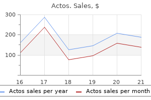
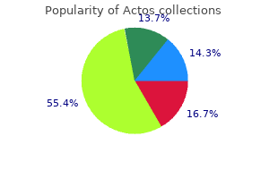
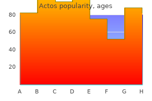
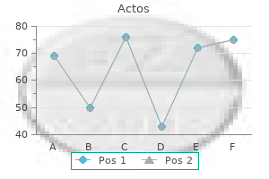
Actos dosages: 45 mg, 30 mg, 15 mg
Actos packs: 30 pills, 60 pills, 90 pills, 120 pills, 180 pills, 240 pills, 360 pills, 270 pills

Cheap actos 45 mg otc
Sometimes these infants may be suspected clinically to be totally different from these with other urea cycle issues due to the magnitude of the hepatomegaly blood glucose 10 30 mg actos order amex. Mutations have been outlined [13�15] diabetic ketoacidosis icd 9 45 mg actos discount mastercard, some of which have led to various splicing. Prior to the event of modern methods of pharmacologic remedy, the tip result of this presentation was uniformly deadly [16]. The picture is indistinguishable from that of the male infant with ornithine transcarbamylase deficiency (Chapter 26). Following a quick hiatus, by which the new child appears regular, anorexia or vomiting and lethargy develop, and these signs are rapidly progressive to deep coma, apnea, and death, except the child is intubated and maintained via mechanical air flow. The toddler might have hypertonia or hypotonia or there may be decerebrate posturing. Patients could have tachypnea and respiratory alkalosis, that are common consequences of hyperammonemia [18]. Clinical abnormalities 235 cerebral hemorrhages have been seen in hyperammonemic infants, as have fatal pulmonary hemorrhages. Such sufferers may present simply with impaired mental improvement [3, 20] or a convulsive disorder. The classic, and most common phenotype is the neonatal form, with speedy progression to coma in the first days of life. In a subacute or late-onset sort, the disease turns into manifest in late infancy or childhood [7, 20]. One affected person reported at 30 years [19], offered at ten years of age with intention tremor. He was described as having mildly impaired mental improvement, but had attended regular faculty, might learn, write, drive a automobile, work in a manufacturing facility, and father a son. Others might have episodes of hyperammonemic encephalopathy and coma thought to be Reye syndrome or encephalitis. Hyperammonemia is usually precipitated by infection and such an episode could additionally be fatal. Serum values of the transaminases are elevated no less than at times of hyperammonemia [21, 23, 24]. Progressive hepatic fibrosis has been identified and there may be ultrastructural abnormalities in hepatocytes. Citrulline and arginine are the precursor of nitric oxide, which is relevant to smooth muscle arteriolar function. Argininosuccinic aciduria has been reported in sufferers, detected by routine neonatal screening, and treated with protein restriction and or/arginine supplementation, in whom no clinical abnormalities have been observed [16, 26]. Plasma concentrations of glutamine and alanine are principally elevated whereas plasma citrulline is slightly elevated as is urinary orotic acid. This compound is so efficiently excreted that it could be missed in the blood, as it can also overlie the peak for leucine or isoleucine. Values for argininosuccinic acid and its anhydride within the urine ranged from 1163 to 6060 mmol/mol creatinine [19]. However, it might typically be missed on routine assays even of the urine for amino acids as a end result of the compound is unstable, the peaks occur in a spot unfamiliar to the operator, or they might overlap these of other amino acids. The greatest way to assay for argininosuccinic acid is to boil the urine; this quantitatively converts the compound to its anhydrides, which are then readily seen on the amino acid analyzer [27]. However, large-scale observational trials within the United States and Europe, as well as implementation of argininosuccinic aciduria in newborn screening applications, have demonstrated that the incidence is as low as 1 in 220,000 newborns [28]. This enzyme is extensively distributed in tissues and may be assayed in erythrocytes, as well as cultured fibroblasts. Enzyme deficiency, due to this fact, not solely ends in endogenous arginine deficiency however, in addition, reduces the flexibility of the cell to use extracellular arginine for nitric oxide synthesis. However, arterial hypertension has not been incessantly reported [21, 25], and it stays to be determined whether this complication remains to be underreported or whether or not impaired systemic nitric oxide manufacturing is restricted to patients with specific mutations. Deficient exercise of the enzyme has been documented in erythrocytes, liver, and fibroblasts [10, 30�33]. In a fraction of its product, it demonstrates different splicing, which could be influenced by regional polymorphisms with relevance to enzyme analysis [15, 34]. The normal enzyme in fibroblasts is immunologically indistinguishable from that of the liver. However, there have been patients reported in whom the activity in fibroblasts was much less deficient than that of liver, and others in whom the activity in the liver was much less poor than that of fibroblasts [33, 35]. The exercise of the enzyme has usually been not directly assayed by willpower of the incorporation of 14C-citrulline into proteins. Assay situations for the enzyme have been improved by the use of the next focus of citrulline. In a group of variant sufferers, this lowered the connection to regular from 18�75 percent to 6�28 p.c [19]. Higher concentrations stimulated incorporation in regular, but not in mutant, fibroblasts. It additionally appeared to correlate with the variant phenotypes, during which greater activity was demonstrable. Heterogeneity in the mutations responsible for poor enzyme activity in argininosuccinic aciduria was demonstrated in complementation research of fibroblasts of 28 sufferers [36], by which there was a single main complementation group, but there have been 12 interallelic complementation subgroups according to 12 allelic mutations. The enzyme is a homotetramer in which the monomeric subunit has a molecular weight of 50 kDa [37]. Immunochemical research of the enzyme after electrophoresis on sodium dodecylsulfate polyacrylamide gel electrophoresis revealed two bands of approximately 49 and fifty one kDa in normal cells [38]. Some mutations seem at a better frequency than others and may characterize sizzling spots with higher susceptibility for alteration [39]. In 4 impartial cell traces, six mutations have been discovered: three missense mutations, one nonsense mutation, and two deletions [13]. The missense mutations had been R111W (arginine 111 to tryptophan), Q286R (glutamine 286 to arginine), and R193Q (arginine 193 to glutamine). A 13-bp deletion inside exon thirteen is the commonest mutation identified to date, occurring in eight % of mutant alleles. The other, a 25-bp deletion, begins at exactly the identical spot, which appears to be a sizzling spot for deletions. In a series of 5 variant patients [19], three novel mutations (R385C in two sufferers, V178M, and R379C) had been detected in homozygous situation. Parents of infants with the disease have been found to have lowered activity of argininosuccinate lyase in erythrocytes and fibroblasts [42]. Prenatal analysis could additionally be carried out by evaluation of the activity of the enzyme 238 Argininosuccinic aciduria in cultured amniocytes [42�44]. Prenatal prognosis in variant households has been achieved by 14C-citrulline incorporation in amniocytes and chorionic villus cells [19].
Actos 45 mg buy free shipping
Therefore diabetes insipidus yeast infection buy 15 mg actos, only 57% to 61% of the population has what is considered the "normal" hepatic arterial perfusion sample uncontrolled diabetes definition discount 15 mg actos visa. An accent artery is a secondary artery that contributes to the perfusion of the vascular arterial bed along with the traditional anatomic arteries (see "Variations of the Hepatic Artery" in subsequent text). Arteries of the Liver Hepatic Artery In the adult individual, the hepatic artery is intermediate in size between the left gastric and splenic arteries. It has a forward direction and curves to the best, beneath the epiploic foramen to the upper facet of the superior part of the duodenum. Crossing anteriorly the portal vein, and roughly parallel, however to the left of the common bile duct, it ascends between layers of the lesser omentum, anterior to the epiploic foramen, to the porta hepatis, where it divides into the best and left branches to the hepatic lobes, accompanying the ramifications of the portal vein and hepatic ducts, within the portal area. Several intra- and extrahepatic branches of the hepatic arteries are recognized, including the right gastric artery, the cystic artery, rami to the bile ducts and connections with the supraduodenal artery, and retroduodenal artery. Other Branches and Connections of the Hepatic Artery Falciform Artery the falciform ligament divides the medial and lateral segments of the left lobe of the liver and connects the liver to the diaphragm and the supra-umbilical part of the anterior belly wall. Its free edge incorporates the ligamentum teres and the small para-umbilical veins, and the falciform artery. The falciform artery originates from the center hepatic artery (when present) and the left hepatic artery. The terminal branches of the falciform artery are linked to the community of the Interlobar Communicating Arcades Redman and Reuter, first demonstrated the existence of interlobar collateral vessels angiographically and described them as preexisting small arterial communications present in the hilum of most cases. Despite the description of interlobar communications by different authors only just lately, Tohma et al. On the left facet, 460 Atlas of Vascular Anatomy phrenic arteries, inside mammary artery, and superior epigastric artery. It can also be the origin of the falciform artery, the proper gastric artery, and the accent left gastric artery. It might come up from the proximal right hepatic artery or from the correct hepatic as an independent artery forming a trifurcation. Portoumbilical Fissure (or Umbilical Fissure) this fissure is marked superficially by the attachment of the falciform ligament, which accommodates the ligamentum teres hepatis in its inferior border. Angled much less generously than the proper fissure, it meets the inferior margin of the liver at an angle of about 50. The liver is divided into proper and left lobes, separated by the primary portal fissure or median fissure (projection of the trail of the center hepatic vein upon the liver surface). The portal fissure runs from the medial aspect of the inferior vena cava to the center of the gallbladder bed. The right lobe is vascularized by the best hepatic artery and the left lobe is fed by the left hepatic artery. The proper lobe has an anterior and a posterior sector (also referred to as anteromedial and posterolateral), separated by the best portal fissure (projection of the trail of the right hepatic vein upon the liver surface). Hepatic�Phrenic Artery Connections the hepatic arteries, significantly the left hepatic artery could develop free communications with the phrenic arterial system. Peripheral hepatic tumors, inflammatory processes of the subphrenic space and base of the lungs, could develop multiple connections, particularly from the bare area of the liver surface. Segmental Anatomy of the Liver the intrahepatic arteries follow a segmental distribution. The division of the liver into segments is delineated by fissures and the distribution of the vascular and ductal buildings. The three primary hepatic veins divide the liver into 4 sectors, each of which receives a portal pedicle, with an alternation between hepatic veins and portal pedicles. Of the four fissures, just one is represented superficially-the portoumbilical fissure. The different three fissures are associated to the three giant hepatic veins, however not apparent in the liver surface. Right Fissure this fissure commences on the proper margin of the inferior vena cava and follows the attachment of the right superior coronary ligament to about 3 to 4 cm from the junction of the latter with the proper inferior layer. The fissure then curves anteriorly to a point on the inferior margin about midway between the gallbladder fossa and the proper margin of the liver. Passing posteriorly, the fissure follows a line that runs parallel to the gallbladder fossa and crosses the caudate course of to attain the right side of the inferior vena cava. Posteroinferiorly, this fissure is represented by a line from the gallbladder fossa to the primary bifurcation of the hepatic pedicle (portal triad) and, then, to the retrohepatic inferior vena cava. Left Fissure (Left Portal Fissure) this fissure runs from the left facet of the inferior vena cava to some extent between the dorsal one third and ventral two thirds of the left margin of Hepatic Segments the caudate lobe is an independent phase, provided by the best and left hepatic artery and portal vein. It is known as section I and has venous drainage directly into the inferior vena cava. It is taken into account to be a half of the left hepatic lobe (segment I of the left lobe) for surgical functions. The caudate lobe is connected to the best lobe by a slender bridge known as the caudate process, behind the porta hepatis. Chapter 18 Abdominal Aorta and Branches 461 the segments of the liver, both at the left or the proper hepatic lobes, observe a clockwise distribution. The hepatic artery, the bile ducts, and the portal vein branches run in the heart of the hepatic segments, whereas the hepatic veins run within the fissures between the segments. The portal vein branches, inside the portal area arise at right angles to the principle axis of the portal vein in a radial style. The artery and bile ducts occupy the periphery of the portal space, however the artery and the bile duct may be a number of. Microscopic Hepatic Structure the traditional hepatic lobules are polyhedral buildings (hexagonal in histologic sections), about 1 mm in diameter, with a small central vein as a central axis, surrounded by portal triads. Each triad accommodates a department of the portal vein, a hepatic artery, a lymphatic vessel, and an interlobular biliary ductule. The portal triad is sheathed by connective tissue, known as the portal canal or perivascular fibrous capsule, surrounded by the limiting plate, having in between the area of Mall. The practical unit is the portal lobule, consisting of parts of a minimum of three neighboring traditional lobules, bile from which drains right into a biliary ductule within the portal canal between three such hepatic lobules. Again, the section shows a portal lobule as a polygonal space, centered on a portal triad, with boundaries passing by way of adjoining central veins. The portal acinus is centered on a preterminal branch of a hepatic arteriole and contains the hepatic tissue served by this, and its boundaries are restricted by the territory of other acini and by two adjoining central veins. The acinus has been divided into three zones: zone 1 (periportal), zone 2, and zone three (close to the central venous drainage). Zone 3 (in the circulatory periphery and close to the central vein) suffers most from harm, creating bridging necrosis. Zone 1, near the afferent vessels, survives longer and may set off the regeneration of the liver. Terminal Hepatic Artery the hepatic artery ramifies parallel with the portal vein branches and bile ducts. Arterioles are released into the lobular parenchyma and terminate at completely different ranges of the lobule, providing arterial blood circulate to zone 1 of the acinus via anastomoses with the portal venule inlet. Much like the bronchial arteries that present bronchial circulation and pulmonary arterial anastomosis within the lungs, within the liver, the principle circulatory arterial circulate is supplied to the periportal area by arterial branches directly to the peribiliary plexus and to the bile duct. Subsequently and to a much lesser extent, the artery provides nutrition to the acinus and to the liver parenchyma itself through the anastomosis of the arterioles with the portal inlet venule.
Actos 45 mg generic online
Eventually the blood passes into the red pulp and the venous sinusoids ymca diabetes prevention program delaware actos 45 mg order with amex, venules diabetes treatment centers of america 30 mg actos free shipping, and small veins. Arteria Pancreatica Magna the body of the pancreas is provided by the arteria pancreatica magna (arteria corporis pancreatica or great pancreatic artery). Arteria Caudae Pancreatis (Caudal Pancreatic Artery) the tail of the pancreas is supplied by multiple branches originated from the splenic artery, proper gastroepiploic artery or splenic branches. These branches anastomose with the transverse pancreatic artery and branches of the arteria pancreatica magna. Segmental Splenic Branches In greater than 80% of circumstances, there are solely two segments, the superior and the inferior. The superior segment is bigger and heavier than the inferior segment in additional than 65% of circumstances. In fewer circumstances, there are three and four splenic segments (superior, intermediate, and inferior). Variations of the Celiac Trunk In 55% to 65% of circumstances, the celiac trunk divides into three branches: the left gastric artery, the splenic artery, and customary hepatic artery. In greater than 55% of cases, the inferior phrenic arteries also arise from the celiac trunk either as a single trunk or individually. The celiac trunk may be absent, and the three arteries come up independently from the aorta. Short Gastric Arteries the quick gastric arteries (rami gastrici breves) arise from the splenic artery, divisional branches, polar arteries for the spleen and splenic parenchyma. Chapter 18 Abdominal Aorta and Branches 465 Couinaud described the following eight several types of variations of the celiac trunk. Classic Celiac Trunk: Hepato-GastroSplenic Trunk that is the basic configuration of the celiac trunk, with the hepatic, left gastric and splenic artery arising as a trunk from the stomach aorta. Three subtypes are described: (1) hepatosplenic trunk with the left gastric artery arising from the trunk. Hepatosplenic Trunk the hepatic and splenic arteries type a trunk, and the left gastric artery arises from the aorta. Hepatogastric Trunk the hepatic and left gastric arteries come up from a standard trunk, and the splenic artery arises instantly from the aorta. Hepato-Splenic-Mesenteric Trunk the left gastric artery arises directly from the aorta and the hepatic, the splenic, and the mesenteric arteries form a single trunk. The anterior department crosses anteriorly to the pinnacle of the pancreas joining the anterior pancreaticoduodenal arcade. The posterior branch crosses posteriorly to the head of the pancreas anastomosing with the posterior pancreaticoduodenal arcade. These branches vascularize the jejunum and ileum, aside from the terminal ileum, forming a collection of arches and anastomoses in three or 4 levels. The straight arteries (vasa recta) and the short arteries (vasa brevia) come up from the 4th- or 5th-level arches and provide the intestinal wall and mucosa. Celiac-Colic Trunk the left colic artery or the center colic artery arises from the celiac trunk. Absent Celiac Trunk the three major arteries arise directly from the abdominal aorta. It is oriented to the right underneath the retroperitoneal layer and vascularizes the terminal ileum, the right colon, the cecum, and the appendix. This artery supplies all of the small intestine, the proper colon, and many of the transverse colon. The descending anastomoses with the ileocolic artery and the ascending with the center colic artery. The proper anastomoses with the right colic artery, the left branch with the left colic artery, a department of the inferior mesenteric artery by way of the marginal arteries. It could also be replaced and arises from the dorsal pancreatic artery or from the celiac trunk. At the tip of the villus, the arteriole is arborescent and divides into a fine subepithelial channel system, which eventually drains into a central venule. There is evidence of arteriovenous anastomoses within the submucous plexus of the gut wall, higher demonstrated within the stomach and colon rather than the small bowel. The regulation of the blood move reaching the intestine is complex and interdependent, enjoying an essential role within the theory of vascular circuits working in parallel, with resistance in sequence. The replaced hepatic artery or accessory hepatic artery follows a path behind the top of the pancreas, in intimate contact with the pancreas tissue, generally actually encircled by pancreatic parenchyma. The arc of B�hler is an uncommon direct pathway between the celiac and superior mesenteric arteries, because of the remaining embryologic ventral anastomosis. The inferior mesenteric artery supplies the left third of the transverse colon, the descending colon, the sigmoid colon, and part of the rectum. It follows a retroperitoneal path in the left colonic branches and enters the sigmoid mesocolon with the rectal arteries. Left Colic Artery this artery programs a retroperitoneal route and divides into ascending and descending branches. The ascending department reaches the transverse mesocolon the place it anastomoses with the center colic artery. Arches that originate from this ascending branch present the blood provide of the distal transverse colon and descending left colon. From the anastomosis between these arteries, the continual marginal artery near the colon wall originates; this artery is recognized as the marginal artery of Drummond. The anastomosis of the marginal artery with the ascending department of the left colic artery and the distal left middle colic artery is frequently made via an extra arcade, the arc of Riolan. Terminal Arteries and Intestinal Villi the intramural vessels of the bowel arise from each vasa recta and the vasa brevia and form the exterior muscular plexus. After crossing the muscle, a wealthy submucosal plexus is shaped from which a number of recurrent branches rim backward into the muscle and should join the external muscular plexus. The submucosal plexus is found over the length of the bowel and is arranged in a coarse rectangular sample. From the submucosal plexus arterioles spring the vertical arterioles, thereby reaching the villi and the mucous membrane. The circulation to the villus is equipped from a single arteriole, about 20 mm in diameter, which originates from the submucosal plexus and runs up to the villi stroma and Chapter 18 Abdominal Aorta and Branches 467 Sigmoid Arteries There are two or three sigmoid arteries throughout the sigmoid mesocolon. There is an upper anastomosis with the descending department of the left colic artery and a decrease anastomosis with the superior rectal artery. Superior Rectal Artery this artery descends into the pelvis in the sigmoid mesocolon and when it reaches the rectum divides into two lateral branches, proper and left, dividing further into smaller branches reaching the sphincter ani internus, forming loops across the lower rectum. There are communications with the middle rectal artery (branch of the inner iliac artery) and with the inferior rectal artery (branch from the inner pudendal artery).
Buy actos 15 mg with mastercard
Corticosteroids diabete omeopatia purchase actos 45 mg line, alkylating brokers diabetes diet education materials actos 30 mg discount otc, and bexarotene can be found for topical use and are incessantly utilized. There are present early clinical trials findings that checkpoint inhibitors are energetic at least in some subsets of T-cell lymphomas and this is an space of active investigation. Recommendations for initial analysis, staging, and response assessment of Hodgkin and non-Hodgkin lymphoma: the Lugano Classification. Rituximab prolonged schedule or retreatment trial for low-tumor burden follicular lymphoma: eastern cooperative oncology group protocol e4402. However, the limits of conventional disease markers, corresponding to serum and urine immunofixation/ electrophoresis, have challenged the ability to precisely quantify the depth of response. Revised international staging system for a number of myeloma: a report from international myeloma working group. International Myeloma Working Group consensus criteria for response and minimal residual illness assessment in multiple myeloma. Common unwanted effects embrace peripheral neuropathy, gastrointestinal toxicity, shingles reactivation, and thrombocytopenia. Studies demonstrate that weekly and subcutaneous dosing of bortezomib yield decrease peripheral neuropathy charges, allowing for increased tolerability and noninferior efficacy outcomes. Carfilzomib Carfilzomib is an irreversible proteasome inhibitor, acting on the chymotrypsin area of the proteasome advanced. Side effects embody fatigue, cytopenias, rash, diarrhea, and increased risk of thromboembolism. Although a number of totally different therapeutics have been tested in the upkeep setting, the most profitable agent to present clinical profit is oral lenalidomide. Of note, there was elevated risk of secondary major malignancies in sufferers receiving lenalidomide upkeep. The various array of mechanistic actions allow for synergistic mixtures that have nonoverlapping toxicity profiles (Table 17. When choosing a salvage regimen, clinicians should think about the degree of aggressiveness a patient is relapsing, preexisting circumstances, and toxicities. It is administered at beginning doses of four mg weekly and combined with lenalidomide and dexamethasone. Common side effects embody diarrhea, vomiting, decreased appetite, and cytopenias. The most common unwanted facet effects embody infusion reactions, fatigue, thrombocytopenia, and anemia. Importantly, two practical issues should be made when performing laboratory testing on patients receiving daratumamab. Additionally, daratumumab can comigrate with the monoclonal band on serum protein electrophoresis, leading to a small overestimation of the m-protein. Initial doses begin at 10 mg/ kg on a weekly foundation for the first two cycles, and then every 2 weeks thereafter. The drug is administered with lenalidomide and dexamethasone, because single-agent exercise is minimal. Similar to daratumamab, elotuzumab might intervene with outcomes of protein electrophoresis. A number of completely different questions stay unanswered, together with the development of recent pathway inhibitors, the position of transplant in fashionable therapeutics, and the sequencing or timing of approved regimens. Patterns of improved survival in sufferers with a quantity of myeloma in the twenty-first century: a population-based study. Risk of plasma cell and lymphoproliferative disorders amongst 14621 first-degree family members of 4458 sufferers with monoclonal gammopathy of undetermined significance in Sweden. Patterns of monoclonal gammopathy of undetermined significance and a number of myeloma in various ethnic/ racial teams: support for genetic factors in pathogenesis. Racial disparities in incidence and consequence in a quantity of myeloma: a population-based examine. Agent orange publicity and monoclonal gammopathy of undetermined significance: an operation Ranch Hand Veteran Cohort Study. Proteomic Characterization of the World Trade Center dust-activated mdig and c-myc signaling circuit linked to a number of myeloma. International Myeloma Working, Group updated criteria for the analysis of a quantity of myeloma. Monoclonal gammopathy of undetermined significance and risk of lymphoid and myeloid malignancies: 728 instances adopted as much as 30 years in Sweden. A long-term research of prognosis in, monoclonal gammopathy of undetermined significance. New standards to identify danger of progression in monoclonal gammopathy of unsure significance and smoldering multiple myeloma primarily based on multiparameter flow cytometry analysis of bone marrow plasma cells. Widespread genetic heterogeneity in multiple myeloma: implications for focused remedy. Intraclonal heterogeneity is a important early occasion in the development of myeloma and precedes the development of scientific symptoms. Genetic plasma cell signatures in high-risk smoldering myeloma versus multiple myeloma patients. The level of minimal residual illness within the bone marrow of patients with multiple myeloma before high-dose therapy and autologous blood stem cell transplantation is an unbiased predictive parameter. High-risk cytogenetics and chronic minimal residual illness by multiparameter circulate cytometry predict unsustained full response after autologous stem cell transplantation in multiple myeloma. Association of minimal residual disease with superior survival outcomes in sufferers with a quantity of myeloma: a meta-analysis. Impact of lenalidomide therapy on stem cell mobilization and engraftment post-peripheral blood stem cell transplantation in patients with newly identified myeloma. Bortezomib with thalidomide plus dexamethasone compared with thalidomide plus dexamethasone as induction therapy earlier than, and consolidation remedy after, double autologous stem-cell transplantation in newly identified multiple myeloma: a randomised part 3 study. Oral melphalan and prednisone chemotherapy plus thalidomide compared with melphalan and prednisone alone in aged sufferers with a quantity of myeloma: randomised managed trial. Treatment with carfilzomiblenalidomide-dexamethasone with lenalidomide extension in patients with smoldering or newly diagnosed a number of myeloma. Safety and tolerability of ixazomib, an oral proteasome inhibitor, together with lenalidomide and dexamethasone in patients with beforehand untreated a quantity of myeloma: an open-label part � research. Carfilzomib, cyclophosphamide, and dexamethasone in patients with newly diagnosed multiple myeloma: a multicenter, section 2 research. Prospective, randomized research of single compared with double autologous stem-cell transplantation for a quantity of myeloma: Bologna 96 scientific research. Second major malignancies with lenalidomide therapy for newly diagnosed myeloma: a meta-analysis of individual patient data.
Buy 45 mg actos fast delivery
Recipients should have an affordable expectation of reaching hematopoietic recovery or engraftment (endogenous granulocyte production) type 1 diabetes xanax buy 45 mg actos visa. Infants with bacterial sepsis diabetes blindness discount actos 15 mg with amex, whose granulocyte counts are lower than three � 109/ L with postmitotic neutrophils comprising lower than 10% of their nucleated marrow cells, might profit from granulocyte transfusions. Pulmonary toxicity could additionally be exacerbated when granulocytes and amphotericin B are administered in shut temporal proximity. Granulocyte transfusion remedy should be evaluated after an initial course of 4 infusions after which periodically. While granulocyte transfusions lower the length of bacterial an infection, proof that granulocyte transfusions lower mortality has been elusive. Ordinarily stored frozen, cryo could be stored at room temperature for as much as 6 hours; on pooling it have to be transfused within four hours. Note: Pathogen-inactivated fibrinogen concentrate (see blood derivatives) is most popular for the therapy of congenital fibrinogen deficiency. Cryo has additionally been used to appropriate the platelet defect of uremic bleeding, although with variable success. The dosage of cryo is decided by the underlying deficiency and the plasma quantity of the patient. To decide the number of baggage of cryo to substitute fibrinogen *The plasma quantity for an average adult = (1 727 % hematocrit/ 100) � patient weight in kg � 70 mL/ kg. For infants and youngsters weighing lower than 40 kg, the plasma volume = (1 727 % hematocrit/ 100) � patient weight in kg � 80 to eighty five mL/ kg. Advances in this field continue to enhance medical outcomes for an growing range of sufferers with malignant and nonmalignant disorders. Stem cell sources now include associated and unrelated bone marrow, peripheral blood, and umbilical twine blood. The factors which have been proven to predict poor mobilization embody superior age, increasing the variety of cycles of prior chemotherapy, prior radiation remedy, and marrow metastasis. At least two collections are ordinary, however massive volumes of 25 to 30 L are extra environment friendly and used increasingly to permit full collections with a single process. Donors must be advised to refrain from contact sports activities for a couple of weeks after the final mobilization. However, given their extremely specialized use and their lifesaving potential, exceptions are made to donor choice criteria normally used for allogeneic blood collections, with the concurrence of the treating doctor and the recipient. Adequate cell dose for engraftment is decided by whether or not the procedure is an autograft, a associated, or an unrelated allograft. In addition, because mismatching is better tolerated, chances improve of discovering an acceptable donor. However, because of decrease numbers of progenitor cells, hematopoietic reconstitution is delayed and consequently recipients of umbilical twine transplants have a better risk of creating fatal infections. Cells are obtained from the placenta through the third stage of supply or postdelivery, with the consent of the mom, and stored in liquid nitrogen. The part quantity is normally 50 to a hundred mL and could also be further decreased by eradicating red cells and plasma. Blood Derivatives Derivatives or blood merchandise are produced commercially by fractionation of plasma and embrace colloids similar to albumin and plasma protein fraction, immune globulins, coagulation issue concentrates, and a selection of orphan proteins similar to -1-antitrypsin and antithrombin. Thromboembolism is a potential opposed effect and will occur much less in 3-factor than 4factor products. Greater than 99% success in stopping Rh alloimmunization in pregnancy34; failure is normally due to missed or inadequate injections. Use of RhIg in Rh-negative men and women without childbearing potential is controversial however might defend from issues of future transfusions. In these circumstances a prophylactic dose of 300 �g of anti-D is given at 28 weeks gestation. After amniocentesis and chorionic villus sampling, with manipulations similar to exterior cephalic model, ectopic pregnancy, abortion, and stomach trauma after 20 weeks gestation. Additional RhIg ought to be administered within the following situations: Expected ongoing danger of fetomaternal hemorrhage. RhIg ought to be given inside seventy two hours but if not feasible it should still be administered as soon as the necessity is recognized for as much as 14 days. Derivative and Recombinant Coagulation Factors Recombinant and plasma-derived coagulation factors provide a concentrated source of the desired issue for prevention and therapy of bleeding episodes in sufferers with issue deficiencies. Recombinant factors contain no different human-derived products and no risk of viral disease transmission (Table 24. Because most transfusion reactions occur within quarter-hour, shut monitoring of vital signs and standing at the beginning of the transfusion might forestall more severe reactions. If a reaction is suspected, the infusion should be halted, the transfusion service notified, applicable samples collected, and the patient monitored. Transfusion reactions could be classified as hemolytic versus nonhemolytic and acute versus delayed. Acute Hemolytic Transfusion Reaction Acute hemolytic transfusion reactions could also be extreme and fatal and may occur with as little as 10 mL of incompatible blood. Presentation: Fever, chills, flank/ back pain, dyspnea, chest pain, anxiety that may progress to hypotension, renal failure, shock, and demise, if severe. Complement activation promotes histamine and serotonin launch that causes wheezing, chest ache or tightness, and gastrointestinal signs. Evaluation: Clerical examine of element bag and examine with affected person identification. Submit to the blood bank: the infusion set, the implicated unit, and any items transfused inside four hours of the reaction. Support of blood strain and renal blood circulate with fluids and pressors if necessary, and induction of diuresis to maintain urine output at higher than 1 mL/ kg/ hr. Prevention: Meticulous clerical examine of the blood unit and affected person identification. Sickle Cell Hemolytic Transfusion Syndrome Patients with sickle cell anemia are at an elevated risk from hemolytic transfusion reactions. A fall in Hb after transfusion of pink cells is suggestive of the hyperhemolytic syndrome. Stopping transfusion is imperative as additional transfusion could result in exacerbation of the syndrome. In addition, sufferers with sickle cell disease are often chronically and closely transfused. Extended red cell phenotyping of patients with sickle cell anemia within the early stages of transfusion remedy reduces the danger of alloimmunization. These reactions not often, if ever, happen because of major immunization and are usually a result of second transfusions. The significance of recognizing these reactions is to document antibody formation and forestall hemolytic reactions with future transfusions.
Syndromes
- Penis cancer
- Infection of the graft
- Angiography of the arteries in the legs (arteriography)
- Loosening or lifting up of the nail
- Urine analysis
- Spreading of a cancerous tumor (metastasis)
- Famotidine (Pepcid)
- MRI
Actos 15 mg generic on-line
Tests for Unstable Hemoglobins Some unstable hemoglobins corresponding to Hb Zurich and Hb Koln are recognized based mostly on precipitation when uncovered to heat (50�C) or 17% isopropanol diabetes type 1 blood test cheap 45 mg actos otc. In such situations diabetes prevention vegan 30 mg actos amex, testing must be repeated several weeks after resolution of the hemolytic episode. Female heterozygotes also sometimes escape detection by screening exams, if the mutated copy of the gene is on the inactive X chromosome. During episodes of acute hemolysis, Heinz bodies (denatured hemoglobin) may be detected in red cells on smears stained with supravital dyes. In the absence of constant hemolysis, restoration to normal ranges will take a minimum of 5 days. Decreased levels can be seen in patients with extreme liver disease, due to decreased synthesis, and occasionally in sufferers with a genetic predisposition for low haptoglobin ranges. Urine Hemosiderin In patients with continual intravascular hemolysis similar to paroxysmal nocturnal hemoglobinuria or cardiac valve hemolysis, renal excretion of hemoglobin results in accumulation of hemosiderin within the renal tubular cells. After staining the urine sediment with Prussian blue, microscopic analysis of the sediment will reveal bluestained particles in renal casts, indicating the presence of iron. D-dimers, however, are measured by turbidometric technique, where elevated clotting of the pattern causes decreased mild permeability. Factor deficiencies may be distinguished from factor-directed antibodies by performing a mixing examine with regular plasma, as discussed in Chapter 20. In this assay, a combination of antithrombin and excess issue Xa is first added to a plasma sample from a heparintreated affected person. Decreased ranges are present in disseminated intravascular coagulation, hemophagocytic syndrome, advanced liver disease, remedy with asparaginase, or hardly ever because of an inherited deficiency. A adverse D- dimer assay is helpful in ruling out thrombosis,35,36 whereas the optimistic worth has a poor specificity for the latter, particularly within the presence of comorbid circumstances corresponding to an infection, irritation, or malignancy. A constructive D-dimer level can be utilized to predict recurrence of thrombosis after discontinuing anticoagulation,37 however the components corresponding to age and sex also wants to be thought-about. They require the patient to be present at the testing laboratory, because samples should be tested rapidly after venipuncture in order to stop in vitro platelet activation and spurious outcomes. In the last decade, automated instruments have been developed to rapidly display screen for platelet function abnormalities33 and monitoring antiplatelet remedy. These assays, in addition to testing for the antiphospholipid antibody syndrome, are discussed in higher detail in Chapter 22. Tests for Hyperfibrinolysis Direct immunochemical assays for proteins regulating clot degradation. The pattern was then clotted with thrombin, and the time required for degradation of the shaped clot was measured. Abnormally brief lysis time was indicative of hyperfibrinolysis (as seen in inherited deficiency of aforementioned regulators or, more commonly, in severe liver disease), however was also current in cases of poor clot formation due to hypofibrinogenemia. The focus of the proteins in each part of the gel may be determined by densitometry. Monoclonal immunoglobulins (M proteins or M paraproteins) kind discrete bands ("spikes") within the or globulin regions, making electrophoresis essential in distinguishing reactive (polyclonal) from monoclonal hypergammaglobulinemias. IgM and IgA paraproteins usually tend to be found close to the globulin area; IgG paraproteins could also be located in any area of the and globulin zones. Falsely adverse outcomes may occur due to decrease sensitivity of the assay (agarose gel is worse than capillary zone electrophoresis, particularly for small paraproteins),forty two or as a outcome of atypical mobility of paraproteins as a result of temperature, pH, or unknown components. Falsepositive outcomes are seen with formation of artifacts, more regularly in agarose gel electrophoresis, and after administration of monoclonal antibodies. To affirm that the suspected monoclonal band certainly accommodates solely a single heavy chain and a single gentle chain, immunofixation studies are needed (see the following discussion). Occasional individuals exhibit two monoclonal proteins ("diclonal" gammopathy), which can represent the merchandise of two separate clones. If both bands comprise the identical heavy and light chain, the two immunoglobulins may originate from a single clone despite differing electrophoretic mobilities, possibly due to multimer formation. The concentration of the serum (or urine) M protein is a marker of tumor burden and can be utilized for serial monitoring and assessing response to therapy. For this willpower to be reliable, the M paraprotein should be quantitated individually from polyclonal immunoglobulins. In case of light chain illness, free light chains are seen solely on serum electrophoresis in myeloma patients with severe renal failure (due to impaired secretion) or in situations the place the sunshine chains form spontaneous tetramers too massive for renal clearance. Urine Protein Electrophoresis Urinary light-chain excretion must be evaluated when plasma cell dyscrasia is suspected. Approximately 15% of myeloma sufferers excrete monoclonal gentle chains within the urine (Bence-Jones proteinuria) without any detectable M protein in the serum. Discrete bands are then assayed by immunofixation to affirm whether or not they symbolize free gentle chains or intact monoclonal immunoglobulins (due to "overflow" of the serum M protein). In sufferers with urinary paraprotein excretion, serial 24-hour urine collections are helpful in monitoring tumor burden and response to remedy. Immunofixation Immunofixation is used to determine composition and make sure monoclonal origin of bands recognized on the serum protein electrophoresis. First, protein electrophoresis is carried out in a number of replicates (usually six) to separate the bands. Then, antibodies in opposition to the heavy (, and �) and lightweight (and) chains are layered individually on each replicate (the remaining replicate is stained with protein-binding dye and used as a control). If solely free mild chain is detected, light-chain illness or the uncommon IgD and IgE myelomas should be suspected (anti-IgD and IgE are routinely not applied in immunofixation). Staining within the remaining lanes signifies the presence of merchandise reacting with particular antibodies directed against IgG (G), IgA (A), and IgM (M) heavy chains, and Ig kappa and lambda light chains. Serum Free Light Chains Concentrations of the free serum and light chains can be decided nephelometrically, by utilizing antibodies specific to gentle chain epitopes that are uncovered in free but hidden in heavy-chain sure molecules. Because of its superior convenience and comparable sensitivity to urine electrophoresis, this assay is being frequently utilized in diagnosis, prognosis, and therapeutic response monitoring in light-chain myeloma, amyloidosis, and smoldering a number of myeloma. For instance, multimer-forming IgA and pentamer-forming IgM paraproteins will be inclined to disassociate into smaller molecules, therefore producing erroneous results. Serum Cryoglobulins Serum cryoglobulins are immunoglobulins that precipitate when the temperature is reduced below 37�C, inflicting obstruction of peripheral small blood vessel move in vivo. A precipitate that dissolves when the tube is rewarmed to 37�C indicates the presence of a cryoglobulin. Redissolved cryoprecipitate is then separated on electrophoresis and subjected to immunofixation, which reveals the offender immunoglobulin. Cryoglobulinemia might because of (1) a monoclonal immunoglobulin, often IgM, (2) a monoclonal IgM with rheumatoid activity binding to polyclonal IgG ("blended cryoglobulinemia"), or (3) polyclonal IgM sure to polyclonal IgG. The latter two are related to quite a lot of lymphoproliferative or autoimmune problems and infections, significantly hepatitis C.
Buy cheap actos 15 mg line
In an expertise with 30 patients [14] diabetes definition fasting blood glucose cheap actos 30 mg visa, 90 p.c introduced with severe acidosis diabetic diet sample cheap 45 mg actos free shipping. They typically follow an infection, and, moreover, a minimum of in infancy, the untreated patient seems to be unusually vulnerable to infection. Episodes are additionally related to diet; sufferers are illiberal of the standard dietary portions of protein. A recurrent sample of sickness follows admission to hospital, correction of acidosis, and a interval of no protein consumption, after which the affected person seems well. Feeding of the standard amount of protein is reinitiated and the affected person despatched house, the place ketosis recurs as soon as toxic portions of intermediates have reaccumulated. Amino acid evaluation reveals the everyday elevation of glycine, as well as of glutamine in the hyperammonemic affected person. Interestingly, episodes of recurrent illness after infancy almost by no means result in clinically vital elevation of ammonia. Infants with propionic acidemia are impressively hypotonic, and this will lead to delay in achieving developmental milestones even in sufferers which are ultimately developmentally regular. Despite mild to average cognitive impairment, focal neurologic abnormalities appear to be rare [17]. Despite a neonatal presentation, at an analysis at 18 years of age she was normal cognitively. Clinical abnormalities eleven We have thought that the cognitive and neurologic sequelae in this illness had been more probably consequences of repeated overwhelming sickness early in life, with attendant shock and diminished perfusion of the brain, than of the metabolic abnormality directly. The sister of the primary affected person was diagnosed previous to the event of any signs, and protein restriction was initiated immediately and carried out effectively [19]. Despite the occurrence of ketoacidosis with an infection, she developed normally and was intellectually fantastic, at most up-to-date report, at over 30 years of age. One was recognized presymptomatically because his brother, whose onset was at thirteen months, had the illness, and the presymptomatically recognized brother was alive and of normal intelligence and neurologic examination at one yr of age. Hyperammonemia over 200 �M was present in four of the early onset group and only one of many late onset group (the brother of the presymptomatically diagnosed patient). Nevertheless, a small population of sufferers with propionic acidemia has had a virtually solely neurologic presentation, sometimes without much ketoacidosis. In two sufferers with an exclusively neurologic presentation [20], the life-threatening episodes of ketoacidosis that often serve as alerting signals were absent. In addition, hyperammonemia was distinguished in late infancy in one and as late as 15 years within the other. An unusual patient [22] was diagnosed at 31 years of age after admission to a psychiatric hospital the place he was admitted for weird conduct and studied further because of involuntary movements. We have also encountered a metabolic stroke in an eight-year-old affected person with propionic acidemia by which there was just about complete infarction of the basal ganglia followed by death [23, 24]. A 15-year-old identified neonatally abruptly developed a stroke of the basal ganglia from which he ultimately recovered [25]. Two previous siblings had died with identical signs to those that she presented with within the early months of life. One sibling was operated on for pyloric stenosis, however at surgical procedure, the pylorus was deemed regular. Patients with propionic acidemia additionally frequently have neutropenia at the time of prognosis. It is aware of therapy of propionic acidemia (vide infra) and should reappear with recurrent metabolic imbalance. These hematological results mirror the results of propionyl CoA on marrow cell improvement, and they respond to metabolic control. This drawback displays the effect of propionyl CoA on T-cell number and performance and notably their response to candida [27, 28]. In our experience, most of those noncandidal skin problems could be attributed to deficiency of protein or a particular amino acid often in a affected person underneath excellent management who all of a sudden developed infection. Osteoporosis is a daily concomitant of this disease and could also be so severe that pathological fractures occur [2]. Diminished bone density may be documented even in sufferers maintained in excellent metabolic management. Acute and recurrent pancreatitis has been observed as a complication of this illness [23], in addition to different organic acidemias. In these patients, vomiting and belly pains are related to elevated ranges of amylase and lipase. Characteristic facial features are: frontal bossing; widened depressed nasal bridge, and an appearance of wide-set eyes; epicanthal folds, and a long filtrum with upward curvature of the lips. Neuropathologic findings [31, 32] in sufferers dying within the neonatal interval have been these of spongy degeneration of the white matter. In sufferers dying later, abnormalities in the basal ganglia had been outstanding [23, 31]. These included gross shrinkage and marbling, in addition to microscopic neuronal loss and gliosis. Among late problems of inherited metabolic diseases, cardiomyopathy is rising as a serious complication of propionic acidemia [32�35]. Fatal hypertrophic cardiomyopathy was found at autopsy in a affected person regardless of therapy with carnitine and absence of an acute episode of decompensation [34]. Similarities in facial look are evident despite appreciable ethnic variations. The sufferers have been: A�C three Saudi Arabs, D and E two Hispanics, and F one Oriental. Activity in extracts of leukocytes and fibroblasts may be very low, usually less than 5 % of control (Table 2. Residual exercise of propionyl CoA carboxylase correlates poorly with severity of disease or outcome [14]. A optimistic indicates heterozygosity, however a unfavorable may not be according to table 2. This is consistent with the expression of 50 % of exercise in heterozygotes. The amount of residual carboxylase exercise measured in sufferers is assumed to reflect the exercise of other carboxylases on the substrate. The tetrapeptide sequence, Ala-Met-Lys-Met within the amino acid sequence of the chain deduced from the gene [7] appears to be a common feature of the binding website of all carboxylases. Among level mutations in this gene, abolition of biotin binding was common [56, 57]. Among mutations in the B gene, there have been numerous missense mutations, corresponding to C to T change, that modified an arginine at residue 410 of the subunit to a tryptophan [52], which was widespread in Japanese patients; and an insertion/deletion (1218del14ins12) with a frameshift and a stop codon, that has been frequent in Caucasian cell traces studied [9, 51]. However, the 1218del14ins12 was found in 31 percent of Spanish and forty four p.c of Latin American alleles [8, 59]. Prenatal prognosis [59�63] has been accomplished by measurement of exercise of propionyl CoA carboxylase in cultured amniotic fluid cells [59] or chorionic villus cells [60], or fixation of 14C propionate in amniocytes [61].
Cheap actos 30 mg amex
The synthesis of biopterin begins with guanosine triphosphate and proceeds via reduced neopterin (-D-erythro-7 diabetes diet underweight actos 45 mg cheap amex, 8-dihydroneopterin triphosphate) to a dihydro-precursor of tetrahydrobiopterin [35 diabetic menu buy actos 45 mg on-line, 36]. The three isozymes have equivalent molecular weights and kinetic constants, however differ in charge [37, 38]. In the presence of a defect in phenylalanine hydroxylase, the primary compound that accumulates is phenylalanine itself. There is a roughly linear relationship between the concentrations of phenylalanine within the blood and the urinary excretion of phenylpyruvic acid [44]. Phenylpyruvic acid is subsequently converted to phenyllactic acid, phenylacetic acid, and phenylacetylglutamine. Phenylpyruvate can be hydroxylated within the ortho position, ultimately yielding orthohydroxyphenylacetic acid. There are quite a lot of secondary results of the buildup of phenylalanine and its metabolites. Decreased pigmentation has been related to the inhibition of tyrosinase by phenylalanine. Decreased ranges of 5-hydroxytryptamine (serotonin) appear to be as a end result of inhibition of 5-hydroxytryptophan decarboxylase by phenylpyruvic, phenyllactic, and phenylacetic acids. Decreased amounts of epinephrine, norepinephrine, and dopamine are presumably attributable to inhibition of dopamine decarboxylase. This is accomplished by the routine screening of all infants for an elevated focus of phenylalanine in the blood. It is usually carried out on discharge from hospital after the initiation of protein-containing feedings. A drop of blood collected from the heel on filter paper is analyzed for phenylalanine by the bacterial inhibition method developed by Guthrie and Suzi [46], or by a quantitative determination of the concentration of phenylalanine. A second constructive is adopted up with quantitative assay of the concentrations of phenylalanine and tyrosine within the blood confirming the phenylalaninemia and excluding transient tyrosinemia of the new child, a typical cause of a optimistic screening test. In the presence of an elevated focus of phenylalanine and regular or lowered tyrosine, the affected person could also be admitted to hospital, where protein and phenylalanine consumption are fastidiously monitored and fresh urine specimens collected. Precise willpower of the concentration of phenylalanine in blood is of main significance. In this regard, the use of the Guthrie test is inappropriate, notably in regions of the world the place neonatal hepatitis and immature births are Diagnosis 121 desk 15. When new child display screen positive: Obtain plasma for quantitative amino acids Begin low phenylalanine diet elevated Repeat Normal stage <150 �mol/L no additional control Plasma phenylalanine over 340 �mol/L (6 mg/dL). Delete Phenylalanine for: Monitor Plasma Quantative Phenylalanine* qd qd q1�3d q1�3d four. Plasma Phenylalanine (mg/dL) Hours (�mol/L) 24 (4�10) 240�605 48 (10�20) 605�1210 seventy two (20�40) 1210�2420 96 (>40) >2420 *To prevent phenylalanine deficiency When plasma phenylalanine reaches the remedy vary phenylalanine is added to the food regimen Individual amino acid necessities range. The following are tips for initial dietary phenylalanine content material depending on the maximum pretreatment plasma ranges: Plasma Phenylalanine (�mol/L) (mg/dL) Dietary Phenylalanine mg/kg 7. Monitor thereafter every week till 6 months old; q 2 weeks until 1 yr old q four weeks till 3 years old q 6 months till 12 years old Yearly thereafter. A protocol for the administration of the newborn detected by a constructive screening test is given in Table 15. A conventional challenge in a three- to six-month-old infant is a 3-day intake of 24 oz of evaporated milk:water (1:1) which offers a hundred and eighty mg/kg of phenylalanine. The challenge can be adjusted to a hundred and eighty mg/kg of phenylalanine for an older, larger youngster. It is important to remember that this problem was developed to be used with infants, and the predominant experience is at the three-month level. In truth, symptomatic hypoglycemia and hyperinsulinemia have been reported in a 15-year-old affected person so challenged [49]. Some variants represent molecular heterogeneity on the phenylalanine hydroxylase locus specifying variant enzymes with partial exercise. Most of the variants have phenylalanine concentrations underneath 1200 �mol/L, and such infants can tolerate more than seventy five mg/kg of phenylalanine per day. A small variety of variant patients have been studied by liver biopsy [39, forty two, 50], and in every a substantial defect in phenylalanine hydroxylase activity was demonstrated. Transient phenylalaninemia could symbolize an isolated delay in the maturation of phenylalanine metabolizing enzymes. It is due to this phenomenon that patients with phenylalaninemia are routinely examined for his or her dietary tolerance to phenylalanine during the first yr of life. The prevention of clinical disease by the restriction of dietary phenylalanine has supplied the strongest evidence for the idea that the scientific manifestations of the disease outcome from the irregular chemical milieu that follows the genetic defect. At a daily dose of 20 mg/ kg, kuvan may find yourself in increased dietary tolerance for phenylalanine or, in rare instances, substitute of the phenylalanine-restricted diet. In a meta-analysis of published information, sufferers handled with sapropterin previous to six years of age have been discovered to have cognitive efficiency no different from controls, regardless of ingestion of considerably more phenylalanine containing foods [55]. This requires the provision of enough phenylalanine to meet the conventional requirements of this essential amino acid for progress. It also requires the frequent quantitative evaluation of the focus of phenylalanine within the blood (see Table 15. However, Smith and colleagues [51] have really helpful a smaller window of between a hundred and twenty and 300 �mol/L. There had been vital differences at school performance as measured by the Wide Range Achievement Test. Mannerisms, hyperactivity, and indicators of anxiety have been reported in eight-year-old kids treated since early neonatal diagnosis, and those whose diet was much less strictly controlled had been twice as prone to display irregular behaviors than those extra strictly controlled [59]. Levels of phenylalanine had been positively correlated to age and inversely associated to dietary adherence. The precise biochemical mechanism underlying the delicate neurocognitive defects stays unknown. The best administration to forestall later appearance of delicate neurologic defects stays early detection and vigorous therapy. Some older sufferers find that their pores and skin feels better with modest restriction of phenylalanine. Patients were advised to proceed dietary restriction for life, but solely ten followed this routine. Intellectual end result appears to have greatest been predicted by the presence or absence of early insult to brain, whereas performance on a test of problem-solving correlated best with concurrent levels of phenylalanine even in maturity. In a recent examine of 95 sufferers treated from the neonatal interval and assessed at 12 years of age, best cognition results have been those of sufferers whose phenylalanine values have been stored consistently under 900 �M. This was the case in those less than two years of age managed with a restricted food regimen. The level of prealbumin has been discovered [68] to correlate nicely with protein adequacy on this illness, and the brink stage is 20 mg/dL. Linear progress may be expected to be impaired in patients with levels lower than 20 mg/dL. Decrease in glomerular filtration fee, proteinuria and hypertension had been present in sufferers 15�43 years of age [69].
Buy actos 15 mg without prescription
Credit is given if an unequivocal attempt is made but not accomplished due to weak spot blood glucose 86 15 mg actos cheap with amex. Patients with trauma diabetic blisters order actos 15 mg online, amputation, or different bodily impediments must be given suitable one-step instructions. If the patient has a conjugate deviation of the eyes that can be overcome by voluntary or reflexive exercise, the rating will be 1. Patients with ocular trauma, bandages, preexisting blindness, or other disorders of visual acuity or fields must be tested with reflexive movements and a selection made by the investigator. Establishing eye contact and then transferring concerning the patient from side to side will often clarify the presence of a partial gaze palsy. Visual: Visual fields (upper and lower quadrants) are examined by confrontation, with finger counting or by visible menace used as appropriate. Patient have to be inspired, but when they appear at the side of the shifting fingers appropriately, this can be scored as regular. Facial Palsy: Ask or use pantomime to encourage the affected person to show his or her tooth or raise the eyebrows and shut the eyes. Score the symmetry of grimace in response to noxious stimuli in a poorly responsive or noncomprehending patient. If facial trauma, bandages, an orotracheal tube, tape, or different physical barrier obscures the face, these impediments ought to be removed to the extent attainable. Motor Arm and Leg: the limb is placed in the applicable position: prolong the arms (palms down) ninety degrees (if sitting) or forty five degrees (if supine) and the leg 30 degrees (always tested supine). An aphasic patient is encouraged by using urgency within the voice and pantomime, however not noxious stimulation. Only in the case of amputation or joint fusion on the shoulder or hip could the score be "9," and the examiner should clearly write the explanation for scoring as a "9. Limb Ataxia: this merchandise is aimed at finding proof of a unilateral cerebellar lesion. In case of a visual defect, ensure that testing is done within the intact visible field. The finger-nosefinger and heel-shin tests are carried out on each side, and ataxia is scored only if present out of proportion to weak spot. Only in the case of amputation or joint fusion might the item be scored "9," and the examiner must clearly write the reason for not scoring. In the case of blindness, check by touching the nostril from an prolonged arm position. Sensory: Sensation or grimace in response to a pinprick when tested or withdrawal from a noxious stimulus in an obtunded or aphasic patient. Only sensory loss attributed to stroke is scored as abnormal, and the examiner should take a look at as many body areas (arms [not hands], legs, trunk, face) as wanted to accurately verify for hemisensory loss. A rating of 2, "severe or total," must be given solely when severe or complete loss of sensation could be clearly demonstrated. A affected person with a brain stem stroke who has bilateral loss of sensation is scored 2. Best Language: A great deal of information about comprehension shall be obtained during the previous sections of the examination. The affected person is requested to describe what is occurring within the hooked up picture, to name the items on the connected naming sheet, and to read from the hooked up listing of sentences. Comprehension is judged from responses here, in addition to to all the commands in the previous general neurologic examination. If visual loss interferes with the exams, ask the patient to determine objects positioned within the hand, repeat, and produce speech. The examiner must choose a rating in a affected person with stupor or limited cooperation, however a rating of 3 must be used provided that the patient is mute and follows no one-step instructions. Dysarthria: If the patient is assumed to be regular, an enough sample of speech must be obtained by asking the affected person to read or repeat phrases from the attached record. If the patient has extreme aphasia, the readability of articulation of spontaneous speech may be rated. Only if the affected person is intubated or has one other bodily barrier to producing speech could the merchandise be scored "9," and the examiner should clearly write an explanation for not scoring. Reduction in speech and/or comprehension, nevertheless, makes dialog concerning the material offered tough or inconceivable. The range of data that can be exchanged is restricted; the listener carries the burden of communication. Extinction and Inattention (Formerly Neglect): Sufficient data to establish neglect may be obtained in the course of the prior testing. If the patient has a extreme visual loss preventing visual double simultaneous stimulation and the cutaneous stimuli are regular, the score is regular. If the affected person has aphasia however does seem to attend to both sides, the rating is regular. The presence of visual spatial neglect or anosognosia may be taken as evidence of abnormality. Does not recognize own hand or orients to just one side of house zero = Normal (no flexion after 5 seconds) 1 = At least some extension after 5 seconds, however not fully prolonged. On cognitive testing she is keenly responsive (0 points), is aware of her age and the present month (0 points), and can follow two-step instructions with the unaffected aspect (0 points). She has a partial gaze palsy without pressured deviation (1 point); partial hemianopia (1 point); unilateral, full paralysis of her face (3 points); no drift on her proper arm (0 points); no movement of her left arm (4 points); no drift on her right leg (0 points); and a few effort of her left leg towards gravity, although it falls to the bed within 2 seconds of elevation (2 points). Her sensation to pinprick Appendix Commonly Used Formulas and Calculations 1541 is decreased, although she is conscious of the testing (1 point). She has a mild expressive aphasia but is definitely comprehensible (1 point), has some slurring of phrases but continues to be comprehensible (1 point), and reveals no proof of neglect (0 points). Knowledge of disease prevalence, combined with the sensitivity and specificity of the take a look at, yields the optimistic (or negative) predictive worth of that take a look at. For a given sensitivity and specificity, predictive worth is immediately proportional to prevalence. Hence, even a test with excessive sensitivity and specificity could not detect a uncommon disease. Rodgers Iron deficiency is the most typical reason for anemia throughout the world, with over 1 billion folks being affected. The presenting signs may also embody appetite for non-nutritional or uncommon food objects (pica), restless leg syndrome,4 or beeturia. Inadequate utilization of iron stores ("useful" iron deficiency) both through elevated demand or impaired bioavailability. Intracellular iron is stored in ferritin, and the circulating level of ferritin usually correlates intently with the intracellular iron stores. Every day, 1 to 2 mg of iron is obtained from dietary intake to compensate for losses in sweat, urine, and feces. During pregnancy and durations of speedy progress, iron balance have to be positive in order to support increased manufacturing of hemoglobin and myoglobin. Negative iron stability outcomes from elevated loss of iron (nearly always due to bleeding), inadequate dietary intake, and elevated utilization of iron.
Generic actos 45 mg visa
Paradichlorobenzene is described as wet and oily blood glucose 60 buy 15 mg actos, whereas naphthalene is described as having a dry appearance diabetes you magazine buy 15 mg actos with amex. Paradichlorobenzene is familiar to many individuals as a cake of disinfectant utilized in urinals and diaper pails. Body Secretion Analysis Careful evaluation of bodily secretions, the odor emanating from poisoned patients, and the color of their urine might help establish certain toxins. Ethylene Glycol Bedside Toxicologic Tests on Urine Evaluation of the urine of sufferers who may have ingested ethylene glycol may be helpful. Urine must be tested for fluorescence (an additive in many industrial antifreeze products) underneath an ultraviolet light and for the presence of calcium oxalate crystals (a metabolic by-product of ethylene glycol metabolism). The presence of calcium oxalate crystals (either envelopeshaped calcium dihydrate or needle-shaped calcium monohydrate) in urine on microscopic inspection is indicative of high oxalate ranges in serum. Calcium monohydrate crystals can easily be confused with sodium urate crystals; subsequently the presence of the dihydrate crystal tends to be more specific for ethylene glycol ingestion. Fluorescein, the actual fluorescing materials, is often positioned in commercially obtainable antifreeze to enable mechanics to detect radiator leaks with a Wood lamp or other ultraviolet gentle source. Therefore high levels of fluorescein in urine suggest important ethylene glycol ingestion. The use of controls may enhance the sensitivity and specificity from 49% and 75%, respectively, to a sensitivity and specificity of 100%. All are rapid, cheap, delicate tests that give a qualitative rather than a quantitative outcome. Acetoacetic acid, acetone, and phenylpyruvic acid will cause false-positive results. Thus, this take a look at could also be falsely positive in patients with diabetic, alcoholic, or hunger ketoacidosis. Phenol-containing medicine similar to diflunisal, sulfasalazine, and salicylamide may produce false positives. To perform this take a look at, add a quantity of drops of 10% ferric chloride to 1 or 2 mL of urine that has been collected in a take a look at tube. The quick appearance of a bluish-purple color signifies that salicylates are current within the urine. The dihydrate form is more particular for ethylene glycol toxicity as a end result of the monohydrate form can simply be confused with urate crystals. It would require 60 to 120 minutes from the time of ingestion for this reaction to become constructive in patients with normal renal function, so early take a look at outcomes may be deceptive. The Trinder check makes use of a mix of mercuric chloride and ferric nitrate in deionized water. Acetoacetic acid and high ranges of phenothiazines may give false-positive outcomes. Bedside Toxicologic Tests on Oral Secretions and Breath: Ethyl Alcohol Several bedside devices have been developed to measure alcohol concentrations in body fluids. Breath alcohol analyzers have been developed since the Fifties and are currently used in law enforcement. These devices sometimes use infrared spectral evaluation to decide the concentration of alcohol in expired air. Almost all of the alcohol found in expired air on the degree of the mouth is secondary to alcohol diffused from the bronchial system somewhat than the alveolar system. Other causes of inaccurate readings embrace the recent ingestion of alcohol-containing products, belching or vomiting, use of inhalers, poor method, or restrictive pulmonary pathology. Alcohol concentrations in saliva have been shown to correlate with serum concentrations. Bedside measurement of salivary alcohol concentrations can also be obtained with a dipstick-like gadget. These gadgets use an enzymatic reaction involving alcohol dehydrogenase to measure alcohol concentrations. Bedside visual inspection of venous or arterial blood could also be useful within the diagnosis of methemoglobinemia. Methemoglobinemia happens when regular Hb is uncovered to an oxidant stress (Fe2+ transformed to Fe3+). To evaluate for methemoglobinemia, place a drop of sample blood on a white background (a white coffee filter is appropriate) in a well-lit setting. Methemoglobin ranges of lower than 10% may alter the colour of blood only barely and thereby trigger a false-negative discovering. Methemoglobin ranges of between 12% and 14% may trigger a false-negative studying 50% of the time. Methemoglobin levels higher than 15% are reported to cause a cyanotic appearance in sufferers. With levels of 35% or higher, identification of methemoglobinemia by visual inspection of the colour of blood is kind of accurate. Invasive Diagnostic Procedures Several invasive diagnostic bedside procedures may be useful within the assessment of possible drug overdoses. Furthermore, the routinely used immunoassay drug display screen could not detect lots of the synthetic opiates, corresponding to fentanyl and fentanyl derivatives, tramadol, meperidine, methadone, and buprenorphine. The conventional problem dose of naloxone in an adult or youngster is 2 mg every 2 minutes intravenously till a response is achieved or 10 mg is given. This could partially reverse the opioid overdose�related symptoms and confirm the diagnosis without precipitating a withdrawal syndrome in sufferers with opioid dependency. Most sufferers with an opioid overdose will exhibit some response to 1 to 4 mg of naloxone, however some massive overdoses might require bigger doses. High doses of naloxone could also be wanted to reverse many synthetic opiates, such as buprenorphine and methadone. Because naloxone has a half-life of between 30 and 60 minutes, a continuous drip of naloxone can be used to avoid resedation. Nalmefene, a longacting opioid receptor antagonist that has a terminal half-life of roughly 11 hours, can be given to sufferers with suspected overdose. Theoretically, a single dose of nalmefene shall be efficient longer than the effects of heroin or most abused opiate substances. Unlike alcohol withdrawal, naloxone-induced opioid withdrawal in adults is short-lived and never often lifethreatening. Withdrawal could be prevented if lower initial doses of naloxone or nalmefene are given after which slowly titrated upward to the specified effect. Reported drug half-lives could have vital variability within the medical setting. If no narcotic impact is clear in 60 to one hundred twenty minutes after standard doses of naloxone (common with heroin, for example), no clinically significant resedation is expected. Larger naloxone doses may prolong the expected antidote impact of naloxone, and longer observation is required. All timing and dose recommendations are tips, and all clinical choices with regard to resedation ought to be individualized. However, flumazenil is now not beneficial as empiric remedy (part of the coma cocktail) of all sedated patients. The most supported use of flumazenil is to reverse extreme physician-initiated aware sedation with benzodiazepines and for kids with suspected benzodiazepine overdose.